Abstract [Objective] The incidence rate of nontuberculous mycobacterial (NTM) infection has been increasing globally in recent years. However, reports of osteoarticular NTM infection are relatively rare. We report the characteristic clinical features of patients with osteoarticular NTM infection. [Patients and Methods] We examined 14 patients with osteoarticular NTM infection (mean age, 68 years) were treated in our hospital in the 20 years between 1995 and 2015. [Results] The rate of osteoarticular NTM infection in whole osteoarticular infection during the same period in our hospital was 2.7%. The NTM species isolated from the 14 patients included Mycobacterium avium (n=7), M. intracellulare (n=5), M. fortuitum (n=1), and M. kansasii (n=1). Twelve patients had spinal involvements, and their levels were categorized as thoracic (n=3), lumbar (n=4), thoracolumbar (n=1), and cervicothoracic (n=4), with an average number of affected vertebra of 4.4. Nine patients had pulmonary lesions, including fibrocavitary (n=5) and nodular/ bronchiectatic types (n=4). Nine patients had lesions in sites other than the spinal and pulmonary regions, including the skin (n=6), rib (n=2), ilium (n=2), humerus (n=2), ulna (n=1), wrist (n=1), knee joint (n=1), femur (n=2), tibia (n=1), toe (n=1), and kidney (n=1). In the initial examination, 11 patients were misdiagnosed, which delayed the final diagnosis in 7 patients. Six patients received chemotherapy with rifampicin, ethambutol, and clarithromycin, and 8 patients received other macrolide-based therapy. Five patients underwent surgical treatments in former hospitals, and 8 patients underwent surgical treatments (including salvage surgeries) in our hospital. With regard to outcome, 9 patients achieved healing, 2 patients with relapse were healed after retreatment, 1 patient was undergoing treatment, 1 patient had interrupted treatment, and 1 patient died during the treatment period. [Discussion] Osteoarticular NTM infection presented widely spread lesions in the spine and other various locations, mostly developed as a part of disseminated infection. Most patients were aged or immunosuppressed, but some patients were healthy individuals with no relevant medical history. Thus, if a patient is diagnosed with disseminated NTM infection, examination for possible lesions in other sites, including the bone and joint, should be performed. Cutaneous lesions, includ-ing subcutaneous abscess, were also characteristic. It is worth mentioninclud-ing that such lesions can develop even under chemotherapy. Given the increasing trend in the overall incidence of NTM infection, awareness that NTM is a causative organism of osteoarticular infection is important.
Key words : Nontuberculous mycobacterial infection, Osteomyelitis, Spondylitis, Arthritis, Disseminated infection
1Departments of Orthopaedic Surgery and 2Respiratory Diseases,
National Hospital Organization Toneyama National Hospital
Correspondence to : Kazutaka Izawa, Department of Orthopaedic Surgery, National Hospital Organization Toneyama National Hospital, 5_1_1, Toneyama, Toyonaka-shi, Osaka 560_8552 Japan.
(E-mail: izawakaz@toneyama.go.jp)
(Received 24 Aug. 2015 / Accepted 20 Oct. 2015)
−−−−−−−−Original Article−−−−−−−−
CLINICAL ANALYSIS OF OSTEOARTICULAR NONTUBERCULOUS
MYCOBACTERIAL INFECTION
1
Kazutaka IZAWA and
2Seigo KITADA
Introduction
The incidence of nontuberculous mycobacterial (NTM) infection has been increasing globally, including Japan. Its prevalence in Japan is estimated to be 33_65 cases per 100,000 individuals, which is considered one of the highest in the world1). The pathophysiological mechanism and treatment
of NTM have not been clarified yet. Especially osteoarticular NTM infection, which is usually reported as a part of the pathology of disseminated NTM infection, is relatively rarely
reported in case series. We report the characteristic clinical features of 14 patients with osteoarticular NTM infection who were treated in our department.
Patients and Methods
We examined 14 patients with osteoarticular NTM infection (age: range, 52_86 years; mean, 68 years) who were treated in our department within the 20 years between 1995 and 2015. Seven of the 14 patients were male. Diagnosis of osteoarticular NTM infection was made based on the detection of bacteria
Table 1 Clinical characteristics of the 14 patients with osteoarticular NTM lesions
Case Age (years)/sex Year of diagnosis Mycobacterium species Spinal lesion Other NTM lesions Comorbidity
1 2 3 4 5 6 7 8 9 10 11 12 13 14 69M 52F 80M 74M 55F 83F 69M 86F 83F 52M 54F 71M 65M 64F 1995 1998 2004 2006 2006, Rec 2009 2010 2011 2011 2013, Rec 2012 2013, Rec 2013, Rec 2015 M. avium M. fortuitum M. intracellulare M. intracellulare M. avium M. intracellulare M. avium M. avium M. avium M. kansasii M. intracellulare M. avium M. intracellulare M. avium T7 _ 9 T8 _ 12, L3 L2 _ 4 C5/6, L5/S1 C1/2, T1, 6, 11, 12, L2 _ 4 T10 _ 12 C6, T2, 3, 9, 10 _ 12, L1 _ 5, S1 L2 _ 4 C6 _ T1 T10/11 − L4, S2 − L1, 3 Fourth rib Kidney, subcutaneous − −
Sacro-iliac joint, ninth rib, cutaneous − Subcutaneous − Humerus, femur, subcutaneous − Knee joint Femur, illium Tibia, ulna, cutaneous Shoulder, wrist, toe, subcutaneous − Diabetes mellitus − Liver cirrhosis − − − − − − −
After lung cancer surgery −
PMR, IP Rec : recurrence, PMR : polymyalgia rheumatica, IP : interstitial pneumonia
had pulmonary lesions (Table 2), including fibrocavitary (n=5) and nodular/bronchiectatic types (n=4). One patient (case 12) had a persistent smear-positive lesion. Nine patients had lesions located in sites other than the spinal and pulmonary region, including the skin (n=6), rib (n=2), ilium (n=2), humerus (n=2), ulna (n=1), wrist (n=1), knee joint (n=1), femur (n=2), tibia (n=1), toe (n=1), and kidney (n=1). The cutaneous lesion showed variations, including multiple subcutaneous abscess (cases 2, 7, and 9), eczema (cases 5 and 13), and large subcutaneous abscess (case 14). Three patients had comorbidities, including diabetes mellitus, liver cirrhosis, and polymyalgia rheumatica (PMR). None of the patients had human immunodeficiency virus (HIV) infection. Additionally, no patient was engaged in an occupation or in hobbies that may have resulted in exposure to the source of NTM infection. Laboratory data at the first examination showed that the mean C-reactive protein (CRP) level was 4.1±4.0 mg/dL, the mean white blood cell count was 6488.6±4293.7/μμL, and the mean lymphocyte count was 1451.8±1096.2/μμL. The expression level of serum IgA antibody to glycopeptidolipid core antigen specific for Myco-bacterium avium complex (MAC)2) was measured by using
Capilia® MAC Ab ELISA before the start of the treatment in 8
of the 12 patients, whose causative bacteria was MAC. Seven patients (87.5%) had positive results. The patient with a neg-ative result did not have a pulmonary lesion. QuantiFERON®
(QFT) was used in 9 patients for differential diagnosis of tuberculosis, of whom 4 showed positive control failure. (2) Diagnostic problems and treatments (Table 3)
In the initial examination, 11 patients were misdiagnosed with, for example, tuberculosis (n=3), osteoporotic fracture (n=2), metastatic tumor (n=1), osteoarthritis (n=1), subcu-taneous abscess (n=1), rheumatoid arthritis (RA; n=1), PMR (n=1), and no lesion (overlooked lesion on radiography; n= in the lesion specimens obtained during surgery or biopsy.
However, in one case (case 12) with evident pulmonary NTM infection, the size and location of the lesions were not suitable for performing a biopsy. Hence, a diagnosis of osteoarticular NTM infection was made based on the clinical course and diagnostic imaging findings. We evaluated the clinical char-acteristics of osteoarticular lesions and their treatment out-comes in order to identify useful diagnostic and therapeutic features.
Results
(1) Patients characteristic (Table 1)
In our review of the database of osteoarticular infection treated in our department between 1995 and 2015, we identi-fied 523 patients who had osteoarticular infection, including all of the pathogenic organisms (excluding postoperative infections). Therefore, the rate of osteoarticular NTM infec-tion (n=14) was 2.7% (14/523 cases). The first seven cases occurred within the 15 years between 1995 and 2010, and the rate of the infection in this period was 2.0% (7/344 cases). While the remaining seven cases occurred within the 5 years between 2011 and 2015, the rate of the infection increased to 3.9% (7/179 cases), indicating an increasing trend in the recent years. NTM species isolated from the patients included M. avium (n=7), M. intracellulare (n=5), M. fortuitum (n= 1), and M. kansasii (n=1). The spine was the most com-monly affected region that was noted in the 12 patients. When assessed radiologically, the level of spinal involvement was categorized as thoracic (n=3), lumbar (n=4), thoracolumbar (n=1), and cervicothoracic (n=4), with an average number of affected vertebra of 4.4 (range, 2_14). Five patients had paravertebral gravitation abscesses. Eight patients presented with neurological involvement, including paralysis due to myelopathy (n=5) and radiculopathy (n=3). Nine patients
Table 3 Diagnostic problem and treatment in the 14 patients with osteoarticular NTM lesions
Table 2 Statuses of the 9 patients with pulmonary NTM lesions
Case Classification Laterality Disease extent
1 3 4 5 7 8 9 10 12 NB FC NB NB FC FC NB FC FC Uni Uni Bil Uni Uni Uni Uni Bil Uni 1 2 2 1 2 2 1 2 2 FC : fibrocabitary, NB : nodular/bronchiectatic Bil : Bilateral, Uni : Unilateral
Disease extent is classified according to the Japanese Tuberculosis Association classification system.
H : isoniazid, E : ethambutol, CAM : clarithromycin, SPFX : sparfloxacin X : levofloxacin, R : rifampicin, AMK : amikacin, MFLX : moxifloxacin, RBT : rifabutin, KM : kanamycin, SM : streptomycin
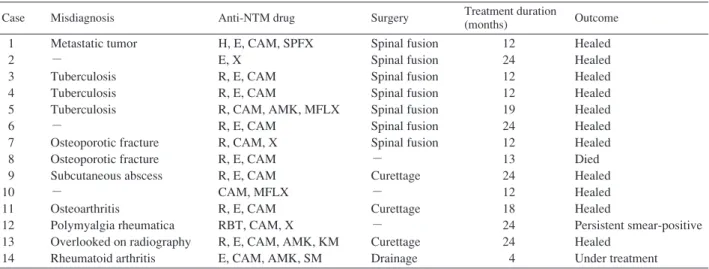
Case Misdiagnosis Anti-NTM drug Surgery Treatment duration
(months) Outcome 1 2 3 4 5 6 7 8 9 10 11 12 13 14 Metastatic tumor − Tuberculosis Tuberculosis Tuberculosis − Osteoporotic fracture Osteoporotic fracture Subcutaneous abscess − Osteoarthritis Polymyalgia rheumatica Overlooked on radiography Rheumatoid arthritis H, E, CAM, SPFX E, X R, E, CAM R, E, CAM R, CAM, AMK, MFLX R, E, CAM R, CAM, X R, E, CAM R, E, CAM CAM, MFLX R, E, CAM RBT, CAM, X R, E, CAM, AMK, KM E, CAM, AMK, SM Spinal fusion Spinal fusion Spinal fusion Spinal fusion Spinal fusion Spinal fusion Spinal fusion − Curettage − Curettage − Curettage Drainage 12 24 12 12 19 24 12 13 24 12 18 24 24 4 Healed Healed Healed Healed Healed Healed Healed Died Healed Healed Healed Persistent smear-positive Healed Under treatment
1). These misdiagnoses delayed the final diagnosis in 7 patients. Although 4 patients underwent retreatment of NTM infection, none of them had a relapse of osteoarticular NTM lesions. Six patients received chemotherapy with rifampicin (RFP), ethambutol (EB), and clarithromycin (CAM), and 8 patients received other macrolide-based therapies. Five pa-tients underwent surgical treatments in former hospitals, including laminectomy (n=1), anterior spinal fusion (n=2), curettage (n=1), and posterior fixation (n=1). Eight patients underwent surgical treatments (including salvage surgeries) in our hospital, including anterior spinal fusion (n=5), curettage (n=2), and drainage (n=1). Three patients were treated conservatively. The duration of chemotherapy ranged from 12 to 24 months (mean, 17.7 months), except in one patient who was undergoing current treatment. Healing of the osteoarticular lesion was judged by integrating findings such as bone formation surrounding osseous lesions on radiography, normalization of the intensity change in magnetic resonance imaging (MRI), closure of fistula in cutaneous lesions, and reduction of the inflammatory response. With regard to outcome, 9 patients achieved healing, 2 patients with a relapse were healed after retreatment, 1 patient was undergoing treatment, 1 patient had interrupted treatment (persistent smear-positive results), and 1 patient died during treatment.
Case presentation
(1) Case 13
A 65-year-old man developed right pretibial pain of un-known origin and visited a local clinic, lower leg radiography revealed no abnormal findings. Fourteen months previously, he developed NTM (M. intracellulare) infection of the right ulnar olecranon and scalp, and completed 6 months of treat-ment with RFP, EB, and CAM in a local hospital. He had been observed for 2 months for the right pretibial pain. Then, he visited a local hospital because of persistent pain. On MRI,
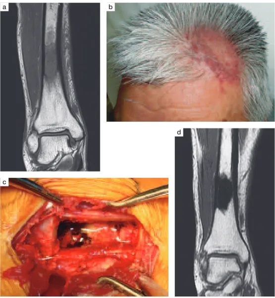
an abnormal intensity area in the diaphysis of the right tibia was detected (Fig. 1a). The lesion was suspected as NTM infection because of his medical history. He was then referred to our department. On physical examination, he had no fever and swelling or tenderness in his right pretibial area, but had reddish hives scattered over both his lower legs. Two large patches of eczema were on his scalp (Fig. 1b), which were relapsed lesions of the previous treatment. The right ulnar lesion did not relapse. Laboratory data showed a CRP level of 0.67 mg/dL, a sedimentation rate of 66 mm/h, a white blood cell count of 4900/μμL, a lymphocyte count of 1000/μμL, and a serum MAC antibody of 2.75 U/mL. Diagnostic imaging revealed no apparent pulmonary lesion, and a sputum test result was smear-negative for NTM. Three months after the onset of pain, curettage was performed by fenestration of the anterior tibial cortex. The lesion was completely confined to the bone marrow cavity, showing yellowish-translucent pus and sequestra (Fig. 1c). After sufficient surgical excision of the diseased tissue and saline irrigation, a defect in the bone marrow was filled with hydroxyapatite paste, and then the
Fig. 1 Case 13
a) Preoperative magnetic resonance image (MRI) showing the intramedullary lesion of the tibia. b) The scalp lesion at initial examination. c) Intraoperative photograph of the intramedullary lesion of the tibia. d) Postoperative MRI of the tibia showing disappearance of the inflammatory change (a low intensity area indicates the hydroxyapatite paste used to fill in the curetted area).
a
c
d b
bone cortex that was excised at first was inlaid in the same location. As M. intracellulare was detected in the intraoper-ative specimen, treatment with RFP, EB, and CAM was restarted in combination with amikacin (AMK) because of the recurrence. AMK was continued for 2 months after the surgery until switching to kanamycin because of the increasing creatinine level. The cutaneous lesions in the scalp and legs were gradually improved after the restart of chemotherapy, the CRP level decreased and turned negative 3 months after the surgery, bone union of the surgical site was achieved 6 months after the surgery, and MRI revealed normalization of the bone marrow intensity 1 year after the surgery (Fig. 1d). Chemotherapy was completed 24 months after the surgery, and so far, no relapse of the NTM infection has been detected. (2) Case 14
A 64-year-old woman developed swelling of her right wrist,
right great toe metatarsophalangeal (MTP) joint, left second toe MTP joint, right ankle joint, and left scapular region 5 years prior. She had histories of interstitial pneumonia (IP) and PMR since she was 40 years old, which were treated continuously with prednisolone. Eight years previously, she was diagnosed with seronegative RA, which caused the right wrist pain and swelling. In order to treat the multiple joint swelling that had developed 5 years previously, which was considered as an exacerbation of the RA, methotrexate, inflix-imab, tocilizumab, and other antirheumatic drugs were used, but no improvement was observed. The swollen left second toe MTP joint and left scapular region were punctured sev-eral times for detection of bacteria, all of which turned out negative results. However, culture for Mycobacterium was not ordered at all. Consequently, the left second toe MTP joint formed a fistula with continuous serous discharge. The
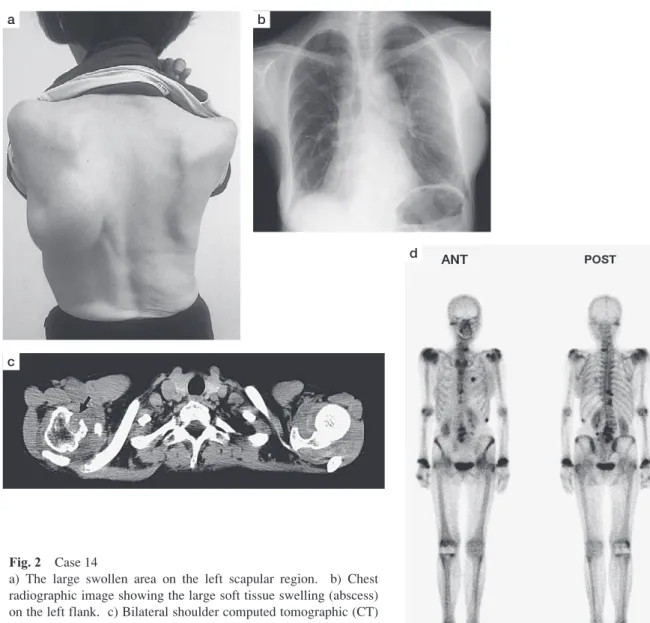
Fig. 2 Case 14
a) The large swollen area on the left scapular region. b) Chest radiographic image showing the large soft tissue swelling (abscess) on the left flank. c) Bilateral shoulder computed tomographic (CT) image showing the swelling of both shoulders and bony destruction of right humeral head (arrow). d) Technetium bone scan showing multiple hot uptake.
a
c
d b
swelling in the left scapular region gradually increased and became a fist-size mass (Fig. 2a). The swollen joint was punc-tured because of tense feeling increased. Finally, mycobacte-rial examination of the aspirated fluid was performed, and the results obtained were positive for M. avium. Thereafter, she was referred to our department. On physical examination, she had no fever, laboratory data showed a CRP level of 1.92 mg/dL, a sedimentation rate of 28 mm/h, a white blood cell count of 8080/μμL, a lymphocyte count of 300/μμL, and a serum MAC antibody of 0.74 U/mL. Diagnostic imaging revealed no apparent pulmonary NTM lesion, but findings showed stable IP (Fig. 2b) and the result of the sputum test was smear-negative for NTM. Computed tomography (CT) revealed obvious bone destruction in the right humeral head (Fig. 2c). Technetium bone scintigraphy revealed many hot uptakes in various locations, including visibly swollen joints (Fig. 2d). M. avium was detected with aspirated fluid from the right wrist, right great toe MTP joint, right ankle joint,
and both shoulder joints. Therefore, she was diagnosed with disseminated MAC infection. Treatment with EB, CAM, and AMK was started. One month after the start of the chemo-therapy, drainage of the left scapular region subcutaneous abscess was performed, thereby 400 mL of yellowish pus with a lot of debris was drained. The abscess was a pure sub-cutaneous abscess rather than a gravitation abscess from the osteoarticular lesion. AMK was discontinued after 4 weeks of administration and switched to streptomycin. The patient’s current treatment is ongoing.
Discussion
Osteoarticular NTM infection mostly develops as a part of disseminated infection and has been known more than a century ago. M. avium, as is clear from its nomenclature, was studied initially as a pathogen in birds. Koch reported on NTM infection evaluated from autopsy samples from a vast number and types of birds in 19073). The lesions in most of
the autopsy cases spread over multiple organs (i.e., dissem-inated infection). As bones and joints were cited as locations of infection, Koch’s report is probably the first report of osteoarthritis caused by NTM. However, NTM as a human pathogen has been described more recently. In 1943, human pulmonary NTM infection was reported in detail for the first time by Feldman4). Human disseminated NTM osteomyelitis
has been reported since the 1950s5) 6). It used to commonly
develop in patients with immunodeficiency condition7) 8), but
an increasing number of patients showed no such back-ground9)_11). In this study, most of the patients were aged or
immunosuppressed but not severely immunocompromised, whereas some of the patients were healthy individuals with no relevant medical history.
In this manner, cases of NTM infection have been emerg-ing gradually in recent years, although its cause is not well understood. In association with its increase, the incidence of osteoarticular NTM infection is also considered to be increasing, but its prevalence remains unclear. McHenry reported a long-term outcome in patients with vertebral osteomyelitis who attended 7 facilities of the region from 1950 to 1994, wherein NTM was detected as a causative organism in 1 (0.4%) of 253 patients12). Given that the data
are limited up to 1994 and the incidence of NTM infection in the United States shows an increasing trend13), the incidence
of osteoarticular lesions should have increased in recent years. Considering that the estimated incidence of NTM infection in New York during the period 2000_2003 was 2.7 cases per 100,000 individuals14) and that in Japan in
2001 was 5.9 cases per 100,000 individuals1), the incidence
rate of NTM infection in our department during the last 5 years (3.9%) is somewhat high. This is probably because our hospital has specialized in mycobacterial infection for years. Furthermore, our region is ranked as having the high-est incidence of tuberculosis in Japan for many years. Most patients in this case series presented disseminated infection. Two patients developed 1 lesion, 3 patients devel-oped 2 lesions, and the remaining 9 patients develdevel-oped three or more lesions. This is clearly a high incidence rate of disseminated lesions compared with osteoarticular infection with Mycobacterium tuberculosis. Spinal lesions were charac-teristically extensive with 4.4 vertebrae affected on average. In addition, a high rate of cervical involvement was also a notable feature (4 [33%] of the 12 patients). The onsets were insidious, and the initial symptoms were relatively mild con-sidering the extent of the lesions, which consequently lead to diagnostic delays. Thus, a patient with disseminated NTM infection should undergo whole-body examination for possible lesions in other sites, including the spine or other osteoarticular regions. In particular, osteoarticular NTM infec-tion in a patient with a collagen disease, such as case 14, is difficult to diagnose, unless bacterial examination by joint puncture is performed. Kanik reported a patient with NTM arthritis who had been treated for seronegative RA for 2.5
years until final diagnosis based on synovial fluid culture find-ings15). Recently, use of immunosuppressive agents such as
biologics to treat such patients has increased. Care should be taken for detecting NTM infection in such patients, as routine screening for tuberculosis is not useful for such cases. Cutaneous lesion was also a distinctive feature (6 [42.9%] of 14 patients). Although NTM species such as M. fortuitum or M. abscessus are commonly detected in the cutaneous lesion, most NTM species can be causative organisms16) 17).
In spite of the rarity of cutaneous tuberculosis, bone and skin development in disseminated NTM infection is one of the common features7) 18)_20). Horsburgh reported 17 patients (46
%) with cutaneous lesions and 5 patients (14%) with osseous lesions in their study of 37 patients with disseminated NTM infection21). Ingram reported 9 patients (17%) with cutaneous
lesions and 40 patients (74%) with osseous lesions in their study of 54 patients with disseminated NTM infection22). The
etiology of localization to the skin and bone in disseminated NTM infection is still unclear. However, Arend speculated that the relative scarcity of macrophages in normal skin and bone tissue could be an underlying part of the tissue-specific susceptibility23).
Recently, interferon (IFN)-γ productivity or autoantibody against IFN-γ has been suspected as a cause of the develop-ment of disseminated NTM infection23)_25). Meanwhile, gene
mutations as the cause of IFN-γγ deficiency have also been reported23) 26). In this study, 4 (44.4%) of 9 patients who had
undergone a QFT test showed positive control failure, suggest-ing the presence of immunoresponse deficiency. Okimoto studied the positive response rate to QFT in patients with MAC infection, and 4.2% of the patients showed positive control failure27), which is a much lower rate than the one in
our study. However, the number of patients in this study was too small, and normal responses to positive control were ob-tained in the remaining 5 patients. Thus, reexamination of this issue with more subjects in the future is necessary. In our literature search, we found no well-documented case series that describes about treatment of osteoarticular NTM infection in recent years. Aberg reported 48 HIV patients with disseminated MAC infection who were treated with 1 year of chemotherapy (including 2 patients with spinal lesion), of whom only 1 patient developed localized osteomyelitis after withdrawal of the therapy28). Marchevsky reported 8 patients
with NTM osteomyelitis, of whom 6 responded well to sur-gical excision alone without recurrence and 2 developed MAC infection and were treated with a combination of sur-gery and chemotherapy29). However, none of the patients
had disseminated NTM infection. In general, surgical out-come of osteoarticular NTM infection has been reported to be favorable17) 30), as is shown in this study. Surgery should
be performed as long as the general condition of the patient permits. Chemotherapy with adequate drug combination and enough duration was considered mandatory for the treatment of osteoarticular NTM infection in its disseminated form. For
example, case 13 had a relapse after the 6-month treatment, and case 9 developed subcutaneous abscesses repeatedly even at 1 year after the start of the chemotherapy, showing a discrepancy with well healed spinal lesions. In addition, if we exclude the 5 patients who received retreatments (cases 5 and 13), the patient whose treatment was interrupted (case 12), the patient who is receiving treatment (case 14), and the patient whose lesions healed recently (case 9), the lesions healed because of the initial treatment in only four out of 9 patients with disseminated NTM infection (case 1, 2, 4, and 7). Furthermore, considering that disseminated NTM infection is generally refractory, the clinical outcomes noted in this study should be carefully interpreted because recurrence may occur subsequently in these four patients. Therefore, the duration of chemotherapy for osteoarticular NTM infection should be carefully determined by considering if all lesions in the body have been healed. In addition, we should always consider the possibility of a relapse even for cases of successful treatment.
Conclusion
The incidence of osteoarticular NTM infection has shown an increasing trend. Thus, it is necessary to keep in mind that it could be complicated with pulmonary NTM infection, as a part of the disseminated infection. The spine was the most common and extensively involved site. Cutaneous lesions, including subcutaneous abscess, were also characteristic fea-tures. Considering the difficulties in diagnosis and treatment, as well as the increasing trend of the overall incidence of NTM infection, awareness of NTM as a causative organism of osteoarticular infection is important.
Acknowledgement
We express cordial gratitude to Dr. Kazuhiko Imoto, Director of Orthopedic Surgery in our hospital, for giving us the opportunity to present this paper.
Confl ict of interest
The authors declare that there is no conflict of interest related to this article.
References
1 ) Ogawa K, Sano C: Strategies for Mycobacterium avium complex infection control in Japan: how do they improve the present situation? Kekkaku. 2013 ; 88 : 355_371. 2 ) Kitada S, Kobayashi K, Ichiyama S, et al.: Serodiagnosis
of Mycobacterium avium-complex pulmonary disease using an enzyme immunoassay kit. Am J Respir Crit Care Med. 2008 ; 177 : 793_797.
3 ) Koch M, Rabinowitsch L: Die tuberkulose der vogel und ihre beziehungen zur sangertiertuberkulose. Virchows Arch Pathol Anat. 1907 ; 190 : 246_316.
4 ) Feldman WH,Davies R,Moses HE,et al.: An unusual mycobacterium isolated from sputum of a man suffering from pulmonary disease of long duration. Am Rev Tuberc.
1943 ; 48 : 82_93.
5 ) Van der Hoeven LH, Rutten FJ, Van der Sar A: An unusual acid-fast bacillus causing systemic disease and death in a child; with special reference to disseminated osteomyelitis and intracellular parasitism. Am J Clin Pathol. 1958 ; 29 : 433_448.
6 ) Weed LA, Karlson AG, Ivins JC, et al.: Recurring migratory chronic osteomyelitis associated with saprophytic acid-fast bacilli: report of a case of 10 years’ duration apparently cured by surgery. Proc Staff Meet Mayo Clin. 1956 ; 31 : 238_246. 7 ) Chicoine L, Lapointe N, Simoneau R, et al.: Anonymous
mycobacterial infection causing disseminated osteomyelitis and skin lesions. Can Med Assoc J. 1968 ; 98 : 1059_1062. 8 ) Lincoln E, Gilbert L: Disease in children due to
mycobac-teria other than Mycobacterium tuberculosis. Am Rev Respir Dis. 1972 ; 105 : 683_714.
9 ) Igram CM, Petrie SG, Harris MB: Atypical mycobacterial vertebral osteomyelitis in an immunocompetent patient. Orthopedics. 1997 ; 20 : 163_166.
10) Kaneshiro K, Takatsuki K, Kanamori K: A healthy adult with disseminated nontuberculous mycobacterial infection with multiple bone lesions. Kekkaku. 2015 ; 90 : 457_461. 11) Myojo M, Fujiuchi S, Matsumoto H, et al.: Disseminated
Mycobacterium avium complex (DMAC) in an immuno-competent adult. Int J Tuberc Lung Dis. 2003 ; 7 : 498_501. 12) McHenry MC, Easley KA, Locker G: Vertebral osteo-myelitis: long-term outcome for 253 patients from seven Cleveland-area hospitals. Clin Infect Dis. 2002 ; 34 : 1342_1350.
13) Khan K, Wang J, Marras TK: Nontuberculous mycobacterial sensitization in the United States: national trends over three decades. Am J Respir Crit Care Med. 2007 ; 176 : 306_313. 14) Bodle EE, Cunningham JA, Della-Latta P, et al.: Epidemi-ology of nontuberculous mycobacteria in patients without HIV infection, New York City. Emerg Infect Dis. 2008 ; 14 : 390_396.
15) Kanik KS, Greenwald DP: Mycobacterium avium/Myco-bacterium intracellulare complex-associated arthritis mas-querading as a seronegative rheumatoid arthritis. J Clin Rheumatol. 2000 ; 6 : 154_157.
16) Falkinham JO 3rd: Epidemiology of infection by nontuber-culous mycobacteria. Clin Microbiol Rev. 1996 ; 9 : 177_215. 17) Piersimoni C, Scarparo C: Extrapulmonary infections asso-ciated with nontuberculous mycobacteria in immunocom-petent persons. Emerg Infect Dis. 2009 ; 15 : 1351_1358. 18) Hofer M, Hirschel B, Kirschner P, et al.: Disseminated
osteomyelitis from Mycobacterium ulcerans after a snake-bite. N Engl J Med. 1993 ; 328 : 1007_1009.
19) Hsueh PR, Hsiue TR, Jarn JJ, et al.: Disseminated infection due to Mycobacterium scrofulaceum in an immunocompe-tent host. Clin Infect Dis. 1996 ; 22 : 159_161.
20) Otaki Y, Nakanishi T, Nanami M: A rare combination of sites of involvement by Mycobacterium intracellulare in a hemodialysis patient: multifocal synovitis, spondylitis, and
multiple skin lesions. Nephron. 2002 ; 92 : 730_734. 21) Horsburgh CR, Mason UG, Farhi DC, et al.: Disseminated
infection with Mycobacterium avium-intracellulare. A report of 13 patients and a review of the literature. Medicine (Baltimore). 1985 ; 64 : 36_48.
22) Ingram CW, Tamner DC, Durack DT, et al.: Disseminated infection with rapidly growing mycobacteria. Clin Infect Dis. 1993 ; 16 : 463_471.
23) Arend SM, Janssen R, Gosen JJ, et al.: Multifocal osteo-myelitis caused by nontuberculous mycobacteria in patients with a genetic defect of the interferon-γγ receptor. Neth J Med. 2001 ; 59 : 140_151.
24) Marazzi MG, Chapgier A, Defilippi AC, et al.: Dissemi-nated Mycobacterium scrofulaceum infection in a child with interferon-gamma receptor 1 deficiency. Int J Infect Dis. 2010 ; 14 : e167_170.
25) Sakagami T: Disseminated nontuberculous mycobacteriosis that is positive for neutralizing anti-interferon-gamma auto-antibodies: a new disease concept based on host factors. Kekkaku. 2015 ; 90 : 561_564.
26) Hirata O, Okada S, Tsumura M, et al.: Heterozygosity for the Y701C STAT1 mutation in a multiplex kindred with multifocal osteomyelitis. Haematologica. 2013 ; 98 : 1641_ 1649.
27) Okimoto N, Nanba F, Kurihara T, et al.: The positive response rate with QuantiFERON-TB GOLD In-Tube in patients with Mycobacterium avium complex. Kekkaku. 2012 ; 87 : 337_339.
28) Aberg JA, Williams PL, Liu T, et al.: A study of discontinu-ing maintenance therapy in human immunodeficiency virus-infected subjects with disseminated Mycobacterium avium complex: AIDS Clinical Trial Group 393 Study Team. J Infect Dis. 2003 ; 187 : 1046_1052.
29) Marchevsky A, Damsker B, Green S, et al.: The clinico-pathological spectrum of non-tuberculous mycobacterial osteoarticular infections. J Bone Joint Surg Am. 1985 ; 7 : 925_929.
30) Shimizu H, Mizuno Y, Nakamura I, et al.: Vertebral osteo-myelitis caused by non-tuberculous mycobacteria: case re-ports and review. J Infect Chemother. 2013 ; 19 : 972_977.