Acta Med. Nagasaki 41: 43-47
Recombination Activating Gene (RAG)-1 and 2 Encoding Proteins
Expressed by the Baculovirus System
Ryoko ARAKI1),2), Mitsuo KAKU1),3), Masahiro ITOH2), Kiyohiro HAMATANI2), Toshiaki USUI1), Masumi ABE2) 1) Department of Laboratory Medicine, Nagasaki University School of Medicine, 1-7-1 Sakamoto, Nagasaki 852, Japan
2) Cell Biology Laboratory, Department of Radiobiology, Radiation Effects Research Foundation, 1-8-6 Nakagawa, Nagasaki 850, Japan 3) Department of Microbiology, St. Marianna University School of Medicine, Kawasaki 216, Japan (Present address).
We have been attempting to obtain mouse recombination activating gene-1 (RAG-1) and RAG-2 protein for biochemi- cal analyses. First of all, we obtained truncated products of these genes expressed and purified using the E. coli expres- sion system and then established the polyclonal and mono- clonal antibodies by means of E. coli expressed peptides as antigens. Subsequently, whole RAG-1 and RAG-2 gene products were expressed the baculovirus expression system.
Since it has been difficult to achieve the significant gene expression of full-lenght cDNA, we employed the glutathione S-transferase (GST)-fused gene-expression system which facilitated the massive expression of gene products. This system was also advantageous in that we could detect the expressed protein molecules not only with anti-RAG anti- body but also with anti-GST antibody.
Introduction
The diversity of immunoglobulins and T-cell receptors for antigens is created by a site-specific recombination of their genes during lymphocyte development, so-called . V (D) J recombination (Tonegawa, 1983). Many researchers have recognized V (D) J recombinase to be a complex and have attempted to identify one of the components of V (D) J recombinase (Aguilera et al., 1987 ; Halligan and Desiderio, 1987 ; Matsunami et al., 1989).
Terminal deoxynucleotidyl transferase (TdT) was shown to be directly involved in V (D) J recombinase, a template-independent DNA polymerase. The function of TdT is the N nucleotide insertion at the recombined sites.
The TdT activity, however, is not essential for the recom- bination step because occurrence of V (D) J recombination without TdT molecules has been detected by the analysis of V (D) J recombination activity-implementing gene trans- fection experiments and TdT knockout mice (Kallenbach et al., 1992 ; Gilf illan et al., 1993 ; Komori et al., 1993).
Melvin Bosma et al. reported in 1983 that the SCID mouse exhibits an aberration in V (D) J recombination during lymphocyte development and then produces aber- rant recombined products (Bosma et al., 1983 ; Schuler et
al., 1986). Recently, the catalytic subunit of DNA depend- ent protein kinase (DNA-PKcs) was proposed as a candi- date for the SCID factor (Blunt et al., 1995 ; Kirchgessher et al., 1995). Further analyses require for understanding the role of the DNA-PK activity to V (D) J recombina- tion.
Recombination activating gene (RAG)-1 and RAG-2 were identified as genes possessing an ability to induce lymphocyte-specific nonhomologous recombination : V (D) J recombination activity into the fibroblast cell line NIH/3T3 (Schatz and Baltimore, 1988 ; Schatz et al., 1989 ; Oettinger et al., 1990). Their vital role in V (D) J recombination in both B and T cells was confirmed by study of RAG-1 and RAG-2 gene knock-out mouse (Mombaerts et al., 1992 ; Shinkai et al., 1992). Recently, the functions of RAG-1 and RAG-2 products are going to be disclosed with truncated RAG-1 and RAG-2 products which exhibits soluble characteristics (McBlane et al., 1995 ; Sadof sky et al., 1995 ; van Gent et al., 1995).
Biochemical analyses of RAG-1 and RAG-2 products seem to be essential for an understanding of the V (D) J recombination machinery. However, the instability and sparsity of RAG-1 and RAG-2 makes their detection in vivo and their purification difficult (Lin and Desiderio, 1993). Furthermore, their expression in other species has also been difficult so far (Gellert, 1992 ; Oettinger, 1992).
To overcome these problems, therefore, we attempted to obtain a large amount of RAG products using the baculovirus system.In this study,we succeeded in mass- expression of RAG-1 and RAG-2 products using the baculovirus expression system with GST-fused RAG genes.
Materials and methods
1. Expression of truncated RAG proteins in E.coli.
M6-BSK+ and MR2-BSK+ contain mouse full-length RAG-1 cDNA and mouse full-length RAG-2 cDNA, respec- tively. Each cDNA fragment is cloned at the Not I site of pBluescript SK+ (Stratagene, La Jolla, Ca., U. S. A).
Amino acid number of the truncated products were shown in figure 1. M6-BSK+ Pvu II -Sac I fragment (216 bp) prepared from M6-BSK+and Pvu II -Hinc II fragment (442 bp) prepared from MR2-BSK+ were transferred to pGEX- 3X (Pharmacia P-L Biochemicals Inc., Milwaukee, Wiscon- sin, U. S. A.). Following the transfection of these vectors into E. coli DH5 a, the cells were grown in LB broth and fusion proteins were purified with anti-GST affinity chromatography to the method reported previously (Smith and Johnson, 1988) .
2. Preparation of antibodies
Polyclonal antibody was prepared by immunizing New Zealand white rabbits with affinity-purified truncated RAG proteins and monoclonal antibodies were prepared by immunizing Dahl R rats with the same antigens according to the method reported previously (Yoshida et al., 1988).
Goat anti-GST polyclonal antibody was purchased from Pharmacia.
3. Cell and virus
Spodoptera frugiperda insect cells (Sf9) and Autographa californica nuclear polyhedrosis virus (AcRP23. LacZ) containing the LacZ gene were used (Possee and Howard, 1987). The insect cells were cultured at 27°C and grown in Grace's medium (Gibco Laboratories, Grand Island, N. Y., U. S. A.) supplemented with 10% fetal bovine serum (FBS) and 0.26% Bacto tryptose broth (Difco Labora- tories, Detroit, Mich., U. S. A.).
4. Expression of recombinant RAG-1 and RAG-2 proteins in insect cells
pAcYM1 was used for baculovirus expression vector (Matsuura et al., 1987). pAcRAG-1-12 was constructed by inserting a Stul-SspI fragment of M6-BSK+ into the Sma I site of pAcYM1. pAcRAG-2-20 was constructed by inserting a Pst I (162)-EcoRV (1963) fragment of MR2- BSK+ into the Sma I site of pAcYM1. The construction of pAcGST-RAG-1, pAcGST-RAG-2, and pAcGST is shown in Figure 2. Virus vector pAc23. LacZ (10-20 ng) linearized by restriction enzyme Eco 811 was mixed with the circular form of recombinant plasmid vectors pAcRAG-1-12, pAcRAG-2-20, pAcGST-RAG-1, pAcGST-RAG-2 or pAc- GST (1 u g). After adding distilled water up to 8 U 1, an equal volume of 50% (v/v) lipofectin (BRL, Gaithersburg, MD, U. S. A.) was added and incubated at room tempera- ture for 15 min. The mixture solution was poured over 1 X 10⁶ of Sf9 cell in a 35-mm dish in which the medium had been replaced by serum-free medium beforehand. After 2 days of culture at 27C, a plaque assay was carried out with the culture supernatant. The plaque assay procedure is as follows : The cultured supernatant of transfectants was diluted with culture medium and inoculated into 0.8
X 106 cells of Sf9 cell without medium. After 1 h of incuba- tion at 271C, the supernatant was discarded. Two millili- ters of 1 % agar and 1 mL of medium were added. Follow- ing 48-72h-incubation at 27°, one milliliter of 0.01% (w/
v) neutral red-0.04% (w/v) X-gal in PBS was added. After
1 810 883 1040 (length)
1 1 RAG
-1 74 a.a.
(HPR1 homologous region)
1 278 425 527
1 1
RAG-2 / 147 a .a.
(acidic stretch)
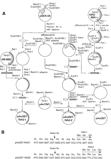
Fig. 1 Expressed regions of RAG-1 and RAG-2 genes using the E. coli system. Open rectangles denote the open reading frames. Areas marked with diagonal lines indicate the regions for expression. The numbers above the genes indicate the amino-acid number.
BamHI SmaI
Eco0109IEcoR i E I Not I
co0109I / PstI
~ RAG
pG GEX-3X MR2-
BSK+Pst I
Not I
BamH I Pst I
Klenow fill in PBluescript11 Pet I partial
self ligation Pst I Pst I EcoR V
EcoO109 I pBluescript 11 Sma I EcoR V
Eco0109 I JBamH I., Eco0109 I JEcoR I linker EcoR I -Pst I
~- BamH I GST adaptor EcoR I
`Eco0109 I Eco0109 I I EcoR I pGEX-3X Pst I /Bgl I Sma I ApaI GST RFC, - EcoR I Sma I Eco0109I A EcoR V
pa I RAG1 EcoR
M6 BSK Eco01091 BamH I
Bgl I Ssp I EcoOl09 I EcoR I
IT4 DNA IEcoRI Ssp I ~
, polymerase Apa Apa I -BamH I I adaptor
BamH I Sma I Eco0109I ~>\ BamH I \\ /EcoR I
EcoR I EcoR I
Eco0109 I BamH I / BamH I
Apa I \
BamH I BarnH I BamH I BamH I I Apa I Apa I -BamH I
~._._~. E~ BamH I adaptor EcoR I
BarnH I Sma I BamH I self ligation I BamH I
pAcYM1 BamH I
BamH I BamH I BamH I ~/ BamH ]
\ \ I \
pAc1BarnH ST pAcGST
pAcGST /
RAG-1 RA-2
- BamH I
B Factor Xa RAG-1
Met Ale Ala
lie Glu Gly Arg Gly Ile Asp Pro Leu Ala Ala
pAcGST-RAG1 ATC GAA GGT CGT GGG ATC GAT CCC CTG GCT GCC
Factor Xa RAG-2
l Met Ser Leu Gin
Ile Glu Gly Arg I Gly Ile Asp Pro Pro Glu Phe Leu Gln
pAcGST-RAG2 ATC GAA GGT CGT GGG ATC GAT CCC CCG GAA TTC CTG CAG
Fig. 2 The construction of the GST-fused RAG-1 and RAG-2 protein-expression vectors for the baculovirus expression system. (A) The RAG-1 gene is denoted by the areas marked with diagonal lines and the RAG-2 gene by the cross-hatched areas. Open boxes denote GST genes. M6-BSK and MR2-BSK contain full-length cDNA segments of RAG-1 and RAG-2, respectively. (B) The amino-acid sequence of the GST-RAG joining regions. The arrows indicate the digestion site by Factor Xa. The amino-acid sequences are shown over the nucleotide sequence.
a 24-h incubation, white plaques were picked up. After three rounds of plaque assay, single plaques were isolated.
The method for large-scale gene-expression with recombi- nant virus is as follows : After infection of recombinant virus at a multiplicity of 5PFU/cell, the cells were cul- tured for 48 h and then collected.
5. Western blot analysis
Western blot was carried out as described previously (Towbin et al., 1979). For secondary reagents, horseradish peroxidase (HRP) labelled protein-A, protein-G and anti-rat IgM (Zymed, So. San Francisco, CA., U. S. A.) were used. The molecules were visualized with the ECL chemiluminescence system (Amersham, Buckinghamshire, U. K.).
6. Preparation of synthesized recombinant RAG-1 and RAG-2 proteins fraction.
The synthesized protein fraction was prepared as de- scribed previously (Cockerill and Garrard, 1986). Briefly, the protocol is as follows : 1 X 10⁸ cells were washed once in phosphate-buffered saline, suspended in two to four volumes of RSB [10 mM NaCl-3 MM MgC12-10 mM Tris HC1 (pH 7.4)-0.5 mM PMSF] and put on ice for 10 min.
Suspended cells were homogenized using the Dounce homo- genizer B pestle with 30 strokes. A pellet was washed twice in RSB-0.25 M sucrose. After suspension in RSB-2 M sucrose and centrifugation at 103700Xg at 4 C for 30 min, the pellet was washed three times in RSB-0.25 M sucrose.
Nuclei were suspended in 100 u L of RSB-0.25 M sucrose-1 mM CaCl2 and digested with DNase I (Takara Co. Ltd., Kyoto, Japan) at a final concentration of 100 u g/ml at 23'C for 2 h. After centrifugation for 10 min at 750 X g at 40C, the pellets were suspended in 2 ml of RSB-0.25 M sucrose, and 2 ml of 4 M NaC1-20 mM EDTA-20 mM Tris HCl (pH 7.4) was added and the same solutions were utilized except for the NaC1 concentration at the protein elution experiment to determine to determine the optimum condition. After incubation (15 min, on ice) and centrifu- gation (1500 X g, 15 min), the pellets were extracted twice by suspension in 1 ml of 2 M NaCl-10 mM Tris HCl (pH 7.4)-0.5 mM PMSF-0.25 mg/ml BSA. The pellets collected by centrifugation (4500 X g, 15 min) were washed with RSB-0.25 M sucrose-0.25 mg/ml BSA. Extracted fractions were stored at -20'C after adding an equal volume of 100% glycerol. Digestion of the extracted fractions with Factor Xa was performed according to the manufacturer's recommendations (Denzyme APS, Aarhus C., Denmark).
Results and discussion
The biochemical analyses of the RAG proteins remain to be elucidated because of following three practical reasons ; first, the absence of high quality antibodies against RAG proteins, secondly, the extremely low quantity of RAG
proteins in vivo, and thirdly, the difficulty of heterologous
expression of RAG genes.
In this study, we prepared polyclonal antibodies and monoclonal antibodies designated as MR-1-1, MR-1-100 and MR-1-130 against RAG-1, and MR-2-10, MR-2-12 and MR-2-32 against RAG-2, repectively. All of these antibod- ies exactly detected full-length nonfused RAG proteins expressed in the insect cells (Figure 3). At the initial step of our study, however, another difficulty was encountered in determining conclusively whether the molecules ex- pressed by the baculovirus of E. coli expression systems were truely RAG morecules, because the anti-RAG anti- bodies did not clearly detect the mammalian RAG mole- cules in vivo (data not shown). This was the greatest obstacle we faced in our experiments.
To overcome this problem, we employed GST-fused RAG proteins in the baculovirus expression system to detect the products only by anti-RAG antibodies but also by anti- GST anitibodies. We also expected a change in the molecu- lar weight of the target molecules by the digestion of the fused products with restriction endopeptidase Factor Xa of which recognition sequence exists between GST and RAG peptides.
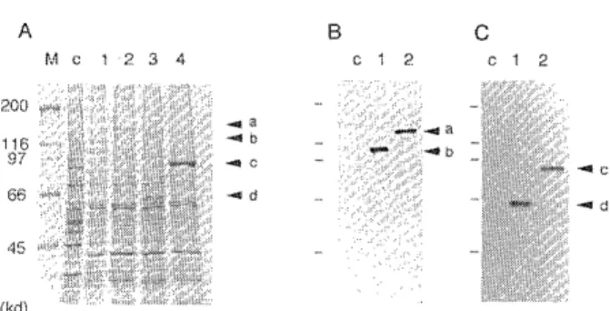
Fig. 3 Expression of the recombinant proteins in insect cells. Sizes are indicated in kilodaltons (kd). (A) Whole-cell extracts of RAG-1-or RAG-2-infected insect cells and GST- fused RAG-1-or RAG-2-infected insect cells were analyzed.
Each lane contains 7.5 X 10" cells. Proteins were separated by SDS-PAGE and stained with Coomassie brilliant blue.
Non-infected Sf9 (lane c) ; RAG-1-infected Sf9 (lane 1) ; GST-fused RAG-1-infected Sf9 (lane 2) ; RAG-2-infected Sf9 (lane 3) ; GST-fused RAG-2-infected Sf9 (lane 4). (B) The expression of RAG-1 proteins was analyzed by Western blot.
Anti-RAG-1 monoclonal antibody MR-1-100 was used for detection. Whole-cell extract prepared from 3.25 X 10" cells was applied on each lane. The loaded samples on each lane are as follows : non-infected Sf9 (lane c) ; RAG-1-infected Sf9 (lane 1) ; GST-fused RAG-1-infected Sf9 (lane 2). (C) The expression of RAG-2 proteins was analyzed by Western blot.Sizes are indicated in kilodaltons (kd). Anti-RAG-2 monoclonal antibody MR-2-10 was used. Whole-cell extract prepared from 3.25 X 10" cells was applied on each lane. The loaded samples on each lane are as follows : non-infected Sf9 (lane c) ; RAG-2-infected Sf9 (lane 1) ; GST-fused RAG-2- infected Sf9 (lane 2). The arrows [a], [b], [c], and [d]
indicate GST-fused RAG-1, RAG-1, GST-fused RAG-2 and RAG-2 and RAG-2, respectively.
The nonfused forms of RAG-1 and RAG-2 proteins were detected faintly by Coomassie staining (Figure 3A), while
clear bands were demonstratable in Western blot analysis
(Figure 3B and C), As shown in Figure 3, RAG-1 molecule
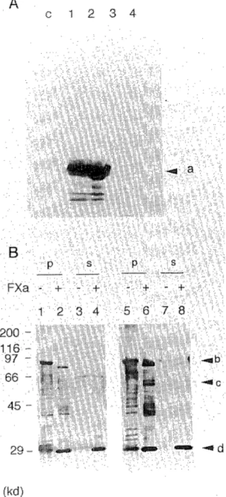
Fig. 4 Confirmation of the recombinant products by Factor Xa digestion. (A) The expression of GST-protein in Sf9 was analyzed with goat anti-GST polyclonal antibody (pharm- acia). The loaded samples of lane c were prepared from whole-cell extract of non-infected Sf9. The other loaded samples were prepared from GST-infected Sf9. The preparing condition is as follows ; whole-cell extract (lane 1) ; extract from cytoplasm (lane 2) ; 2.0 M NaCl eluate (lane 3) ; residual pellet-fraction after 2.0 M NaCl eluate (lene 4).
Samples of lane c and 1 were prepared from 2.5 X 10⁵ cells and samples of alnes 2, 3 and 4 were prepared from extraction of 2.5 X 10⁵ cells. The arrow [a] indicates GST. (B) The GST- fused RAG-2-product was digested with Factor Xa. Anti- GST polyclonal antibody and anti-RAG-2 polyclonal anti- bod were used to detect the sample lanes 1-4 and lanes 5-8, respectively. The residual pellet-fractions of 2.0 M NaCI extraction were used for Factor Xa digestion were prepared by centrifugation. Mock experiment samples without Factor Xa digestion were also loaded as controls : pellet-fractions [P] (lane 1 and 5) ; supernatant-fractions [S] (lane 3 and 7).
Sizes are indicated in kilodalton (kd). The arrows [b], [c]
and [d] indicate GST-fused RAG-2, RAG-2 divided by Factor Xa and GST divided by Factor Xa, respectively.
was detected at approximately 125 kd and RAG-2 molecule at 65 kd. The morecular weight of RAG-1 molecule is equal to that predicted by amino-acid sequence. However, that of RAG-2 was higher than that predicted (56 kd). The in- crease in molecular weight of RAG-2 was also observed in the GST-fused RAG-2 molecule, as described below. In the case of RAG-1, we cannot definitely conclude that the RAG-1 molecule does not undergo a molecular-weight change because the resolution of SDS-PAGE is not suffi- cient at the region where the RAG-1 molecule is detected.
The moleculer weight of GST-fused RAG-1 and GST-fused RAG-2 (155 kd and 94 kd,respectively) was higher than that predicted (148 kd and 85 kd, respectively).
The quantity expressed GST-RAG-1 and GST-RAG-2 was estimated on the basis of SDS-PAGE pattern to be about 130 u g/108 cells 1 mg/108 cells, respectively.
These fused proteins could be digested into the expected molecular size by restriction endopeptidase Factor Xa.
Factor Xa recognizes the amino-acid sequence Ile-Glu-Gly- Arg and digests the C-terminal side of Arg. Both of our GST-fused RAG genes contain the DNA suquence encoding the Factor Xa recognition sequence at the joining region between GST and RAG genes. In order to digest the recombinant protein, we have been attempting to obtain purified products. When cells infected with GST-fused products of RAG-1 and RAG-2 were fractionated with 2M NaCI solution, the products existed in insoluble nuclear fractions.Using these fractions, each half of the molecule divided with Factor Xa was detected by anti-RAG antibod- ies and anti-GST antibody, respectively (Figure 4B). The molecular weight of divided molecules was appropriate. It is confirmed that the commercially available anti-GST antibody could detect the GST region expressed in insect cells (Figure 4A). These observations show that the molecules detected in our system are definitely GST-RAG fused proteins.
The expression of a large number of genes has been reported to date in studies using the baculovirus expression system (Luckow and Summers, 1988). Unlike the E. coli system, this baculovirus system allows proteins to main- tain their original function. Although our RAG-1 and RAG-2 products were detected in an insoluble fraction of the nuclei, they will undoubtedly prove to be useful for research on V (D) J recombinase when we succeed in making them soluble.
Ackknowledgements
We thank Dr. K. Yokoro and Prof. S. Kamihira for encouragement.
We also thank Dr. Shiku for providing NS-1 cell and advice in preparing for anti-RAG antibodies, and Dr. Y.
Matsuura for providing pAcYM1, AcRP23. LacZ, Sf9 and advice, Dr. D. Baltimore for M6-BSK+ MR2-BSK+, Dr. A.
Sarai for computer analysis of RAG-1 and RAG-2 pro- teins, Dr. T. Maita for determination of amino-acid sequence. We are grateful to Dr. Y. Hashimoto and Dr. D.
Schatz for critical reading of the manuscript. The animals used for preparation of antibodies were maintained in the Laboratory Animal Center for Biomedical Research, Nagasaki University School of Medicine. This publication is based on Reserch Protocol 7-91 and manuscript MS 19-94 concerning research performed at the Radiation Effects Research Foundation (RERF), Hioroshima and Nagasaki, Japan. RERF is a private nonprofit foundation funded equally by the Japanese Ministry of Health and Welfare and the United States Department of Energy through the Natioinal Academy of Sciences.
We also thank Mr. B. F. Burke-Gaffney for editing the English manuscript, Mr. Y. Inada for technical assistance and Ms. M. Hirose for secretarial assistance.
Refferences
1) Aguilera, R. J., Akira, S., Okazaki, K. and Sakano, H.: A pre-B cell nuclear protein that specifically interacts with the immunoglobulin V-J
recombination sepuences. Cell 51 : 909-917, 1987.
2) Blunt, T., Finnie, N. J. Taccioli, G. E., Smith, G. C. M., Demengeot, J., Gottlieb, T. M., Mizuta, R., Varghese, A. J., Alt, F. W., Jeggo, P. A.
and Jackson, S. P.: Defective DNA-dependet protein kinase activity is
linked to V (D) J recombination and DNA repair defects associated
with the murine scid mutation. Cell 80 : 813-823, 1995.
3) Bosma, G. C, Custer, R. P, Bosma, M. J.: A severe combined immuno- deficiency mutation in the mouse. Nature 301 : 527-530, 1983.
4) Cockerill, P. N. and Garrard, W. T.: Chromosomal loop anchorage of the kappa immunoglobulin gene occurs next to the enhancer in a region
containing topoiomerase II sites. Cell 44 : 273-282, 1986.
5) Gellert, M.: Molecular analysis oof V (D) J recombination. Annu.
Rev. Genet. 22 : 425-446, 1992.
6) Gilfillan, S., Dierich, A., Lemeur, M., Benoist C. and Mathis. D.: Mice lacking TdT : Mature animals with an himmature lymphocyte
repertoire. Science 261 : 1175-1178, 1993.
7) Halliga, B. D. and Desiderio, S. V.: Identification of a DNA binding protein that recongnizes the nonamer recombinatonal signal sequence
of immunoglobulin genes. Proc. Natl. Acad. Sci. USA 84 : 7019-7023,
1987.
8) Kallenbach, S., Doyen, N., d'Andon, M. F. and Rougeon, F.: Three lymphoid-specific factors account for all junctional diversity character-
istic of somatic assembly of Tcell receptor and immunoglobulin genes.
Proc. Natl. Acad. Sci. USA 89 : 2799-2803, 1992.
9) Kirchgessher, C. U., Patil, C. K., Evans, J. W., Cuomo, C. A., Fried, L.
M., Carter, T., Oettinger, M. A. and Broiwn, J. M.: DNA-dependent
kinase (p350) as a candidate gene for the murine SCID defect. Science, 267 :1178-1183,1995.
10) Komori, T., Okada, A., Stewart, V. and Alt, F. W.: Lack of N regions in antigen receptor variable reggion genes of TdT-deficient lympho-
cytes. Science 261 : 1171-1175, 1993.
11) Lin, W. C. and Desiderio, S.: Regulation of V (D) J recombination
activvator protein RAG-2 by phosphorylation. Science 260 : 953-959, 1993.
12) Luckow, V. A., and Summers, M. D.: Trends in the development of baculorivus expression vectors. Bio Technology 6 : 47-55, 1988.
13) Matsunami, N., Hamaguchi, Y., Yamamoto, Y., Kuze, K., Kangawa, K., Matsuo, H., Kawaichi, M. and Hoinjo, T.: Aprotein binding to the
J kappa recombination sequence of immunoglobulin genes cointatins a
sepuence related to the integrase motif. Nature 342 : 934-937, 1989.
14) Matsuura, Y., R. D. Dossee, H. A. Ovorton and Bishop, D. H. L.:
Baculkovirus expression vectors : the requirements for high level
expression of proteins, including Glycoproteins. J. Gen. Virol. 68
1233-1250, 1987.
15) McBlane, J. F. van Gent, D. C. Ramsden, D. A. Romeo, C., Cuomo, C.
A. Gellert, M. and Oettinger, M. M.: Cleavage at a V (D) J
recombinatioin signal repuires only RAG-1 and RAG-2 proteins and occurs in two steps. Cell 83 : 387-395, 1995.
16) Mombaerts, P., Iacomini, J., Johnson, R. S., Herrup, K., Tonegawa, S.
and Papioannou, V. E.: RAG-1-deficient mice have no mature B and T
lymphocytes. Cell 68 : 869-877, 1992.
17) Oettinger, M.A.,Schatz, D.G., Gorka, C. and Baltimore, D.: RAG-2, adjacent genes that synergistically activate V (D) J recombination.
Science 248 : 1517-1523, 1990.
18) Oettinger, M. A.: Activation of V (D) J recombination by RAG-1 and RAG-2. Trends Genet. 8 :413-416, 1992.
19) Possee, R. D and Howard, S. C.: Analysis of the pollyhedrin gene promotor of the Autographa californica nuclear polyhedrosis virus.
Nucleic Acid. Res. 15 : 10233-10248, 1987.
20) Sadofsky, M. J., Hesse, J. E., van Gent, D. C. and Gellert, M.: RAG-1 mutations that affect the target specificity of V (D) J recombination:
a possible direct role of RAG-lin site recognition. Genes & Dev. 9 2193-2199, 1995.
21) Schatz, D. G. and Baltimore, D.: Stable expression of immunoglobulin
gene V (D) J recombinase activity by gene transfer into 3T3 fibroblasts. Cell 53 : 107-115, 1988.
22) Schatz, D. G., Okettinger, M. A. and Baltimore, D.: The V (D) J recombination activating gene, RAG-1. Cell 59 : 1035-1048, 1989.
23) Schuler, W., Weiler, I. J., Schuler, A., Phillips, R. A. Rosenberg, N. T., Mak, W., Kearney, J. F., Perry, R. P. and Bosma, M. J.:
Rearrangement of antigen receptor genes is defective in mice with
severe combined immune dificiency. Cell 46 : 963-972, 1986.
24) Shinkai, Y., Rathbun, G., Lam, K. -P., Ollz, E. M., Stewart, V., Mendelsohn, M., Charon, J., Datta, M., Young, F., Stall, A. M. and
Alt, F. W.: RAG-2-deifcinet mice lack mature lymphocytes during to
inability to initiate V (D) J rearrangement. Cell 68 : 855-868, 1992. . 25) Smith, D. B. and Johnson, K. S.: Single-step purification of
polypeptides expressed in Escherichia coli as fusions with glutathione
S-transferase. Gene 67 : 31-40, 1988.
26) Tonegawa, S.: Somatic generation of antibody diversity. Nature 302 575-581, 1983.
27) Towbin, H., Staehelin, B. L. and Gordoin, J.: Electrophoretic transfer of proteins from polyacrylamide gels to nitrocellulose sheets: Procedure
and some applications. Proc. Natl. Acad. Sci. USA 76: 4350-4354,1979.
28) van Gent, D. C., McBlkane, J. F., Ramsden, D. A., Sadofsky, M. J., Hesse, J. E., and Gellert, M.: Intitiation of V (D) J recombination in
a cell-free system. Cell 81 : 925-934, 1995.
29) Yoshida, K., Hamatani, K., Koide, H., Ikeda, H., Nakamura, N., Akiyama, M., Tsuchiyama, H., Nakayama, E.. and Shiku, H.:
Preparation of anti-ras Mr 21,000 protein monoclonal antibodies and
immunohistochemical analyses on expression of ras genes in human
stomach and thyroid cancers. Cancer Res. 48 : 5503-5509, 1988.