Fukushima Medical University
This document is downloaded at: 2021-11-07T23:33:42Z
Title Cryptorchidism after the Fukushima Daiichi Nuclear Power Plant accident:causation or coincidence?
Author(s) Kojima, Yoshiyuki; Yokoya, Susumu; Kurita, Noriaki; Idaka, Takayuki; Ishikawa, Tetsuo; Tanaka, Hideaki; Ezawa,
Yoshiko; Ohto, Hitoshi
Citation Fukushima Journal of Medical Science. 65(3): 76-98
Issue Date 2019
URL http://ir.fmu.ac.jp/dspace/handle/123456789/1054
Rights © 2019 The Fukushima Society of Medical Science. This article is licensed under a Creative Commons [Attribution- NonCommercial-ShareAlike 4.0 International] license.
DOI 10.5387/fms.2019-22
Text Version publisher
Vol. 65, No. 3, 2019
[Review]
Cryptorchidism after the Fukushima Daiichi Nuclear Power Plant accident : causation or coincidence?
Yoshiyuki Kojima
1), Susumu Yokoya
2), Noriaki Kurita
3,4,5), Takayuki Idaka
6), Tetsuo Ishikawa
7), Hideaki Tanaka
8), Yoshiko Ezawa
9)and Hitoshi Ohto
10)1)Department of Urology, Fukushima Medical University School of Medicine, Fukushima, Japan, 2)Thy- roid and Endocrine Center, Fukushima Medical University School of Medicine, Fukushima, Japan,
3)Department of Clinical Epidemiology, Graduate School of Medicine, Fukushima Medical University, Fukushima, Japan, 4)Department of Innovative Research and Education for Clinicians and Trainees (DiRECT), Fukushima Medical University Hospital, Fukushima, Japan, 5)Center for Innovative Re- search for Communities and Clinical Excellence (CiRC2LE), Fukushima Medical University, Fukushi- ma, Japan, 6)Medical Research Center, Fukushima Medical University School of Medicine, Fukushima, Japan, 7)Department of Radiation Physics and Chemistry, Fukushima Medical University, Fukushima, Japan, 8)Department of Pediatric Surgery, Fukushima Medical University Hospital, Fukushima, Japan,
9)Medical Affairs Division, Fukushima Medical University Hospital, Fukushima, Japan, 10)Radiation Medical Science Center for the Fukushima Health Management Survey, Fukushima Medical University, Fukushima, Japan
(Received September 30, 2019, accepted November 14, 2019)
Abstract
Cryptorchidism (undescended testes) is among the most common congenital diseases in male chil- dren. Although many factors have been linked to the incidence of cryptorchidism, and testicular androgen plays a key role in its pathogenesis, the cause remains unknown in most cases. Recently, a Japanese group published a speculative paper entitled, “Nationwide increase in cryptorchidism af- ter the Fukushima nuclear accident.” Although the authors implicated radionuclides emitted from the Fukushima accident as contributing to an increased incidence of cryptorchidism, they failed to establish biological plausibility for their hypothesis, and glossed over an abundance of evidence and expert opinion to the contrary. We assessed the adequacy of their study in terms of design setting, data analysis, and its conclusion from various perspectives. Numerous factors must be considered, including genetic, environmental, maternal/fetal, and social factors associated with the reporting of cryptorchidism. Other investigators have established that the doses of external and internal radia- tion exposure in both Fukushima prefecture and the whole of Japan after the accident are too low to affect testicular descent during fetal periods ; thus, a putative association can be theoretically and empirically rejected. Alternative explanations exist for the reported estimates of increased crypt- orchidism surgeries in the years following Japan’s 2011 earthquake, tsunami, and nuclear cri- sis. Data from independent sources cast doubt on the extent to which cryptorchidism increased, if at all. In any case, evidence that radionuclides from the Fukushima Daiichi Nuclear Power Plant could cause cryptorchidism is lacking.
Key words: Cryptorchidism, radiation, Fukushima Daiichi Nuclear Power Plant accident, androgen
Introduction
Cryptorchidism, the failure of one or both tes-
tes to descend into the scrotum, is among the most common congenital diseases in boys. In utero, tes- ticular descent occurs in two phases : transabdomi- Corresponding author : Yoshiyuki Kojima, M.D. E-mail : [email protected]
https://www.jstage.jst.go.jp/browse/fms http://www.fmu.ac.jp/home/lib/F-igaku/
76
nal and inguinoscrotal1). The testis finishes trans- abdominal descent by 10 weeks’ gestation, and passes through the inguinal canal toward the scro- tum between 20 and 28 weeks’ gestation (inguino- scrotal phase). Testicular descent is usually com- pleted before birth (around 35 weeks’ gestation)2,3). Androgen levels, especially testosterone secreted by Leydig cells, peaks between 14 and 17 weeks’ gesta- tion2). In most cases of cryptorchidism, the testis locates between the internal inguinal ring and the scrotum, owing to some disturbance in the andro- gen-dependent inguinoscrotal phase. Indeed, an- drogen blockage during the inguinoscrotal phase in- duces cryptorchidism in animal studies1). Recent reports have described the association between pathogenetic mechanisms for cryptorchidism and genetic, environmental, and maternal/fetal fac- tors3); despite this, the cause of cryptorchidism re- mains unknown in most cases.
Environmental endocrine disrupters (EEDs), a group of compounds with potentially adverse health effects, are thought to be associated with cryptorchi- dism. EEDs can be found in foods, packaging, and pesticides, and some EEDs inhibit the normal func- tion of androgen in animal studies. Although ex- perimental studies using animal models have provid- ed support for the hypothesis that EEDs impact on human fetal testicular descent and cryptorchidism, as yet there is insufficient evidence to conclusively implicate EEDs in cases of human cryptorchidism3).
A recent publication by Murase et al., “Nation- wide increase in cryptorchidism after the Fukushima nuclear accident,” suggests that radionuclides emit- ted from the Fukushima Daiichi Nuclear Power Plant could be a major cause of increased cryptorchidism4). However, numerous limitations of their study impair the authors’ credibility. Herein, we first summarize global trends pertaining to cryptorchidism. We then review known effects of radiation exposure on testicular function, spermatogenesis, and steroido- genesis in order to determine if and when radiation could plausibly interfere with testicular descent.
Finally, we consider whether or not cryptorchidism anywhere in Japan increased after Japan’s earth- quake, tsunami, and nuclear crisis, and whether the dispersion of radionuclides could be implicated.
1. Trends in the rates of cryptorchidism around the world
Various epidemiological surveys have reported changes in the prevalence of cryptorchidism. Some older surveys, before the 1980s, reported prevalenc-
es of cryptorchidism increasing year by year. Chil- vers et al. reported that the annual number of hospi- tal patient discharges including a diagnosis of cryptorchidism had risen by a factor of 2.3 in the Ex- amination of Hospital Inpatient Enquiry data for England and Wales over the years 1962-19815). Campbell et al. studied trends in hospital discharges of patients with cryptorchidism in Scotland from 1961 to 1985 and found substantial increases in the discharge of boys with cryptorchidism in different age groups6). The John Radcliffe Hospital Cryptor- chidism Study Group examined a total of 1,849 boys born to mothers residing in a defined area around Oxford and showed that the cryptorchidism rate had apparently increased by 65% over two decades, in contrast to a concurrent doubling of the national or- chidopexy rate7).
Other surveys before and after the 1980s re- ported that the prevalence of cryptorchidism did not always increase. Paulozzi examined data from a larger number of countries participating in the Inter- national Clearinghouse for Birth Defects Monitoring Systems to address the question of whether cryptor- chidism increased worldwide between the 1960s and the 1990s8). They reported clear increases in two U.S. systems and in the South American system, but not in seven other systems elsewhere8). Richiardi et al. analyzed Swedish trends in orchiopexy rates using data from the Swedish Hospital Discharge Register between 1977 and 1991 and reported that the prevalence of cryptorchidism decreased in Swe- den starting from the early 1980s9). Toledano et al.
examined the number of orchiopexy procedures us- ing routine hospital admission data for England, Wales, and Scotland for the fiscal years 1992-1993 through 1998-1999 and showed that the orchiopexy rates for boys 0-14 years old fell by 33% between 1992 and 199810). Cappello et al. examined all or- chiopexies performed between 1984 and 2002 using the New York State Statewide Planning and Re- search Cooperative System database and demon- strated that although the annual number of orchio- pexies exhibited some fluctuation, there was no significant trend during the study period11). A more recent report showed year-to-year variation, but no increase in the prevalence of cryptorchidism over a 26-year period, using data obtained from the Nova Scotia ATLEE Perinatal Database of all live births in Nova Scotia, Canada since 198812). Thus, some previous studies have reported an increasing preva- lence over time, whereas others have not, or have reported no significant trends of prevalence. Such variation calls attention to difficulties of design and
execution in epidemiological surveys of cryptorchi- dism. Indeed, the use of orchiopexy rates to mea- sure the prevalence of cryptorchidism has funda- mental limitations because the number of orchiopexies does not always directly correlate with the incidence of cryptorchidism.
Murase et al. cited an incidence of cryptorchi- dism at 1 year of age as 1.0%-1.7% according to Jap- anese guideline for diagnosis and treatment of crypt- orchidism published in 2005. They also estimated that 5,000-8,500 surgeries will be required annually, given that approximately 500,000 males are born in Japan every year4). However, the data do not nec- essarily align with the actual incidence of cryptorchi- dism in Japan. While citing Japanese guidelines13), the incidence range of 1.0%-1.7% is from referenc- es14-18) pertaining to Western people rather than Jap- anese. There are no similar reports on the inci- dence of cryptorchidism based on a national survey of Japanese people.
2. Effects of radiation on testicular function
Rodent studies indicate that mammalian testes are sensitive to radiation19). Testicular function in- cludes spermatogenesis and steroidogenesis, and the impairment of testicular function can result in male infertility and abnormality of both internal and external development, such as cryptorchidism and hypospadias. Therefore, when the effects of radia- tion on testicular function are discussed, spermato- genesis and steroidogenesis should be considered individually.
Although high-dose radiation exposure in ani- mals, and radiation therapy in humans, can affect testicular function, both spermatogenesis and ste- roidogenesis are dose-dependent to different de- grees. Although high-dose radiation exposure can impair steroidogenesis in adults, there is no evi- dence that it can affect testicular descent during the fetal period.
1) Radiation and spermatogenesis
(1) Effects of radiation on spermatogenesis
Radiation deletes germ cells and can result in permanent azoospermia, suggesting that it can lead to male infertility20). There is also some evidence of association between radiation exposure and the impairment of spermatogenesis in rodents and hu- mans.
Animal studies
Delic et al. reported that spermatogenic damage was observed after doses of 3-5 Gy but not after doses of 0-2 Gy to adult rat testes21). Furthermore, Shetty et al. reported that irradiation with 6 Gy caused a complete block of spermatogonia differenti- ation in LBNF1 rats22). Among non-human pri- mates, macaques have the most histologically simi- lar testes to humans, and showed a disturbance of spermatogenesis after 2 or 4 Gy that lasted 6 months until recovery, with incomplete recovery even after 18 months23-25). Foppiani et al. evaluated the effect of bilateral testicular irradiation (2 Gy) on testicular volume and sperm parameters in adult cy- nomolgus monkeys26) and reported a decrease in testicular volume and sperm count after irradiation.
Human studies
Testicular injury is common following radio- therapy in humans, and patients often suffer from azoospermia or male infertility. Moreover, testicu- lar x-irradiation results in the suppression of sperm counts in humans27). Compared to the mouse, spermatogenesis in man is approximately 3.1 times more sensitive to ionizing irradiation27). A single exposure of the testis to ionizing radiation at dose levels of 6 Gy or below can cause significant distur- bance of spermatogenesis28). A dose of 0.15 Gy leads to a significant decrease in semen volume, and 0.3-0.5 Gy causes temporary oligospermia29). Speiser et al. assessed ten patients who received daily testicular doses of 0.12 Gy for a total dose of 1.4-3.0 Gy ; all such patients had azoospermia, but only two of them for longer than 16 months30). De Felice et al. reported that doses of irradiation > 0.35 Gy caused azoospermia, which was reversible in some cases31). The time taken for recovery in- creases with larger doses ; complete recovery takes place within 9-18 months following radiation with
< 1 Gy, but doses in excess of 2-6 Gy may result in permanent azoospermia31). According to the Inter- national Atomic Energy Agency (IAEA), a dose of 1.0 Gy leads to a temporary reduction in the number of spermatozoa, that of 1.5 Gy leads to temporary ste- rility, and that of 5.0 to 6.0 Gy (acute) can produce permanent sterility in males32).
Male germ cells mainly consist of spermatogo- nia, spermatocytes, and spermatids. Doses that cause death of spermatocytes are higher than those for spermatogonia (2-3 Gy) ; while spermatids are not damaged by such doses, after 4-6 Gy a notice- able decrease in the sperm count can be observed33). De Felice et al. reported that the susceptibility of
spermatogonia, spermatocytes, and spermatids to a single dose irradiation was < 1 Gy, 1-3 Gy, and > 3 Gy, respectively31).
(2) Effects of radiation on germline DNA damage and minisatellite mutations
Spermatogonia are also less susceptible to DNA damage after exposure to ionizing radiation, and the minimum dose that causes detectable DNA damage in male germ cells is 30 Gy34).
Tawn et al. investigated minisatellite germline mutation rates in childhood and young adult cancer survivors who received radiotherapy and reported no significant difference between subsequent pater- nal mutation rates of 5.6% in exposed fathers with a mean preconception testicular dose of 1.23 Gy ver- sus 5.8% in unexposed fathers. Furthermore, the maternal mutation rates of 1.6% in cancer-surviving mothers with a mean preconception ovarian dose of 0.58 Gy versus 2.1% in unexposed mothers were also not significantly different. These results indi- cate that preconception radiotherapy for childhood or early adulthood cancer does not increase the germline minisatellite mutation rate35). The same groups also investigated germline minisatellite mu- tation rates in male workers who were occupational- ly exposed to radiation at the Sellafield nuclear facil- ity and showed no significant difference between the paternal mutation rate of 5.0% for control fathers with a mean preconception testicular dose of 9 mSv and that of 5.8% for exposed fathers with a mean preconception testicular dose of 194 mSv. These results indicate that such exposures did not destabi- lize the germline passed on to future generations36). 2) Radiation and steroidogenesis
In considering the association between radia- tion exposure and incidence of cryptorchidism, the effects of radiation exposure on steroidogenesis, es- pecially testosterone production, is most important, because testosterone induces testicular descent in utero. Leydig cells of the testis, which secrete tes- tosterone, are remarkably more radio-resistant than the germinal epithelium and are only injured by high therapeutic radiation doses37). Furthermore, Ley- dig cell function is usually preserved up to 20 Gy in prepubertal boys and 30 Gy in sexually mature men31).
Animal studies
(1) Effect of local radiation exposure to the testes on steroidogenesis
Delic et al. reported that local radiation expo- sure to the testes induced Leydig cell dysfunction,
as indicated by increased serum luteinizing hormone (LH, to a maximum of 385% of control after 5 Gy) and decreased serum testosterone (to a minimum of 30% of the control after 10 Gy) at 8 weeks post-irra- diation21). The dysfunction, with a threshold of ap- proximately 4 to 5 Gy, was associated with a loss of Leydig cells from the testis21). This group also ex- amined dose- and time-response relationships mea- sured after local X irradiation of 1 to 20 Gy to puber- tal rat testes and demonstrated that the threshold dose for Leydig cell dysfunction was approximately 5 Gy38). Dysfunction after higher doses was ob- served by 2 weeks post-irradiation as a dose-depen- dent decrease in serum testosterone concentrations, and the levels were undetectable after 15 or 20 Gy38). Foppiani et al. demonstrated that bilateral testicular irradiation (2 Gy) increased follicle-stimu- lating hormone (FSH) levels and, to a lesser degree, testosterone levels, within several weeks of irradia- tion in adult cynomolgus monkeys25). Laporte et al.
reported that the plasma FSH, LH, prolactin, and testosterone levels of adult rats with testicles ex- posed to radiocobalt (0.8 Gy) did not change signifi- cantly compared to controls39).
(2) Effect of whole-body radiation exposure on ste- roidogenesis
Pinon-Lataillade et al. examined the effect of continuous whole-body low-dose gamma irradiation at a dose-rate of 0.07 Gy/day for 92 days on plasma LH, FSH, and testosterone concentrations and tes- ticular histology in rats40). They demonstrated no significant changes in LH and testosterone concen- tration, although a significant increase in plasma FSH concentration occurred after the numbers of spermatogonia and preleptotene spermatocytes had been reduced40).
Human studies
Some previous studies have shown effects of direct radiation exposure to the testis on steroido- genesis in humans. Acute testicular irradiation to healthy normal men at doses between 0.08 and 6 Gy resulted in no statistical change in plasma testoster- one level, although increased plasma LH levels and disturbance of spermatogenesis were observed after high dose radiation exposure (0.75 and 6 Gy)41). Furthermore, high dose radiation to the remaining testis after unilateral orchiectomy (27.5-30 Gy) dur- ing childhood (1-4 years) greatly increased FSH and LH levels, with a median basal testosterone level that was significantly lower than that observed in adulthood42).
Silvakumar et al. demonstrated that fractioned
gamma radiation exposure (2, 4, 6, 8, and 10 Gy) caused adverse effects on cultured human Leydig cell steroidogenesis in vitro in a dose-dependent manner43). While lower doses (2 and 4 Gy) were ineffective, higher doses (6 Gy and above) drastically decreased LH receptor levels, basal and LH-stimu- lated cAMP generation, and basal, LH-, and cAMP- stimulated steroidogenesis ; as a result, they con- cluded that higher doses of radiation impair Leydig cell steroidogenesis by affecting LH signal transduc- tion at the level of both pre- and post-cAMP genera- tion43).
Radiotherapy of rectal carcinoma causes collat- eral damage to testes44,45). The mean cumulative radiation exposure to testicles during a course of pelvic radiotherapy (50 Gy) is 3.56 Gy44). Mean LH and FSH levels significantly increased after therapy (350% and 185% of pre-treatment values, respec- tively), while testosterone levels decreased to 78%44). After radiotherapy, the absolute risk in- crease was 0.17-0.30 for posttreatment testosterone levels below 8 nmol/L45). Shapiro et al. examined serum LH, FSH, and testosterone levels in 27 males with soft-tissue sarcoma who were treated with high-dose radiation (from 0.01 to 24 Gy) to the tu- mor bed and showed a dose-dependent increase in serum FSH and LH values following irradiation, al- though no significant changes in total testosterone values were observed46). They concluded that sub- tle Leydig cell dysfunction and germ cell depletion may occur at exposures greater than 2 Gy. Littley et al. studied the endocrine sequelae of total body ir- radiation for hematological malignancy in 21 patients (11 male) who were treated with 10 Gy in five frac- tions or 12 to 13.2 Gy in six fractions over 3 days47). In their study, serum testosterone levels (12.4-35 nmol/L) were normal, although gonadotro- phin-releasing hormone-stimulated gonadotrophin levels were elevated in all patients.
3) Effects of exposure of Cesium 137 (137Cs) on steroidogenesis : Lessons learned from the Cher- nobyl nuclear plant accident
The nuclear plant accidents at Chernobyl and Fukushima caused environmental dispersion of ra- dionuclides including noble gases, short-lived radio- nuclides (radioactive iodine and tellurides), and ra- dioactive strontium and cesium. The release of
137Cs, which has a half-life of 30.1 years, may result in its widespread distribution in plants and ani- mals. The associated radiation exposure is domi- nated by external gamma irradiation as 137Cs decays, and by soil-to-plant-to-human transfer of 137Cs in
the food chain48). The presence of environmental
137Cs after the nuclear power plant accidents at Chernobyl and Fukushima raises many health issues for the affected populations, especially if they are chronically exposed through the food chain49).
137Cs contamination levels exceeding 1,480 kBq/m2 were observed over 3,100 km2 and 272 km2 in Chernobyl and Fukushima, respectively50). The area where 137Cs contamination exceeded 6,500 Bq/
L − corresponding to the maximum concentrations found in Belarusian milk immediately after the Chernobyl accident51) − was 13,000 km2 around the Chernobyl nuclear plant, versus 600 km2 around the Fukushima nuclear power plant52-54). When the en- vironmental impacts of the nuclear accidents of Chernobyl and Fukushima are compared, the conse- quences of the Chernobyl accident clearly exceed those of the Fukushima accident in the amount of ra- dionuclides emitted and the relative size of highly contaminated and/or evacuated areas55). Food safe- ty controls and evacuations were quickly and effi- ciently implemented after the Fukushima Daiichi Nuclear Power Plant accident55), so the projected health effects in Fukushima are significantly lower than those of Chernobyl55). Thus, data from areas around the Chernobyl nuclear plant cannot be arbi- trarily extrapolated to areas around the Fukushima Daiichi Nuclear Power plant.
Animal studies
Several animal studies have simulated the ef- fects of radiation exposure from the Chernobyl nu- clear plant accident on testicular function. Grignard et al. examined the effects of chronic contamination with low doses of 137Cs on testicular steroidogenesis in adult rats56). In their study, rats were exposed to
137Cs doses of 6,500 Bq/L in drinking water, to mimic radiological exposure in the territories around Cher- nobyl. They showed that testosterone level was not affected following 137Cs ingestion, whereas the concentration of 17β-estradiol decreased threefold in adult rats exposed to 137Cs56). The same group examined the effects of chronic contamination with low doses of 137Cs (6,500 Bq/L) in utero or from birth on testicular steroidogenesis in rats57). They showed that chronic exposure of growing rats to
137Cs at doses found in the Chernobyl area exerted few effects on testicular steroidogenesis and histolo- gy despite the presence of 137Cs in the testis, and that growing organisms appeared less sensitive to
137Cs exposure than adults. They concluded that testicular steroidogenesis was not altered by chronic exposure to 137Cs Chernobyl fallout, whether in ute-
ro or post-natal57). In their study, the testis weight was identical between control and contaminated groups, and although the testicular location was not described in their paper57), this was presumably nor- mal due to its lack of mention. Manens et al. also examined the effect of exposure by chronic ingestion of 137Cs (6,500 Bq/L) and tested for 9 months in ex- posed adult, neonatal, and fetal rats. In their study, the serum testosterone level was identical between control and contaminated adult, neonatal, and fetal rats49), although the blood level of 17β-estradiol only decreased in the adults49). These animal studies show that the likelihood of effects on adult testoster- one production from chronic exposure to 137Cs after the Chernobyl nuclear plant accident is extremely low. Furthermore, these findings also suggest that the effect on neonatal and fetal testosterone produc- tion is low, given that the radio-sensitivity in neo- nates and fetuses is lower than that in adults.
Therefore, chronic contamination with 137Cs (6,500 Bq/L) is not considered to affect testicular descent.
Human studies
Reproductive disorders, such as altered sperm parameters, were observed among liquidators (nu- clear plant cleanup workers) in 137Cs-contaminated regions after the Chernobyl nuclear power plant accident ; these workers were estimated to have re- ceived high doses of radiation exposure58-61). Fisch- bein et al. reported significant changes in the ultra- morphology of sperm heads in Chernobyl liquidators versus controls of similar age, but no significant dif- ferences were observed between liquidators and controls with respect to sperm density, viability, morphology observed by light microscopy, semen volume, or biochemical markers59). On the other hand, Goncharov et al. examined hormone and se- men parameters in liquidators exposed to 0.001-0.33 Gy60). They showed that such exposure did not cause major long-lasting disruption of endocrine sta- tus or spermatogenesis 7 ± 9 years later. Further- more, there were no significant differences in plasma FSH levels between the liquidators and controls, and the testosterone levels in the liquidators were even significantly higher than in controls60).
3. Did cryptorchidism really increase after the Fukushima Daiichi Nuclear
Power Plant accident in Japan?
On 11 March 2011, a magnitude 9.0 earthquake off the Pacific coast of Northeastern Japan provoked
a tsunami that breached more than 500 km of shore- line, with run-up heights to 40 meters. The Fuku- shima Daiichi Nuclear Power Plant suffered major damage from the earthquake and tsunami. All op- erating reactors went into automatic shutdown, but a triple backup failure (grid, generator, and battery power) interrupted emergency core cooling. Melt- downs led to hydrogen gas explosions that released large volumes of radioisotopes into the environ- ment. Thereafter, radiation exposure emerged as the most serious concern to residents living in the affected area.
According to Murase et al., “Nationwide in- crease in cryptorchidism after the Fukushima nucle- ar accident,” a 13.4% increase in cryptorchidism, based on hospital discharge rates, followed the Fu- kushima Daiichi Nuclear Power Plant accident4). Their title implies that a nuclear accident directly in- duced an increased incidence of cryptorchidism throughout Japan. Casual readers might readily be- lieve that there was a direct relationship between radiation exposure and cryptorchidism, but as the authors admit, “A large amount of radionuclides emitted from the Fukushima was suspected to be a major cause but currently no evidence is available.4)” Thus, their provocative title does not reflect scien- tific reality. In 2011, other numerous disasters and events, such as the explosive eruptions of Mt. Shin- moedake and the avian influenza epidemic, happened in Japan. In spite of no evidence, why did the au- thors only focus on the Fukushima Daiichi Nuclear Power Plant accident? We can grant that these other disasters and events are also exceedingly un- likely to be associated with the incidence of cryptor- chidism. In a subsequent paper, Murase et al. also reported a nationwide increase in complex congeni- tal heart diseases after the Fukushima Daiichi Nu- clear Power Plant accident62). The major problem in their prior paper is that a direct causal relation- ship between the increased incidence of cryptorchi- dism and the Fukushima Daiichi Nuclear Power Plant accident was not actually proven, as the au- thors themselves admit. We strongly doubt that cryptorchidism actually increased after the accident in Japan ; as described below, we challenge the va- lidity of their cryptorchidism analysis through six major points.
Prior to our six points, we call attention to Ko- bashi et al., “Unambiguous evidence is required to accurately understand the health impact of nuclear accident”63) that challenges the report by Murase et al. on a putative increase in complex congenital heart diseases after the accident62).
1) By what mechanism could the accident increase the incidence of cryptorchidism?
Androgens, especially testosterone from Leydig cells, are involved in the completion of testicular de- scent in utero ; therefore, impaired fetal androgen action can result in cryptorchidism1). A number of reports suggest that this anomaly can be associated with genetic, environmental, and maternal/fetal fac- tors. Many researchers have examined the associ- ation between cryptorchidism and genetic altera- tions of Homeobox A10 (HOXA10), insulin-like factor 3 (INSL3), INSL3 receptor (LGR8/GREAT), andro- gen receptor (AR), estrogen receptor α (ESR1), and Ad4BP/SF-1 genes, and have suggested that some of these genes may be implicated in cases of crypt- orchidism1). In addition, several reports suggested an association between estrogen-mimicking EEDs and the incidence of cryptorchidism, because of the negative impact increased estrogen has on testicular descent in utero1). Maternal exposure to EEDs may be associated with abnormal migration of the testes in the male fetus64), although clear evidence in humans remains ambiguous3,65).
Other maternal and fetal factors may be associ- ated with cryptorchidism, including family history of cryptorchidism, parameters such as birth weight, gestational age, and size for gestational age, and ma- ternal cola consumption or smoking during pregnan- cy3,66,67). In spite of many such maternal and fetal risk factors, Murase et al. focused only on under- weight and preterm births, which were almost con- stant during the study period4); however, they did not exclude the effects of other factors in their pa- per.
Quoting Murase et al., “...emotional stress and radioactive material would be considered as 2 main possible factors contributing to the increase. How- ever, in human no association has been observed be- tween cryptorchidism and severe emotional stress...
although prenatal maternal stress has proved to be a risk factor for cryptorchidism in rats.”4) They con- tinue, “In contrast, radioactive material released from the Fukushima nuclear accident may be con- cerned considering its amount and known toxici- ty4).” How such “toxicity” could induce cryptorchi- dism is neither explained by Murase et al. nor supported by any of their citations. To the best of our knowledge, there is no report that shows a di- rect association between radiation exposure and tes- ticular descent during fetal periods.
Assuming that a large quantity of radionuclides emitted from the Fukushima Daiichi Nuclear Power
Plant was suspected to be a major cause of the in- creased incidence of cryptorchidism, as asserted by Murase et al., a possible mechanism may be that ra- diation exposure disturbed steroidogenesis, espe- cially testosterone production, during periods critical for testicular descent. As described above and be- low, the effects of radiation exposure to the testes are dose-dependent, and the possibility of the effects of low-dose radiation exposure on testosterone pro- duction during fetal periods is extremely low.
(1) Possibility of external radiation exposure in Fu- kushima Prefecture affecting the incidence of cryptorchidism
According to the IAEA report on the Fukushima Daiichi Nuclear Power Plant accident, the release of radioactive materials from the Fukushima accident were approximately one-tenth of those from Cher- nobyl68). In terms of cesium, the effective dose was also approximately one-tenth (Fig. 1)69). The Fuku- shima Health Management Survey reported results for 460,408 residents in Fukushima Prefecture dur- ing the first 4 months after the accident : 66.3% re- ceived doses < 1 mSv, 94.9% received < 2 mSv, 99.7% received < 5 mSv, and the maximum dose was 25 mSv70). Fujimura et al. also reported that children between the ages of 0 and 15 years (n = 4,571) had a mean radiation dose of 1.5 mSv/year 6 months after the disaster, 1.5 mSv/year in 2012, 1.0 mSv/year in 2013, and 0.65 mSv/year in 2014, in Ni- honmatsu City in Fukushima Prefecture71). Bedwell et al. assessed the doses received by members of the public in Japan following the nuclear accident at the Fukushima Daiichi Nuclear Power Plant and demon- strated that across most of Japan the estimates of the dose were very low, and were estimated to be less than the annual average dose from natural back- ground radiation in Japan72). Even in regions clos- est to the Fukushima Daiichi Nuclear Power Plant, the maximum lifetime effective dose is estimated to be well below the cumulative natural background dose over the same period72).
The exposure doses resulting from the Fuku- shima Daiichi Nuclear Power Plant accident, as men- tioned above, are shown in a unit of mSv. Since the gamma radiation exposure due to cesium was almost uniform throughout the whole body, it can be inter- preted as corresponding to organ-specific exposures in units mGy. While the doses due to the Fukushi- ma Daiichi Nuclear Power Plant accident were most- ly on the order of a few mSv (mGy) or less, the dos- es likely to cause effects on testicular function of humans/animals are at least on the order of a few hundred or thousand mGy, as reviewed above.
Therefore, the dose of radiation exposure resulting from the Fukushima Daiichi Nuclear Power Plant ac- cident is considerably lower than that in animal stud- ies and radiation therapy in humans that can affect steroidogenesis. According to the Guidelines for Diagnostic Imaging During Pregnancy by the Ameri- can College of Obstetricians and Gynecologists, fetal risks of anomalies, growth restriction, or abortion have not been reported with radiation exposure less than 50 mGy73,74). Furthermore, the International Commission on Radiological Protection determined that 100 mGy is the threshold above which malfor- mations may occur in newborns75), indicating that exposure of fetuses in utero to < 100 mGy should not cause teratogenicity63). According to these data, the possibility of external radiation exposure in Fukushima Prefecture inducing cryptorchidism is in- finitesimally close to zero.
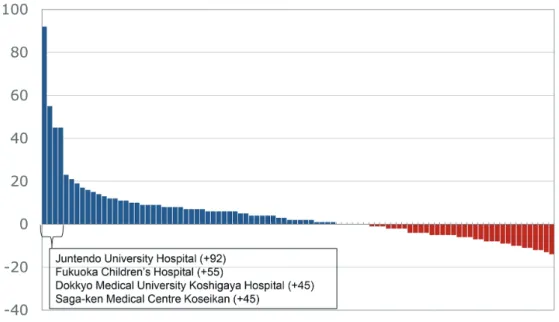
Murase et al. wrote that “the increase between FY (fiscal year) 2011 and FY2012 is obvious, and a gradual increase is observed from FY2012 to FY2014”4). As shown in Figure 2, the air dose of radiation in Fukushima Prefecture rapidly decreased with time76). For reference, typical air doses of ra- diation in major cities such as Shanghai, Seoul, Sin- gapore, Munich, Paris, and New York are 0.59, 0.09,
0.17, 0.12, 0.10, 0.06 μSv/h, respectively77). The authors did not account for why the incidence was maintained after 2012, despite the rapid decrease in radiation levels in Fukushima Prefecture. Consid- ering other possible mechanisms that may induce cryptorchidism after 2012, could the continued in- crease be due to DNA damage in germ cells by the exposure, causing abnormalities such as cryptorchi- dism in the next generation? In general, DNA damage in the male germline is associated with poor fertilization rates following in vivo fertilization, de- fective preimplantation embryonic development, and high rates of miscarriage and morbidity in the off- spring, including childhood cancer78). Physical fac- tors, such as radiation exposure, can induce DNA damage in the mammalian germline ; however, to the best of our knowledge, there is no report to indi- cate that DNA damage in the germline induces cryptorchidism. Boice et al. showed that radiother- apy for cancer (gonadal dose : over 100 mSv) did not carry much, if any, risk for inherited genetic disease in offspring conceived after exposure79). Indeed, the rate of congenital anomalies did not increase in the children of atomic bomb survivors in Hiroshima and Nagasaki80). These data imply that radiation doses with potential transmissibility of germline Fig. 1. Effective radiation doses after the Fukushima Daiichi Nuclear Power Plant accident in 35 prefectures of Ja-
pan, compared to the dose for evacuees in three countries due to the Chernobyl accident.
*35 prefectures are those that Murase et al. showed in Figure 3 of their paper4). Distance between each prefec- tural office and Fukushima Daiichi Nuclear Power Plant is also shown. First-year effective dose (averaged over each prefecture) after the accident86).
#Effective dose for evacuees in 1986 (for ~8 months after the accident) 69).
*#
damage to offspring would have to be quite high. As described above, spermatogonia are also less sus- ceptible to the occurrence of DNA damage after ex- posure to ionizing radiation, and the minimum dose that causes detectable DNA damage in male germ cells is 30 Gy34). In addition, the germline minisat- ellite mutation rate was not affected in exposed fa- thers with a mean preconception testicular dose of 1.23 Gy, nor in exposed mothers with a mean pre- conception ovarian dose of 0.58 Gy35). As shown in Figure 2, the air dose of radiation in Fukushima City at 10 : 00 pm on the 15th of March, April, May, June, July, and August in 2011 was 20.7, 1.83, 1.43, 1.40, 1.24, and 1.15 μSv/h, respectively. Supposing that these mid-month doses were received 24 hours a day, each for 30 days, the cumulative radiation expo- sure for 6 months could be estimated as shown : • (20.7+1.83+1.43+1.40+1.24+1.15) (μSv/h) ×
24 (hours) × 30 (days) = 19.98 mSv/6 months
Judging from previous data34-36), even this degree of chronic exposure − in fact, an overestimate because rapid exponential decay was the dominant term in March76) − is considered not to induce germline DNA damage. Therefore, the hypothesis that germline DNA damage could be caused by external radiation exposure in Fukushima Prefecture and in- duce cryptorchidism after 2012 can be dismissed. It defies reason why “a gradual increase is observed from FY2012 to FY2014,” even if “a large amount of radionuclides emitted from the Fukushima was sus- pected to be a major cause” as Murase et al. assert4). For reference, the average background dose re- ceived by the general population is around 2.4 mSv/
year, which can vary depending on geology and altitude ; this ranges from 1 to 10 mSv/year, but can be more than 50 mSv/year. Exposure of airline crews flying a New York-Tokyo polar route amounts to 9 mSv/year, and the effective dose from abdominal
& pelvic CT scans is 10 mSv81). Table 1 summariz- es the external radiation exposure levels in each sit- uation.
(2) Possibility of internal radiation exposure in Fu- kushima Prefecture affecting the incidence of cryptorchidism
As described above, the maximum 137Cs con- centrations measured in milk in Belarus immediate- ly after the Chernobyl accident was 6,500 Bq/
L51). On the other hand, provisional regulation val- ues for radioiodine were exceeded for ≤ 13 days (16-28 March 2011) in Fukushima Prefecture.
While the maximum radioiodine level detected in tap water was 965 Bq/kg (equal to Bq/L) on 20 March 2011, no tap water samples exceeded the provisional regulation value (200 Bq/L) for radioactive cesium (134Cs and 137Cs) after 31 March 201282).
Consumer Co-operative in Fukushima Prefec- ture measured radioactive cesium concentrations in the daily meals of 100 families using a gamma ray spectrometer (low detection limit < 1 Bq/kg), and showed that only 10% and 2% of families, respec- tively, had received detectable radio-cesium concen- trations from November 2011 to March 2012 (maxi- mum 6.7 Bq/kg of 137Cs) and from June 2012 to September 2012 (maximum 1.9 Bq/kg of 137Cs, re- spectively)83). Continuous consumption of such food for 1 year would result in an annual committed effective dose of 0.037 mSv70). Fukushima prefec- ture measured the internal exposure dose of 184,208 residents using a whole body counter between June 2011 and February 2014, and showed that 99.986%
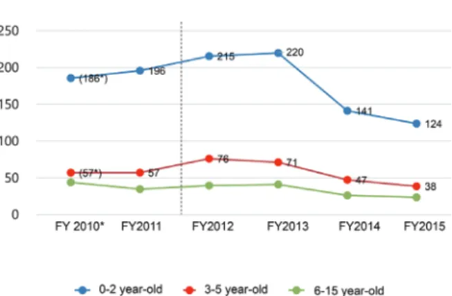
received < 1 mSv, with the maximum dose being 3 mSv70,84). Screening at Minamisoma City, which is Fig. 2. Changes in air doses of radiation in six cities
of Fukushima Prefecture from 2011 to 201576). Fukushima City, Koriyama City, Shirakawa City, Minami Soma City, and Iwaki City are located ap- proximately 64 km northwest, 58 km east, 81 km southeast, 98 km east, 24 km north, and 43 km southeast, respectively, of the Fukushima Daiichi Nuclear Power Plant. Reference values of radia- tion levels in these cities are 0.04, 0.04-0.06, 0.04- 0.05, 0.04-0.05, 0.05, and 0.05-0.06 μSv/h, respec- tively.
located 23 km north of the Fukushima Daiichi Nu- clear Power Plant, showed that approximately 17%
of children and 38% of adults showed detectable in- ternal exposure, with a concentration of 2.8 to 57.9 Bq/kg (median, 11.9 Bq/kg) and 2.3 to 196.5 Bq/kg (median, 11.4 Bq/kg), respectively85). The commit- ted effective dose by cesium was < 1 mSv, with the exception of one person at 1.07 mSv85).
In the animal study outlined above, the effect of chronic contamination (6,500 Bq/L) with 137Cs fol- lowing the Chernobyl nuclear plant accident on adult testosterone production is low, and that on neonatal and fetal testosterone production is estimated to be extremely low, because radio-sensitivity in the neo- nate and fetus is lower than that in an adult49,56,57). According to the data shown above, a dose of 137Cs found in Fukushima Prefecture after the accident is considered to be too low to affect testosterone pro- duction. Therefore, the possibility that internal ra- diation exposure in Fukushima prefecture induced cryptorchidism is also infinitesimally close to zero.
Ionizing radiation can arise from human activi- ties or from natural sources81). Most radiation ex- posure is from natural sources ; this includes radio- activity in the rocks and soil of the Earth’s crust, radon (a radioactive gas given out by many volcanic rocks and uranium ore), and cosmic radiation. For example, radioactivities of around 4,500 Bq, 1,000
Bq, 15 Bq, 3,000 Bq, and 30,000 Bq can be ascribed, respectively, to one adult human (65 Bq/kg), 1 kg of coffee, 1 banana, the air in a 100 m2 Australian home (radon), and 1 household smoke detector (containing americium)81). Table 2 summarizes internal radia- tion exposure levels in each situation.
(3) No evidence to support that both external and internal radiation exposure induces cryptorchi- dism
The 2013 United Nations Scientific Committee on the Effects of Atomic Radiation (UNSCEAR) re- port on the Fukushima Daiichi Nuclear Power Plant accident stated that prenatal exposures from the ac- cident were not expected to increase the incidence of spontaneous abortions, miscarriages, perinatal mortality, congenital effects, or cognitive impair- ment86). Fukushima Health Management Survey data also showed that the incidence of stillbirth, pre- term birth, low birth weight, and congenital anoma- lies were similar to recent averages elsewhere in Ja- pan, and concluded from a pregnancy and birth survey that no significant adverse outcomes were observed over the whole of the Fukushima Prefec- ture after the disaster87).
In summary, there is no evidence to support that external and/or internal radiation exposure could induce cryptorchidism by a disturbance of ste- roidogenesis or germline DNA damage after the Fu- Table 1. External radiation exposure levels in various situations
Received doses of 460,408 residents in Fukushima Prefecture during the first 4 months after the accident70) 66.3% < 1 mSv, 94.9% < 2 mSv, 99.7% < 5 mSv ; the maximum dose was 25 mSv
Mean radiation dose of children between the ages of 0-15 years (n = 4,571) in Nihonmatsu City of Fukushima Prefecture71)
1.5 mSv/year 6 months after the disaster, 1.5 mSv/year in 2012, 1.0 mSv/year in 2013, 0.65 mSv/year in 2014 The average dose received from background radiation81)
Around 2.4 mSv/year
Exposure of airline crews flying a New York-Tokyo polar route81) 9 mSv/year
Effective dose from abdominal & pelvic CT scans81) 10 mSv
Fetal risk of anomalies, growth restriction, or abortion reported in the Guidelines for Diagnostic Imaging During Pregnancy by ACOG73,74)
Have not been reported with a radiation exposures < 50 mGy
Possibility of occurrence of malformations in newborns suggested by ICRP75) Threshold > 100 mGy
A unit of mSv reported as an exposure dose due to the Fukushima Daiichi nuclear plant accident can be interpreted as corre- sponding individual organ/tissue exposures in mGy, based on nearly uniform exposure to cesium.
kushima Daiichi Nuclear Power Plant accident.
Furthermore, the quantity of radionuclides emitted from the power plant was too low to influence testic- ular descent. Thus, at the very least, Murase et al.
should have detailed how such low-level radionu- clides might induce cryptorchidism, theoretically or empirically, if they wanted to invoke any mechanism other than the ones we have shown to be implausi- ble.
2) Did the incidence of cryptorchidism increase throughout Japan?
Murase et al. reported that a nation-wide in- crease in cryptorchidism was observed after the ac- cident4). They showed increases in the rate of hos- pital discharge after orchiopexy in 35 prefectures of Japan4). Since there are 47 prefectures in Japan, al- most three quarters of the prefectures were includ- ed in their study. The 35 prefectures included those in Southern Japan, including Fukuoka, Ka- goshima, and Okinawa Prefectures, that are 1,052
km, 1,161 km, and 1,773 km from the Fukushima Daiichi Nuclear Power Plant, as shown in Figure 1, which also shows radiation exposures in 35 prefec- tures86). For comparison, the average doses for Chernobyl evacuees in three countries are also shown69). In general, a dose-response relationship is the effect on an organism or, more specifically, the risk of a defined outcome, produced by a given amount of an agent or a level of exposure88). A dose-response relationship exists when increasing levels of exposure are associated with either an in- creasing or a decreasing risk of the outcome. Dem- onstration of a dose-response relationship is consid- ered strong evidence for causality88). Thus, if radiation exposure affects the incidence of cryptor- chidism, the incidence in various prefectures should decrease with distance from the source of exposure, even if the decrease is not linear or uniform. How- ever, Murase et al. failed to show any dose-response relationship in their paper. In fact, the analytical formula (2) described in their supplementary appen- Table 2. Internal radiation exposure levels in various situations
Values for radioiodine in Fukushima Prefecture82)
Provisional regulation values were exceeded for ≤ 13 days (16–28 March 2011).
The maximum detected level was 965 Bq/kg (equal to Bq/L), detected on 20 March 2011.
Values for radio-cesiums (134Cs and 137Cs) after the Fukushima Daiichi nuclear plant accident82)
No tap water samples exhibited radioactivity that exceeded the provisional regulation value (200 Bq/L) as of 31 March 2012.
Radioactive cesium concentrations in the daily meals of 100 families examined by Consumer Co-operative in Fukushima Prefecture83)
10% of families received detectable (1 or more Bq/kg) radio-cesium (maximum 6.7 Bq/kg of 137Cs), from November 2011 to March 2012.
2% of families received detectable (1 or over Bq/kg) radio-cesium concentrations (maximum 1.9 Bq/kg of 137Cs), from June 2012 to September 2012.
Annual committed effective dose assuming continuous consumption of these contaminated foods : 0.037 mSv.70) Screening for internal exposure at Minamisoma City85)
17% of children showed detectable internal exposure of 2.8 to 57.9 Bq/kg (median, 11.9 Bq/kg).
38% of adults showed detectable internal exposure of 2.3 to 196.5 Bq/kg (median, 11.4 Bq/kg).
The committed effective dose from cesium was < 1 mSv except for one person with 1.07 mSv.
Exposure dose of 184,208 residents using whole body counting conducted by Fukushima Prefecture from June 2011 to February 201470,84)
99.986% received < 1 mSv, with a maximum dose of 3 mSv.
Radioactivity of some natural and other materials81) 15 Bq in 1 banana
1,000 Bq in 1 kg of coffee
4,500 Bq in 1 adult human (65 Bq/kg)
30,000 Bq in 1 household smoke detector (with americium)
The maximum 137Cs concentrations measured in milk in Belarus immediately after the Chernobyl accident49) 6,500 Bq/L
dix S1 explicitly showed that they omitted parame- ters indicating a decreased difference in the hospital discharge rate with increasing distance from the Fu- kushima Daiichi Nuclear Power Plant4).
For reference, the whole of Japan and Fukushi- ma Prefecture cover 378,000 km² and 13,783 km2, respectively, whereas the area affected by evacua- tion orders was 370 km2, which is only 0.098% of Ja- pan and 2.7% of Fukushima Prefecture. Thus, the majority of people in Japan and Fukushima Prefec- ture were not significantly affected by accident-re- lated radiation89). Bedwell et al. reported that in the north, south, and west of Japan, all estimates of the lifetime effective dose received by members of the public following the accident are very low, and in all cases are less than 1 mSv72). Furthermore, the es- timated dose received in these regions is less than the typical Japanese and worldwide average annual doses from natural background of 2.1 mSv90) and 2.4 mSv91), respectively. Across all regions of Central and Eastern Japan, including Fukushima Prefecture, they are below 10 mSv. These findings indicate that the majority of people in Japan were not signifi- cantly exposed to radiation from the Fukushima Dai- ichi Nuclear Power Plant, although Murase et al.
concluded that “a large amount of radionuclides emitted from the Fukushima was suspected to be a major cause.”4) Such a declaration undoubtedly contributes to misunderstanding.
In addition, Murase et al. also state : “A straightforward comparison between disaster-affect- ed area and non-affected area seemed to be difficult because many residents migrated from the affected area4).” Figure 3A shows the actual number of peo- ple who migrated from Fukushima Prefecture to other prefectures after the Fukushima Daiichi Nu- clear Power Plant accident. The total number throughout Japan in fiscal year 2011 (FY2011) was 62,70092). The population of Fukushima Prefecture in 2011 was 1,988,99593). Thus, only ~2% of peo- ple living in Fukushima Prefecture migrated to other prefectures in 2011. Figure 3B shows specific mi- gration patterns from Fukushima Prefecture follow- ing the earthquake, tsunami, and nuclear crisis.
Most people migrated from Fukushima Prefecture to Japan’s capital city, Tokyo (7,627 in 2011), or neigh- boring prefectures such as Yamagata (12,944 in 2011) and Niigata (6,762 in 2011) 92). The number of people who migrated to further south or west was extremely low. For example, the number of people who migrated to Kyoto, Osaka, Hiroshima, and Oki- nawa in 2011 was 767, 838, 310, and 701, respec- tively92). The population in Kyoto, Osaka, Hiroshi-
ma, and Okinawa in 2011 was 2,632,496, 8,865,448, 2,855,734, and 1,401,93393), respectively, indicating that only 0.029, 0.0094, 0.0035, 0.005%, respectively, of the population in each prefecture migrated from Fukushima Prefecture in 2011. How could such a small number of people migrating from Fukushima Prefecture affect the incidence of cryptorchidism elsewhere? Such publically available data could have informed the analysis of Murase et al., but by their own admission, even a straightforward com- parison was too difficult for them to undertake.
As described above, radiation exposure is not only external exposure but also internal via the food chain48). Murase et al. described it as follows : “Af- ter the nuclear accident, radionuclides may have been distributed through the ecosystem through food contamination.”4) That may have been true af- ter Chernobyl, but Japan has the world’s strictest standards for managing radioactive contamination of food, and anything exceeding those standards cannot be distributed. According to the Japan Food Sanita- tion Act, derived intervention levels for radioactive cesium, which are the upper limits allowed for food to be distributed in the supply chain, are 10, 50, 50, and 100 Bq/kg (~Bq/L) in drinking water, milk, in- fant foods, and general foods, respectively89). EU Council Regulations (Euratom) are more le- nient : 1,000, 1,000, 400, and 1,250 Bq/kg in drink- ing water, milk, infant food, and other food, respec- tively. American Guidance Levels for Radionuclides in Domestic and Imported Foods allow 1,200 Bq/kg in general89). Results of Japan’s thorough monitor- ing of agriculture, forestry, and fishery products pri- or to shipment are announced, and very few foods have exceeded the standard limit (100 Bq/kg). Nec- essary measures are in place to ensure that foods are not distributed in the market if they are found to have exceeded the standard limit. Because of in- tense and immediate monitoring, and the rapid de- crease of initially high contamination levels94), foods in Fukushima Prefecture were very safe and remain so, making the possibility of internal exposure from food extremely low in Fukushima and throughout Ja- pan.
Murase et al. cited two papers94,95) in their de- scription as follows : “After the nuclear accident, ra- dionuclides may have been distributed through the ecosystem through food contamination4).” Howev- er, these two papers dispute what Murase et al.
sought to imply. Mert et al. wrote as follows : “It seems very unlikely that more than very few mem- bers of the public in Japan exceeded the maximum permissible internal exposure of 1 mSv/year. This
observation is in agreement with the results of pre- vious studies94).” Furthermore, Shozugawa et al.
wrote as follows : “we can conclude that 137Cs de- tected in remote areas 300 km or more from Fuku- shima nuclear power plant contained activity from Pre-Fukushima events such as Chernobyl accident (1986) and atmospheric nuclear explosions (from 1945).” 95) Therefore, Murase et al. seem to have misrepresented the two papers to suit their own ideas. In addition, Murase et al. referred to another two papers in the context of “...incineration of debris
as part of the treatment of disaster waste.”96,97) In fact, per Iwami and Sasai97), if a dose of 1.4 Bq/m3(N) of 137Cs was emitted into the air by the incineration of debris, and people inhaled the air every day, 24 hours a day, 365 days a year, the dose of radiation exposure from the air by the incineration of debris in a year is calculated as below. The intake air vol- umes per day are 20 m3 and 5 m3 for an adult and a 1-year-old child, respectively, and the inhalation dose coefficients of the radionuclides (effective dose per unit intake) are 4.6×10⁻⁹ and 5.4×10⁻⁹ Sv/Bq, Fig. 3. Changes in the number of emigrants from Fukushima Prefecture after the Fukushima Daiichi Nuclear Power
Plant accident. A. Total number of Japanese emigrants. B. The number of emigrants in each prefecture.
respectively98).
• Adult : 1.4 Bq/m3×20 m3×4.6×10-9 (Sv/
Bq)×365=0.047 mSv/year
• 1-year-old child : 1.4 Bq/m3×5 m3×5.4×10-9 (Sv/Bq)×365=0.014 mSv/year
These data indicate that the dose of radiation expo- sure emitted into the air by the incineration of debris is extremely low and is unlikely to affect the inci- dence of cryptorchidism.
In summary, for the reasons described above, the hypothesis that radiation provoked an increase of cryptorchidism throughout Japan has no support.
3) Was the study design appropriate? Is it correct to use a DPC survey database to evaluate the num- ber of cases of cryptorchidism?
Murase et al. counted the number of orchiopex- ies using the Diagnosis Procedure Combination (DPC) database, which is a national administrative claims and discharge database covering acute-care inpatients in Japan. All 82 academic hospitals in Ja- pan are obliged to adopt this system, while commu- nity hospitals participate on a voluntary basis. The DPC database contains discharge abstract and ad- ministrative reimbursement claims data for inpatient episodes collected from participating hospitals.
This database system started in Japan in 2003, and its use has recently spread to acute care hospitals and is in transition. The ratio of hospitals involved in the DPC survey per total hospitals is very low (Figure 4). This system is unsuitable to assess year-on-year comparisons in the number of surger- ies performed all over Japan, and thus represents a serious flaw in the design setting of the research by Murase et al..
In the event that we assess the number of sur- geries using the DPC system, as Murase et al. did, the majority of hospitals in Japan are exclud- ed. Murase et al. obtained cryptorchidism dis- charge data collected over a 6 year period from the DPC survey database in Japan in order to estimate the discharge rate after cryptorchidism surgery be- fore and after the accident4). Only 94 hospitals in Japan that participated in the DPC system and re- ported 10 or more discharges after cryptorchidism surgery within 6 years covering before and after the accident were involved in their survey. However, there are many more hospitals (7,528 as of 2011)4) in Japan, some of which are considered to have per- formed orchiopexy, even though their number of or- chiopexies per year is under 10. In 2008, the Min- istry of Health, Labour, and Welfare started to
construct a database of all electronic health insur- ance claim data, the so-called National Database of Health Insurance Claims, and Specific Health Checkups of Japan (NDB), which currently covers approximately 98% of the healthcare services pro- vided by health insurance99-101). The NDB has grown to become one of the largest medical databas- es in the world100,102,103). The NDB can be a power- ful tool to survey the status of Japanese medical care in the future100). According to NDB open data104), the total number of orchiopexies (open orchiopexy [segment number : K836] + laparoscopic orchio- pexy [segment number : K836-2]) was 9,810 (9,658 + 152) and 9,497 (9,368 + 129) in 2014 and 2015, respectively, in the whole of Japan. Murase et al.
showed, in Supplementary Table S3 of their paper, that the number of orchiopexies was 6,404 and 6,042 in 2014 and 2015, respectively, in the DPC hospi- tals. Therefore, because the NDB has recently covered approximately 98% of healthcare services provided by health insurance, the highest estimate of the number of orchiopexies in DPC hospitals per those of all hospitals in Japan in 2014 and 2015 was only 65.3% (6,404/9,810) and 63.6% (6,042/9,497), respectively. The authors claim as follows : “The discharge number in the DPC data reflects the actual number of surgeries in Japan well4)”, but their de-
Fig. 4. Changes in the number of all Japanese hospi- tals and those involved in the DPC survey (DPC hospitals).