Mume Leaf Margin Necrosis, a New
Disease of Japanese Apricot Caused by
Multiple Infection with Cucumber Mosaic
Virus and Prunus Necrotic Ringspot
Related Ilarvirus
By
Jun K
URIHARA*, Keiichi T
OMARU**, Takayuki O
TSUBO***,
Keiko T. N
ATSUAKI**** and Yutaka A
RIMOTO*****
(Received May ,-, ,**//Accepted October ,1, ,**/)Summary : A new disease of Japanese apricot (Prunus mume Sieb. et Zucc.) (cv. Nankou) occurred in the many orchards in southern part of Wakayama prefecture, Japan. Disease symptoms were high frequency of abortive flower occurrence, leaf emergence delay, leaf mottling, leaf margin necrosis and early defoliation. Fruit yields decreased markedly. Two viruses, Cucumber mosaic virus (CMV-Um) and prunus necrotic ringspot related ilarvirus (PNRLV) were isolated from diseased trees. Electron microscopy of purified CMV-Um and PNRLV indicated spherical particles ,2 nm size in CMV-Um and ,/ῌ,2 nm in PNRLV and in both viruses of purified preparation. CMV-Um RNA was resolved to be five distinctive species. It was revealed that the CMV-Um contained satellite RNA with the same size of CMV-Y satellite RNA. The coat protein subunits of CMV-Um had a Mr of approximately ,0,***. PNRLV was serologically related to the American isolate of Prunus necrotic ringspot virus (PNRSV) but neither to the European isolate nor to Prune dwarf virus with ELISA and western blot analysis. Graft inoculation of diseased buds to “Shirofugen” flowering cherry (P. serrulata Lindl.) caused necrosis at the grafted sites. Graft inoculation of diseased Nankou scion onto healthy one showed the same symptoms as naturally infected trees in the stock shoots. Similar symptoms were caused by slashing inoculation with surgical knife onto the stem of young healthy Nankou seedling with mixed inoculum of both viruses of CMV-Um and PNRLV. The disease is named mume leaf margin necrosis (formerly leaf edge necrosis). For the detection of viruses from the diseased Prunus trees by sap inoculation or ELISA, the season for leaf sampling was best in June, followed by May and July, the stage of young green leaves with soft tissues.
Key words : Prunus mume, Cucumber mosaic virus, Prunus necrotic ringspot virus, Mume leaf margin necrosis
ῌῌῌῌῌῌῌῌῌῌῌῌῌῌῌῌῌῌῌῌῌῌῌ
Introduction
A previously unreported disease of Japanese apricot (Prunus mume Sieb. et Zucc.) was first reported in +323
in the southern part of Wakayama prefecture, one of the major production areas of Japanese apricot in Japan. The disease has been known by farmers as ‘Chagasu’, in Japanese, which means dried tea leaf
* ** *** **** *****
Formerly : Department of Agricultural Science, Graduate School of Agriculture, Tokyo University of Agriculture, Present address : Nagano Vegetable and Ornamental Crop Experiment Station Saku Branch
Formerly : Tokyo University of Agriculture
Green Academy School, Tokyo University of Agriculture
Department of International Agricultural Development, Faculty of International Agriculture and Food Studies, Tokyo University of Agriculture
RIKEN
論 文
waste,+, ,.ῌ
. The first symptoms of this disease appeared as malformation of blossoms in February (Plate I-+-a). Leaves exhibiting a slight interveinal chlorosis in early summer and marginal necrosis of leaves on dis-eased trees in autumn were observed as an additional symptom (Plate I-+-b). Because of the great reduction in the fruit yield, the disease is economically impor-tant+,ῌ+., +1, +2ῌ
. This paper describes the causal viruses of the new disease, Mume leaf margin necrosis. The symptom expression by sap-inoculation to young grafted Nankou plantlet3, ,,ῌ
and detailed data for the identification of Cucumber mosaic virus were published separately (KURIHARAet al., ,**/)+0ῌ.
Materials and methods
Virus isolation and host range
Leaves showing interveinal chlorosis were collected from P. mume (cv. Nankou) in June at the southern part of Wakayama prefecture and usually stored in freezer at῍2*῎. Leaf samples were homogenized with *.+ M sodium phosphate bu#er, pH 1.*, containing ,ῌ nico-tine and *.,ῌ sodium diethyl- dithiocarbamate (Na-DIECA) (extraction bu#er) with cold mortar and pestle. Then the sap filtered through double hold gauze was rub inoculated to carborundum-dusted leaves of herba-ceous indicator plants. For the single lesion isolation, the virus from a necrotic local lesion of Chenopodium quinoa leaves was inoculated to Physalis floridana in which the virus caused systemic symptoms. On the other hand, from systemically infected C. quinoa leaves the virus did not cause infection in P. floridana. It was considered that two di#erent viruses were involved in the P. mume showing leaf margin necrosis symptom. Infected leaf samples collected from three di#erent Japanese apricot orchards showed the same reactions to several indicator plants in sap inoculation, respec-tively. Isolates of viruses from one of the samples were used in further experiments. After ELISA testing, isolate inducing systemic symptoms on C. quinoa tentatively designated as prunus necrotic ringspot like Ilarvirus virus (PNRLV) and that producing local necrotic lesions on C. quinoa without systemic infec-tion as Cucumber mosaic virus mume isolate (CMV-Um). CMV-Um and original mixed viruses was maintained in Nicotiana tabacum L. cv. Samsun NN and PNRLV was maintained in C. quinoa.
Leaves infected with each virus isolate were homo-genized in extraction bu#er with cold mortar and pestle. They were then rubbed onto carborundum-dusted leaves of indicator plants. The inoculated plants were kept in plant growth chamber at ,/῎ and observed for more than one month after inoculation.
Recovery tests of all inoculated plants were carried out by inoculation to C. quinca and other di#erential hosts and/or serological tests.
Electron microscopy
Leaf extracts and purified virus preparations were stained with ,ῌ uranyl acetate (UA) or ,ῌ phos-photungstic acid (PTA) (pH 1.*) and observed by elec-tron microscope (JEM-+**CX).
Virus purification
CMV-Um was purified using modified procedure of FRANCKIet al.,ῌ. Detailed procedure of the methods was
described in a separate paper (KURIHARAet al., ,**/)+0ῌ.
PNRLV was purified following the protocol by ONG
and MINK,*ῌ
. Leaves of infected C. quinoa frozen at ῍2*῎ were triturated in warring blender for / min with ,* mM sodium phosphate (pH 2.*), containing +* mM ,-mercaptoethanol and *.*+ M DIECA (, ml/g of tissue). The extract was clarified by centrifugation at +*,*** rpm for -* min in Kokusan type IN rotor. The supernatant was filtered through two layers of Kim-wipes paper tissues, and the pH was adjusted to ../ with /ῌ acetic acid. The slurry was allowed to stand for -* min and clarified by centrifugation at +3,/** rpm for +/ min in Beckman type ./Ti rotor. The superna-tant was concentrated at -3,*** rpm for - hr in the same rotor and the pellet was resuspended in ,* mM phos-phate bu#er, pH 2.*. After the second cycle of di#eren-tial centrifugation, further purification was achieved by sucrose density gradient (+*ῌ.*ῌ) rate zonal cen-trifugation at ,+,*** rpm for 0 hr in Beckman type SW ,2rotor. All procedures were carried out at .῎.
For mechanical inoculation of each virus to healthy C. quinoa plants, the final pellets from the purification procedure were resuspended in / mM sodium borate bu#er, pH 2.* containing + mM EDTA for CMV-Um and ,*mM sodium phosphate, pH 2.*, containing + mM EDTA for PNRLV. The inoculated plants were kept in the plant growth chamber at ,/῎ and monitored for symptom development.
Serology
Antisera to CMV-Um or PNRLV were prepared by immunization for rabbit with each purified virus. One New Zealand white rabbit for each virus was im-munized three times with intramusucular injections, once a week, of *.,/ mg of purified CMV-Um or PNRLV in *./ ml of bu#er emulsified with *./ ml of Freund’s complete adjuvant. After , months, two additional intramusucular injections, and *.+,/ mg of virus intra-venous injection as booster, were administered.
Anti-serum was obtained from blood collected + week after booster injection. The titer was determined by indirect ELISA.
To determine relationships with previously reported ilarviruses isolated from Prunus plants, ELISA was conducted. PNRSV, Prune dwarf virus (PDV),2ῌ
and Apple mosaic virus (ApMV),2ῌ
antisera kits prepared by Agdia (Elkhart, IN, USA) and PNRSV antiserum kit prepared by Bioreba (Switzerland) were used.
SDS-PAGE of coat protein subunit and Western blot analysis
The relative molecular weight of the coat protein was determined by sodium dodecyl sulfate (SDS) poly-acrylamide gel electrophoresis (PAGE) on vertical +,./ῌ gel with .ῌ stacking gel in ATTO AE-0,** (ATTO, Tokyo, Japan) as previously described+3ῌ
. Western blot analysis for CMV-Um and PNRLV was conducted following the method described by TOWBIN
et al.,1ῌ
. For identification of PNRLV, antisera to PNRSV American type culture collection (As-PNRSV-G, ATCC ,,) and PDV (As-PDV, ATCC PVAS-,3*) were used. Antiserum to Spinach latent virus (SPLV)+, ,2ῌ
kindly given by Dr. L. BOS(Institute of Phy-topathological Research ; IPO, Wageningen, The Neth-erlands) and PDV infected peach tissue supplied by Dr. KANO (National Institute of Fruit Tree Science,
Tsukuba, Ibaraki, Japan) were also used. To facilitate determination of relative molecular mass, low range biotinnilated SDS-PAGE molecular weight marker (Bio Rad, USA) was used.
Detection of virus and sampling season
As the virus detection from P. mume was not always easy, we performed the detection tests in di#erent seasons to determine the best season for sampling. During May +33. to April +33/, leaf samples of the same diseased mume plants showing the symptoms were collected from two orchards in Minabe several times. The samples were tested by sap inoculation, rubbing method and ELISA to detect the viruses as described in other paragraphs.
Graft inoculation to Shirofugen flowering cherry and Nankou plantlets
Grafting with budding chip to twig of Shirofugen flowering cherry (P. serrulata) was commonly used for the detection of Prunus necrotic ringspot virus (PNRSV) from Prunus plant-*ῌ
. Shoots of diseased Nankou were collected in June, and were graft-inoculated onto Shirofugen twigs by chip budding method-*ῌ
.
Graft inoculation on 0 Nankou plantlets of three
years old were carried out with diseased scion. At the same time healthy Nankou scions were grafted onto 0 Nankou plantlets as controls.
Virus inoculation to Japanese apricot (Nankou) seed-lings
Japanese apricot (cv. Nankou) seeds and seedlings were obtained from the southern part of Wakayama. Plants were raised in green house for , months follow-ing stratification at .῍ in moist peat soil. Two di#er-ent mechanical inoculation procedures, rub inoculation with carborandum and slashing inoculation onto green stem were used. Samsun NN tobacco leaves infected with CMV-Um and C. quinoa leaves infected with PNRLV were homogenized in each / volume of extrac-tion bu#er. These homogenates were used as inocu-lums for each test. A surgical knife was dipped into the inoculum, and up to -* slashes per seedlings were made onto about -* cm length of young green stems. Single inoculation with CMV-Um or PNRLV and mixed inoc-ulation with inoculum of both viruses (+ : +) were con-ducted onto stem of / seedlings each in May. At the same time, extraction bu#er was inoculated to / seed-lings as negative control. Plants were maintained in a plant growth chamber (,/ῌ,*῍/day-night) until au-tumn and transferred to outside open-air through win-ter.
Results
Symptomatology of P. mume (cv. Nankou)
The first symptom in diseased plant appeared as higher frequency of abortive flower occurrence than in healthy plants in February (Plate I-+-a). Delay of leaf sprouting in spring and slight interveinal chlorosis of leaves in early summer were also typical symptoms. Marginal necrosis of leaves and early defoliation were observed on diseased trees in autumn (Plate I-+-b). These symptoms were observed in naturally infected trees with these two viruses and artificially inoculated plants with both the grafting and slashing methods (Plate I-,).
Host ranges of the viruses
Twenty-four plants of ,/ species in +* families were susceptible with CMV-Um and many of them were infected systemically showing mild symptoms (Table +). CMV-Um locally infected on legume plants and tobacco (cv. Samsun NN) showed only systemic faint vein clearing.
In C. quinoa, irregular chlorotic spots on the in-oculated leaves and the systemic symptom with vein clearing on young leaves were observed . days and +.
days after the inoculation with PNRLV, respectively (Plate I--). These symptoms were not observed on the plants mock-inoculated with bu#er. However, PNRLV did not infect Cyamopsis tetragonoloba and Cucumis sativus, which were reported as the di#erential hosts of the common isolate of PNRSV.ῌ0, ,2ῌ
(Table ,). Electron microscopy
In the purified preparation of tobacco plants infected with CMV-Um isometric particles with average diame-ter ,2 nm were observed (Plate I-0), but not seen in comparable healthy material. In leaf dips of C. quinoa plants infected with PNRLV virus like particles could not be observed when the samples were stained with PTA (pH 1.*). After formalin fixation of specimens before staining, however, quasi-isometric virus like particles with ,/ῌ,2 nm diameter could be seen (plate II-+). Observation of virus particles in leaf dips from diseased P. mume plants were not successful, but in the purified specimens from inoculated tobacco plants, two types of viral particles were observed (Plate II-,). Virus purification
After , hrs of sucrose density gradient centrifugation
of CMV-Um, single visible band in gradient was ob-tained. Fractions collected at the peak absorbance were resuspended and ultracentrifuged. The isometric particles with average diameter ,2 nm were observed by the electron microscopy of the pellet fixed with +ῌ formalin and stained with PTA pH 1.* (Plate I-0). The purified virus preparation had a maximum absorption at ,0* nm and minimum at ,., nm ; the A,0*/A,., and A,0*/A,2* ratios were +./+ and +.2, (uncorrected for light scattering), respectively. All C. quinoa plants inoculated with purified virus preparation developed necrotic local lesions on inoculated leaves within -days.
After 0 hrs of rate zonal centrifugation of PNRLV, two visible bands in gradient were obtained. Thirty-six fractions were separated and measured for ance at ,0* nm. The fractions showing peak absorb-ance (Fig. +) were collected and concentrated at -3,*** rpm for - hr in Beckman type ./Ti rotor.
The pellets were resuspended and fixed with +ῌ formalin and stained with PTA pH 1.*. In the electron microscopy, quasi- isometric particles with average di-ameter ,/ῌ,2 nm were seen (Plate II-+). The virus parti-cles had a maximum absorption at ,0* nm and mini-mum at ,.. nm ; the A,0*/A,.. and A,0*/A,2* ratio ranged from +.,/ to +.-0 and +..0 to +.0. (uncorrected for light scattering), respectively. The local chlorotic spots on the inoculated leaves (Fig. +) and the systemic vein clearing and yellowing typical of PNRLV (Plate I--) were shown in C. quinoa plants inoculated with the bottom band fraction within +. days, but no symptom was seen on the plants inoculated with any other frac-tions including the top band.
Table + Reactions of various plants to sap inoculation with CMV-Um
Table , Reactions of various plants to sap inoculation with PNRLV
Serology
Serology for CMV referred to a separate report+0ῌ
. CMV-Um antiserum showed titer +**,*** by indirect ELISA. To determine the serotype of the virus isolate, CMV-Y,0ῌ
(subgroup I) and CMV-P,/ῌ
(subgroup II) were used as standard. In agar gel double di#usion test, data was shown in the separate report+0ῌ
. PNRLV antiserum indicated titer /*,*** by indirect ELISA as shown in Fig. ,. In DAS ELISA using PNRSV antibody (As-PNRSV, Agdia and ATCC), PNRLV infected leaf tissues indicated positive reactions as shown in Table -. SDS-PAGE of coat protein subunit and western blot analysis
The Mr of the coat protein subunits of CMV-Um was shown as approximately ,0,*** by +,./ῌ PAGE. It was similar to the ones of CMV-O or CMV-P (Plate II-.). The coat protein subunits of PNRLV migrated into , com-ponents in +,./ῌ PAGE containing the major compo-nent with the Mr ,-,*** which was within the range among previously reported values/, ,2ῌ
.
In Western blotting, PNRLV was reacted to antise-rum of American isolate of PNRSV (plate II--) and SPLV antiserum (data not shown) but not to antiserum of PDV (plate II--) and European isolate of PNRSV antiserum (Bioreba), respectively.
RNAs of CMV-Um
In agarose gel electrophoretic analysis, CMV-Um RNA was resolved as five distinctive species. The fastest one was a satellite RNA. It was also observed in dsRNA diagnosis of this isolate (data not shown) and its Mr was the same as CMV-Y satellite RNA+/, +0ῌ
. Mi-gration pattern of RNA species of CMV-Um (Plate II-/) were the same as those of CMV-O but RNA , and - were slightly di#erent in +./ῌ agar gel.
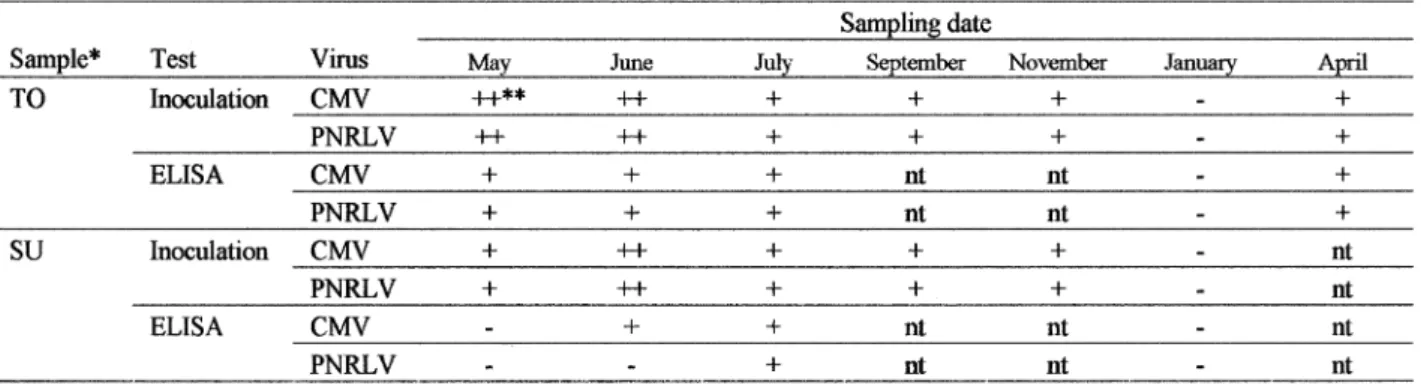
Virus detection and sampling seasons
The results were shown in Table .. According to the experimental results, June followed by May and July was the best season for the detection of two viruses by sap-inoculation to di#erential host plants and ELISA test.
Graft inoculation to Shirofugen flowering cherry and Nankou plantlet
One month after the graft inoculation of diseased buds to Shirofugen flowering cherry, necrosis on bark and inside wood tissues were observed at the diseased
Fig. + Sedimentation profile of PNRLV-Um and its infectivity by rate zonal centrifugation. The top and bottom visible bands are de-signated as T and B, respectively. Sedi-mentation is left to right.
Fig. , Reaction of As-PNRLV by indirect ELISA.
Purified PNRLV (+ mg/ml) (῏), PNRLV infected C. quinoa (῎), healthy C. quinoa (῍)
Table - Serological reactions of diseased Prunus mume leaf samples by DAS-ELISA using Pathoscreen (Agdia)
bud grafted sites (Plate I-., /) but not in the healthy one grafted sites.
Eight months after graft inoculation of diseased scion to Nankou plantlets, leaf margin necrosis symp-tom in new developing leaves of the stock shoots were observed. In addition, frequency of abortive flower occurrence was 2*..ῌ on the stock grafted with dis-eased scion in the spring of next year, but was ,..-ῌ on the stock grafted with healthy one (Table /).
Virus inoculation to Prunus mume seedlings
Stem slashing inoculation resulted in successful transmission of viruses. Four weeks after the inocula-tion, some of the CMV-Um inoculated P. mume (cv. Nankou) seedlings showed vein yellowing in newly emerged leaves, and PNRLV or mix inoculated seed-lings showed mottle. Some of the seedlings mix in-oculated with CMV-Um and PNRLV showed systemic leaf margin necrosis / months after the inoculation in November (Plate I-,-b), but the other plots did not show the symptom. In the next spring, all seedlings in-oculated with mixed inoculum of PNRLV and CMV-Um showed delay of leaf emerging about one week (Plate I-,-a), and in the following autumn, defoliation was about one week earlier than the other plants in-oculated with CMV-Um or PNRLV only. The symptom severity including delay of leaf emergence and earlier
defoliation was most pronounced in the case of mixed inoculation followed by single inoculation of PNRLV and CMV-Um. Mock inoculated plants showed no sym-ptoms at all. Two years after inoculation, P.mume seedlings inoculated with mixed inoculum of two viruses also showed delay of leaf emergence in spring, severer yellowing and earlier defoliation symptoms in autumn as same as in the first year. The symptoms observed in the inoculated seedling were similar to the ones in original diseased plants at the orchards in Wakayama prefecture (Plate I-+-b). The recovery test of two viruses from mix inoculated plants was succeeded by sap inoculation and ELISA.
Discussion
These results showed that two distinct viruses are associated with leaf margin necrosis disease in Japa-nese apricot. The viruses were identified as Cucumber mosaic virus (CMV) and a kind of ilarvirus which is serologically related to Prunus necrotic ringspot virus (PNRSV). This is the first report of CMV and Ilarvirus isolated from P. mume. Grafting of naturally infected P. mume scion onto healthy Nankou plantlets showed the same symptoms in the stock shoots indicating that the disease was graft transmissible. Mixed inoculum of CMV-Um and PNRLV were able to back to P. mume seedlings by stem slashing inoculation onto green stem Table . Detection of CMV-Um and PNRLV in various times through the year by sap inoculation and ELISA
Table / Reactions of - years old Prunus mume cv. Nankou graft inoculated with diseased cv. Nankou in February, +33.
and resulted in similar symptoms. The further result of the symptoms in Nankou plantlets,,ῌ
and comparison of CMV RNA - nucleotide sequences reported in the separate report+/, +0ῌ
indicated that CMV-Um belonged to CMV subgroup I in molecular base. However, CMV-Um was distinct from CMV subgroup I and II in serol-ogy.
An example of CMV strain which indicates it is serologically distinct from subgroup I and II was also reported in CMV from soybean2ῌ
, which is di#erent from infectivity of CMV-Um to soybean. Molecular weight of CMV-Um coat protein was similar to CMV-O. Migration pattern of RNA species of CMV-Um were the same as those of CMV-O except for a slight di#erence in RNA , and -. Systemic symptoms of indicator plants i.e. Samsun NN tobacco infected with CMV-Um were very mild. CMV-Um had a satellite RNA, but we did not test about the function of the satellite RNA in this study.
PNRLV was transmitted mechanically to 3 of ,, species tested and was purified from systemically in-fected C. quinoa. Purified preparation of PNRLV was separated into two visible bands by the rate zonal centrifugation with +*ῌ.*ῌ sucrose density gradient. The shape and the size of PNRLV particle was quasi-isometric and approximately ,/ῌ,2 nm in its diameter and Mr of its coat protein was ,-,***. The Mr value is within the category of previously reported PNRSVs0ῌ
. PNRLV was serologically related to American isolate of PNRSV. It is not possible to determine if PNRLV was a new member of Ilarvirus or a strain of an existing member of this genus. However, PNRLV can be distin-guished from previously reported PNRSVs because of its narrow herbaceous host range and di#erent reac-tion on PNRSV indicator plants including cucumber and Cyamopsis tetoragonoloba. RNA sequence analysis of the isolate will be useful for further classification.
It was reported that CMV causes only latent infec-tion on Prunus species,3ῌ
. KISHIet al.++ῌ
described that P. mume plant grafted with peach scion showing line pattern symptoms developed line pattern and stunted. P. mume plant inoculated with PNRSV or PDV showed no symptom or slight growth inhibition, respectively, and recovered , years after inoculation.
The symptoms of Japanese apricot leaf margin ne-crosis disease are similar to those of plum leaf scald disease (PLS) reported in Argentina, Brazil, Paraguay and southeastern United States-, ,-ῌ
, but PLS did not induce abortion of flower. PLS was reported to be caused by the gram-negative, xylem-limited, fastidious bacterium Xylella fastidiosa Wells. In the present study, following the method of FRENCHand KITAJIMA-ῌ
,
we could not find the bacteria from petiole tissue of P. mume a#ected by leaf margin necrosis disease.
As for the leaf margin necrosis symptoms were most prevalent in autumn at the orchard of southern part of Wakayama prefecture but not so severe in the experi-mental condition in Tokyo except the only one case of typical symptoms development. So, we made addition-al experiment using the same kind of originaddition-al diseased plant in Wakayama, grafted-Nankou plantlet instead of Nankou seedling. The results are reported in the sepa-rate report,,ῌ
. In this experiment, leaf necrotic lesions were observed, however, the typical leaf margin necro-sis was not observed. Examination of fungi from and around the leaf necrosis were checked and -ῌ. kinds of fungi were isolated but Glomerella spp. which causes similar leaf necrosis was not found,,ῌ
. KANSAKOet al.
(,***)+*ῌ
reported that periodic spray of fungicide (Manzeb) decreased the occurrence of interveinal chlo-rosis of leaves and leaf margin necchlo-rosis of infected plants. It may be possible that the applications of fungicide a#ect the symptoms, severity of the inter-veinal chlorosis and the leaf margin necrosis, through the fungicidal activity against various microbes on the leaves, and/or the physiological changes of the plants. It needs further verification that the infection of two viruses, described above, themselves can cause leaf margin necrosis under certain conditions.
Detection of CMV and Ilarvirus from diseased P. mume leaves is not so facile, especially Ilarvirus is quite labile, as genus name is derived from isometric labile ringspot virus, and quite unstable in the macerated leaf tissues. It needs careful arrangements of inoculation techniques and to raise the indicator plants for inocula-tion with higher susceptibility. It will be useful to use molecular technology for the detection and identific-ation of viruses, such as reverse transcriptase- poly-merase chain reaction and hybridization techniques. In the best season, the diseased tree has young leaves with soft tissues and the quantities of viruses may be greater than other seasons, and extraction of viruses are much easier than other seasons, because of the soft leaf tissues.
Acknowledgments
This research was supported by Tanabe city, Farmers Association of Minabe, and Kinan, Wakayama Prefecture. We express our gratitude for this support. We thank Drs L. BOS and D.Z. MAAT for kind gift of SPLV antiserum, A. YAMAGUCHI for providing
Shiro-fugen flowering cherry, D.V.P.Reddi for seeds of C. tetragonoloba, and T. KANO for PDV and PNRSV
Note
A part of the paper was reported at the +1th Interna-tional Symposium on Virus and Virus-like Diseases of Temperate Fruit Crops, Bethesda, MD. USA. +331+2ῌ
.
Literature cited
+ῌ BOS, L., HUTTINGA, H. and MAAT, D.Z., +32*. Spinach
latent virus, a new ilarvirus seed-borne in Spinacia ole-racea. Neth. J. Pl. Path. 20 : 13ῌ32.
,ῌ FRANCKI, R.I.B., MOSSOP, D.W. and HATTA, T., +313. Cu-cumber mosaic virus. in : Descriptions of Plant Viruses. No. ,+-. CMI/AAB, Kew, Surrey, England.
-ῌ FRENCH, W. J. and KITAJIMA, E.W. +312. Occurrence of
plum leaf scald in Brazil and Paraguay. Plant Dis. Rep. 0,: +*-/ῌ+*-2.
.ῌ FULTON, R.W. +3/1. Properties of certain mechanically transmitted viruses of Prunus. Phytopathology .1 : 02-ῌ 021.
/ῌ FULTON, R.W. +31*. Prunus necrotic ringspot virus. CMI/
AAB Desc. Plant Viruses. No. /.
0ῌ FULTON, R.W. +32-. Ilarvirus group. in Descriptions of
Plant Viruses. No. ,1/ CMI/AAB, Kew, Surrey, England. 1ῌ HANADA, K. and TOCHIHARA, H. +32*. Genetic analysis of
cucumber mosaic, peanut stunt and chrysanthemum mild mottle viruses. Ann. Phytopath. Soc. Japan .0 : +/3ῌ +02.
2ῌ HANADA, K. and TOCHIHARA, H. +32,. Some properties of
an isolate of the soybean stunt strain of cucumber mosaic virus. Phytopathology 1, : 10+ῌ10..
3ῌ IEMURA, H., SHIMAZU, K. and NATSUMI, K. +33/. Occurrence of a graft transmissible disease of Mume. Ann. of Phy-topath. Soc. Japan 0+ : 0-+ (Abstract in Japanese) +*ῌ KANSAKO, M., IEMURA, H., SHIMAZU, K. and NATUMI, K.
,***. Studies on Japanese apricot leaf-edge necrosis dis-ease. Report of the Fruit Tree Experiment Station, Wakayama Research Center of Agric. Forestory and Fisheries. (in Japanese).
++ῌ KISHI, K., ABIKO, K. and TAKANASHI, K. +31-. Studies on the virus diseases of stone fruit VIII. cucmber mosaic virus isolated from Prunus trees. Ann. Phytopathol. Soc. Japan -3 : ,31ῌ-*. (in Japanese).
+,ῌ KURIHARA, J., TOMARU, K., NATSUAKI, K.T., OTSUBO, T. and ARIMOTO, Y. +33/. Characterisitics of cucumber mosaic
virus and prunus necrotic ringspot virus obtained from mume (Prunus mume Sieb. et Zucc.). Ann. Phytopathol. Soc. Jpn. 0+ : 0*- (abstr. in Japanese).
+-ῌ KURIHARA, J., TOMARU, K., NATSUAKI, K.T., OTSUBO, T. and
IKEDA, F. +33.. On the viruses isolated from ‘Chagasu’ disease of mume (Prunus mume Sieb. et Zucc.). Ann. Phytopathol. Soc. Jpn. 0* : --3 (abstr. in Japanese). +.ῌ KURIHARA, J., TOMARU, K., OTSUBO, T., NATSUAKI, K.T. and
ARIMOTO, Y. +33/. Mume Leaf-edge necrosis, a new dis-ease caused by mixed infection with cucumber mosaic and prunus necrotic ringspot viruses. Ann. Phytopathol. Soc. Jpn. 0+ : 0*- (abstr. in Japanese).
+/ῌ KURIHARA, J., TSUDA, S., KIRITA, M. and TOMARU, K. +330. Serology and nucleotide sequence of RNA - CP coding region of cucumber mosaic cucumovirus isolated from Prunus mume. Ann. Phytopathol. Soc. Jpn. 0, : --+ (abstr. in Japanese).
+0ῌ KURIHARA, J., TSUDA, S., KIRITA, M., NATSUAKI, K.T. and
TOMARU, K. ,**/. Cucumber mosaic virus isolated from
Prunus mume belongs to subgroup I but is distinct in serology. Jour. Agri. Sci., Tokyo University of Agriculture /*. (in Japanese) in Press.
+1ῌ KURIHARA, J., TSUDA, S., KIRITA, M., OTSUBO, T. and T O-MARU, K. +331 Cucumber mosaic cucumovirus isolated from Prunus mume belongs to subgroup I but is distinct in serology. Abstr. Xth Inter. Congress of Virology +/1 (Jerusalem, Israel, +331).
+2ῌ KURIHARA, J., TOMARU, K., OTSUBO, T., SAKAKIBARA, M., NATSUAKI, K.T., ARIMOTO, Y., TSUDA, S. amd KIRITA, M. +332. A new disease of Prunus mume (Japanese apricot) caused by multiple infection with cucumber mosaic cucumovirus and prunus necrotic ringspot related il-arvirus. Proc. +1th Int. Symp. on fruit tree virus diseases. Ed. HADIDI, A. Acta Hort. ISHS.+2-ῌ+3-.
+3ῌ LAEMMLI, U.K. +31*. Cleavage of structural proteins during the assembly of the head of bacteriophage T.. Nature ,,1 : 02*ῌ02/.
,*ῌ ONG, C.A. and MINK, G.I. +323. Evaluation of agarose gel electrophoresis for resolving nucleoprotein components of prunus necrotic ringspot virus. Phytopathology 13 : 0-+ῌ0+3.
,+ῌ OTSUBO, T., HARANO, H., NATSUAKI, K.T. and TOMARU, K. +33+. Mume Chagasu syndrome. Horticulture 0* : ++.ῌ++/ (Abstr. in Japanese).
,,ῌ OTSUBO, T., KURIHARA, J., SAKAKIBARA, M. and TOMARU, K. ,**,. Confirmation of symptom reproduction in mume leaf margin necrosis disease on Prunus mume (cv. Nankou) by sap inoculation with causal two viruses, Cucumber mosaic and Prunus necrotic ringspot-like viruses. Jour. Agri. Sci., Tokyo University of Agriculture. .1(+) : ++ῌ+0 (In Japanese).
,-ῌ RITCHIE, D.F. +33/. Plum leaf scald. In : OGAWA, J.M, eds. Compendium of Stone Fruit Diseases. APS Press. St. Paul, MN, USA, /.ῌ//.
,.ῌ SHIMAZU, Y. and NATSUMI, K. +323. Tests of causal factors of ‘mume chagasu’ syndrome. Annual Report of Waka-yama Fruit Tree Exp. Sta. ++.ῌ++/ (In Japanese). ,/ῌ TOCHIHARA, H. and TAMURA, M. +310. Viruses in Japanese
butterbur (Petasites japonicum Miq.). Ann. Phytopathol. Soc. Japan ., : /--ῌ/-3 (in Japanese).
,0ῌ TOMARU, K. and HIDAKA, Z., +30*. Strains of cucumber mosaic virus isolated from tobacco plants. I : Ordinary strain, II : Mild strain III : Yellow strain. Bulletin of the Hatano Tobacco Experiment Station .0 : +,/ῌ+.3 (in Japanese).
,1ῌ TOWBIN, H., STAEHELIN, T. and GORDON, J., +313. Electro-phoretic transfer of proteins from polyacrylamide gels to nitrocellulose sheets. Procedure and some applica-tions. Proc. Natl. Acad. Sci. USA 10 : .-/*ῌ.-/-.
,2ῌ Van REGENMORTEL, M.H.V., FAUQUET, C.M., BISHOP, D.H.L., CARSTENS, E.B., ESTES, M.K. LEMON, S.M., MANILOFF, J., MAYO, M.A., MCGEOCH, D. J., PRINGLE, C.R. and WICKNER, R.B. ,*** Virus Taxonomy, Seventh Report of the Inter-national Committee on Taxonomy of Viruses, Academic Press. ++0,.
,3ῌ WILLSON, R.S. and WEINTRAUB, M. +3/1. Properties of a strain of cucumber mosaic virus isolated from prunus hosts. Can. J. Botany -/ : 10-ῌ11+.
-*ῌ YAMAGUCHI, A., PATRAKOSOL, P. and ISHII, H., +32*. Some improvements of Shirofugen-test for stone fruit viruses. Bull. Fruit Tree Res. Stn. A 1 : 0-ῌ1* (in Japanese).