1 Long-term ureteroscopic management of upper tract urothelial carcinoma: 28-year single-center experience
Yuki Maruyama1), Motoo Araki* 1), Koichiro Wada1), Kasumi Yoshinaga1), Yosuke Mitsui1), Takuya Sadahira1), Shingo Nishimura1), Kohei Edamura1), Yasuyuki Kobayashi1), Masami Watanabe1), Toyohiko Watanabe1), Manoj Monga2), Yasutomo Nasu1), and Hiromi Kumon3)
1) Department of Urology, Okayama University Graduate School of Medicine, Dentistry and Pharmaceutical Science, 2-5-1, Shikata-cho, Kita-ku, Okayama, 700-8558, Japan
2) Department of Urology, The Cleveland Clinic, 9500 Euclid Ave, Cleveland, OH 44195, USA
3) Niimi University, 1263-2, Niimi, Okayama, 718-8585, Japan
Corresponding author contact information:
Motoo Araki M.D., Ph.D.
2
E-mail address: [email protected]
3 Abstract
Background: Long-term survival outcomes of patients who undergo endoscopic
management of non-invasive upper tract urothelial carcinoma remain uncertain. The longest mean follow-up period in previous studies was 6.1 years. This study reports the long-term outcomes of patients with upper tract urothelial carcinoma who underwent ureteroscopic ablation at a single institution over a 28-year period.
Methods: We identified all patients who underwent ureteroscopic management of upper
tract urothelial carcinoma as their primary treatment at our institution between January 1991 and April 2011. Survival outcomes, including overall survival, cancer-specific survival, upper-tract recurrence-free survival, and renal unit survival, were estimated using Kaplan-Meier methodology.
Results: A total of 15 patients underwent endoscopic management, with a mean age at
diagnosis of 66 years. All patients underwent ureteroscopy, and biopsy-confirmed pathology was obtained. Median (range; mean) follow-up was 11.7 (2.3-20.9; 11.9) years. Upper tract recurrence occurred in 87% (n=13) of patients. Twenty percent (n=3) of patients proceeded to nephroureterectomy. The estimated cancer-specific survival rate was 93% at 5, 10, 15, and 20 years. Estimated overall survival rates were 86%, 80%, 54%, and 20% at 5, 10, 15, and 20 years. Only 1 patient experienced cancer-specific
4
mortality. The estimated mean and median overall survival times were 14.5 and 16.6 years, respectively. The estimated mean cancer-specific survival time was not reached.
Conclusions: Although upper tract recurrence is common, endoscopic management of
non-invasive upper tract urothelial carcinoma provides a 90% cancer-specific survival rate at 20 years in selected patients.
Mini Abstract
The long-term (mean, 11.9 years) survival outcomes of selected patients who underwent endoscopic management of upper tract urothelial cell carcinoma was excellent with 90% cancer-specific survival rate at 20 years.
Keywords: urothelial carcinoma; urinary tract cancer; ureteroscopy; long-term survival;
5 1. Introduction
The standard treatment for upper tract urothelial carcinomas (UTUC) is radical nephroureterectomy with excision of the bladder cuff, due to reported durable outcomes and high recurrence rates experienced by patients with UTUC due to tumor aggressiveness [1-3]. However, interest in renal preservation and the local management of UTUC with ureteroscopy and laser ablation therapy has increased in recent years.
Ureteroscopic management was historically limited to very select patients who have a solitary kidney, bilateral UTUC, end-stage chronic kidney disease, or comorbidities preventing radical surgery [4, 5]. Beyond such imperative reasons, the role of ureteroscopic management has recently extended to otherwise healthy patients with low-grade unifocal disease with a normal contralateral kidney. The recently updated European Association of Urology Guidelines indicate that conservative management of UTUC can be considered in patients with clinically low-risk cancer in elective situations [1].
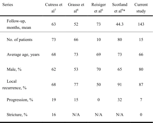
Although ureteroscopic management is a recognized option for selected patients, information about long-term survival outcomes and risks has been based on the results of relatively small series, without long-term follow-up. To date, no institution has reported outcomes beyond a mean follow-up of 10 years; the longest mean follow-up
6
period has been 6.1 years (Table 1) [6-9].
Our institution has over 28 years of experience in minimally invasive management of UTUC, with excellent follow-up. Herein, we report on the natural history of UTUC patients treated by ureteroscopic management, with a mean follow-up period of nearly 12 years.
7 2. Materials and Methods
2.1. Patients and procedures
We retrospectively collected data from all UTUC patients who underwent ureteroscopic management at Okayama University Hospital between January 1991 and April 2011. All patients were followed up for >6 years, except for 2 who died within 6 years. Ureteroscopic management was selected based on imperative indications, which were defined as a solitary kidney, compromised renal function, or poor general condition (primary indications), or individual surgeon and patient preference (elective indications) for patients with low grade and low stage UTUC, with consent obtained from each patient. Before ureteroscopy, all patients considered possible candidates underwent computed tomography (CT), cystoscopy, and cytology of voided urine. Patients with positive urine cytology were encouraged to undergo nephroureterectomy, however, 3 patients elected to undergo ureteroscopic management. Clinical and pathological information was acquired from hospital medical records to assess outcomes. Pathological verification was obtained via ureteroscopic cold-cup biopsy. Tumor staging was based on the 2002 American Joint Committee of Cancer TNM classification, and tumors were graded according to the World Health Organization International Society of Urological Pathology consensus classification [10, 11]. This retrospective study complied with the standards of the Declaration of Helsinki and current ethical
8
guidelines, and was approved by the Okayama University Institutional Review Board (Approval number; 1906-014).
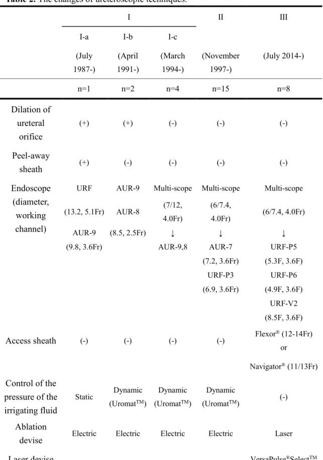
The ureteroscopic procedures and techniques used at our institution for observation of the upper urinary tract have been described previously [12-14]. Briefly, the distal to
middle portion of the ureter was inspected using a 6.9 Fr semi-rigid ureteroscope without a guide wire. An initial guide wire was then placed up to the middle ureter in order to avoid any inadvertent injury to the collecting system. The semi-rigid ureteroscope was removed and a flexible ureteroscope (Table 2) was passed over the guide wire into the ureter under fluoroscopic guidance. The guide wire was removed. The proximal ureter and the entire collecting system were inspected. Any suspicious lesion was biopsied with forceps and ablated.
Our technique has been evolved over 28-years, and the transition is summarized (Table 2). Flexible or semi rigid ureteroscopic therapy was performed with either 2/3 Fr electrocautery (100W) or Nd/Ho: YAG laser energy, removing all signs of visible tumor. At the end of the procedure, a single-J stent (6 Fr in diameter, external stent) or double-J stent (6 Fr in diameter) was placed for several days or until the next ureteroscopy, respectively, in cases that required ureteral dilation, ureteral access sheath, and/or ablation. Concurrent bladder tumors were treated with standard therapies. Bacillus
9
Calmette-Guerin therapy (BCG) for UTUC was used selectively in cases with positive cytology and frequent recurrence of UTUC. BCG (240 mg in normal saline [120 mL] at 1 mL/min for 2 hours) was administered weekly for a total of six times via a percutaneous suprapubic single-J stent placed in the renal pelvis. Suprapubic single-J stent is capped between instillations and kept during administration period, which is usually 6 weeks.
2.2. Follow-up
Ureteroscopy was generally conducted every 3 months with cystoscopy and urine cytologic analysis for at least the first 2 years. Additional ureteroscopy and cystoscopy were performed at 6-month intervals for the first 3 to 5 years. Then, these procedures were continued at 6-month intervals thereafter for up to 10 years. Whenever tumors recurred, the follow-up schedule was reset to the initial intervals. Evaluations of additional upper tract tumors or metastasis were conducted at 6-month intervals for the first 5 years, and at 12-month intervals for the next 5 years, by retrograde ureteropyelography intravenous urogram or CT urography depending on renal function. Intravenous urogram or CT urography was avoided when retrograde ureteropyelography is scheduled in next ureteroscopy.
10 2.3. Statistical analysis
The Kaplan-Meier method was used to estimate survival data, including overall survival (OS), cancer-specific survival (CSS), recurrence-free survival (RFS), and nephron-sparing survival (NSS). Tumor recurrence was defined as ipsilateral or contralateral upper tract recurrence determined ureteroscopically or radiologically, or the appearance of metastasis. CSS was determined when deaths could be directly attributed to UTUC by registry data and a review of clinical case notes. Statistical analyses were performed using EZR version 1.36 (Saitama Medical Center, Jichi Medical University), a graphical user interface for R [15].
11 3. Results
3.1. Patient demographics and tumor characteristics
From January 1991 to April 2011, 15 patients were newly managed with ureteroscopy for UTUC. The mean follow-up for all patients was 143 months (11.9 years), with a median follow-up of 140 months (11.7 years; range, 27-251 months [2.3-20.9 years]). All patients were followed up for >6 years, except for one who died of UTUC at 4.9 years, and one who died of lung cancer at 2.2 years, after initial ureteroscopic management.
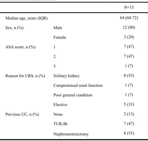
The patient demographics are listed, and the detailed ureteroscopic procedures for each patient are illustrated in a swimmer survival plot (Table 3) (Figure 1). The patient anesthetic fitness was generally classified according to American Society of Anesthesiologists (ASA) grade; only 1 patient was classified as ASA grade 3 (patient No. 13). Two patients underwent neither transurethral bladder tumor resection (TUR-Bt) nor nephroureterectomy before initial ureteroscopic management (patient No. 1 and 3), and 8 patients had been previously treated with nephroureterectomy, resulting in a solitary kidney (53%). Five patients (33%) were indicated as elective cases, including those with small, low-grade UTUC.
12
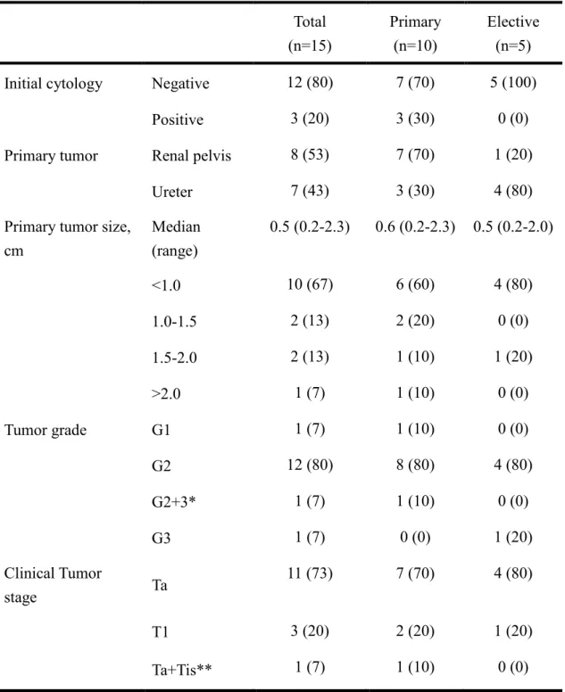
Fifty-three percent of tumors were in the renal pelvis, while 47 percent were in the ureter (Table 4). One patient had a tumor >2.0 cm in diameter (2.1 cm) that was over the limit of our indication for ureteroscopic management; nevertheless, ureteroscopic management was chosen because the patient had previously undergone nephroureterectomy and strongly wished to spare his solitary kidney to avoid aneuric dialysis (patient No. 12). One case had bilateral tumors managed with ureteroscopy; the left tumor was Ta (G2) and was treated ureteroscopically, while the right tumor was Tis (G3) and was treated with upper tract BCG (patient No. 14).
3.2. Procedure outcomes
During the follow-up period, 15 patients underwent a total of 268 (range, 5 to 35 per patient) ureteroscopy procedures and 66 (range, 1 to 11 per patient) laser ablation therapy procedures. Of the 268 procedures, two postoperative complications (1%) occurred, both of which were pyelonephritis. No patients developed ureteral stricture. Evaluation was not completed in one procedure because the ureteroscope could not pass a torturous ureter; a ureteral stent was placed and a second procedure was performed successfully 1 month later (patient No. 12).
13
Three patients underwent nephroureterectomy due to the recurrence which was not managed by ureteroscopy (patient No 5, 12 and 13). After nephroureterectomy, no recurrences were observed; however, 1 patient proceeded to hemodialysis due to loss of bilateral kidneys (patient No. 12); this was the only case in the cohort that required dialysis. Note this is the patient who had an initial tumor size of 2.1 cm, which is outside the indication, as previously described. He received two courses of neoadjuvant gemcitabine, cisplatin, and paclitaxel (GCP) combination chemotherapy, and the tumor decreased from 2.1 cm to 1.3 cm. Then, laser ablation was performed, followed by one additional course of GCP chemotherapy. The recurrent tumor was too extensive to be managed ureteroscopically; thus, the patient underwent right nephroureterectomy and initiated dialysis. Two cases received chemotherapy, including this case. The other case received tegafur-uracil for lymph node metastasis (patient No 14), and died of UTUC 4.9 years after initial ureteroscopy.
BCG perfusion therapy for UTUC was performed in 7 patients (patient No. 1, 3, 6, 8, 11, 14, and 15) (Figure 1). The purpose of BCG was prevention of recurrence of UTUC in 6 patients (patient No. 1, 3, 6, 8, 11, and 15) and treatment of CIS in the contralateral side in 1 patient (left side of patient No. 14). Of these, 4 patients did not experience recurrence following upper tract BCG.
14 3.3. Survival outcomes
Overall, 8 (53%) patients died during a mean follow-up of 11.7 years. Only 1 patient (7%) experienced cancer-specific mortality, as described previously. The other causes of death were lung cancer (2/15 [14%]), pneumonia (2/15 [14%]), prostate cancer (1/15 [7%]), small intestine perforation from ileus (1/15 [7%]), and aging at 85 years (1/15 [7%]). In Kaplan-Meier estimates of survival outcomes, the estimated CSS rates were 93% at 5, 10, 15, and 20 years, respectively (Table 5) (Figure 2). The estimated OS rates were 86%, 80%, 54%, and 20% at 5, 10, 15, and 20 years, respectively. The estimated mean and median OS were 14.5 and 16.6 years, respectively, while the estimated median CSS time was not reached. A total of 54 upper tract recurrences were experienced over a mean follow-up of 11.0 years, with an incidence of 3.6 per patient for the entire cohort. The location of recurrences is described in Table 6. Two patients did not experience recurrence after initial ureteroscopic ablation (patient No. 3 and 4). Estimated RFS was 13% at 5 years, and median RFS was 12 months. The estimated NSS was 79% at 5 years and the median NSS time was not reached. Eight patients developed 26 bladder recurrences after initial UTUC, and TUR-Bt was performed along with ureteroscopic management in these cases.
15 4. Discussion
The present study illustrates the natural history of UTUC patients undergoing ureteroscopic management. This study has the longest follow-up duration (mean, 11.9 years) accompanied by complete and detailed descriptions of each patient course compared to previous studies (Table 1) [6-9]. During nearly 12 years of follow-up, only 1 patient experienced disease-specific mortality. The present results provide further reassurance to both clinicians and patients regarding the safety of ureteroscopic management as a treatment option for select patients with UTUC.
Endoscopic management of UTUC has generally been limited to solitary low-grade disease in high-risk patients. However, in recent years, the indications for this type of management have begun to be applied in carefully selected patients with a normal contralateral kidney [1]. Even in the present study, 5 (33%) patients electively underwent ureteroscopic management. Among these patients, survival outcomes compared to nephroureterectomy are the biggest concern. The UTUC Collaborative reported the outcomes of 1363 patients who underwent nephroureterectomy, with 5-year CSS rates of 93.5% for patients with pT0/Ta/Tis disease and 91.0% for patients with pT1 disease, and 10-year CSS rates of 89.6% for patients with pT0/Ta/Tis disease and 85.4% for patients with pT1 disease [16]. The estimated CSS rates in the present series
16
compared favorably, with 5-, 10-, and 20-year CSS rates of 93% overall. These survival outcomes are comparable to those reported for similar patients in the literature (Table 5). However, 15- and 20-year CSS rates are lacking in other studies. Our long observation period provides new and excellent information, leading to further support of this indication [8, 17].
OS was also favorable in the present study, with estimated 5- , 10-, 15- , and 20-year OS rates of 86%, 80%, 54%, and 20%, respectively. In previous studies of ureteroscopic management, estimated 5-year OS rates of 57-75% and 10-year OS rates of 40-56% have been reported [4, 8, 18]. No previous studies have provided 15- or 20-year OS data. The patient backgrounds in the present study were comparable to those in previous studies (Table 1). The observed favorable OS might have been influenced by favorable CSS. The 20-year OS rate of 20% appears to be relatively low. However, considering that the mean age at diagnosis is 66 years and more than half of the present patients had a solitary kidney, favorable OS is expected. The average Japanese life span is 84 years. High CSS and low long-term OS is characteristic of cancers of low malignant potential, such as low-risk prostate cancer, in which radical prostatectomy is not always the best management option [19]. The present results suggest that ureteroscopic management of UTUC is a reasonable option in select patients compared to nephroureterectomy. Our
17
favorable outcomes seem to be associated with high adherence of our patients to strict follow-up protocol. We conducted ureteroscopic management for only patients who consent the frequent ureteroscopy instead of preserving renal function.
Despite the favorable survival outcomes observed herein, ureteroscopic treatment is associated with a high overall upper tract recurrence rate of 87%, with 5- and 10-year RFS rates of 13% and 13%, respectively. These low RFS rates also emphasize the importance of strict follow-up and patient compliance as a requirement for successful endoscopic management. Despite the high upper tract recurrence rate, 2 patients who electively underwent ureteroscopic management had no recurrences and avoided unnecessary nephroureterectomy. In addition, the NSS was 79% at 5, 10, and 20 years. These data suggest that ureteroscopic management can yield a high long-term nephron-sparing rate despite a high rate of upper tract recurrence in select patients.
Preventing unnecessary nephroureterectomy reduces the risk of complications associated with surgery or chronic renal failure, especially in cases with a solitary kidney. Ureteroscopic management is associated with fewer complications than nephroureterectomy [20]. In addition, we previously reported that lower renal function due to a solitary kidney influences the risk of cardiovascular events [21]. It has been well documented that once placed on dialysis, elderly patients have limited survival,
18
even in Japan. The survival rates of dialysis patients in 2012 were 73% at 3 years and 60% at 5 years; [22] the 5-year survival rate was worse in elderly patients at 27% [22, 23].
Fifty-three percent of patients in the present study developed intravesical recurrence, which is similar to the percentages in other ureteroscopic reports [6, 7, 17]. However, all intravesical recurrences were noninvasive papillary tumors, and were resected endoscopically without total cystectomy.
BCG is currently the most common drug for upper urinary tract perfusion; however, its utility is still regarded as investigational because of the small sample size of most previous studies [1, 24, 25]. A report with the largest experience showed no significant benefit associated with adjuvant BCG perfusion after ablation or percutaneous resection of UTUC [26]. However, BCG perfusion therapy for UTUC was effective in the present study: 4 of 6 patients (67%) with G2 UTUC did not experience upper tract recurrence after BCG. The role of BCG perfusion after ablation of Ta/T1 tumors is not as apparent, and should be evaluated on an individual basis.
Almost all procedures were performed in the present study without complications. A major complication of ureteroscopic treatment is ureteric stricture, which occurs in 3% to 23% of patients; fortunately, no patients in the present study developed stricture [7,
19
17, 27]. In general, stricture is treated by stenting, balloon dilatation, or laser incision; however, rare cases require nephroureterectomy [28]. Additional concerns include undergrading and the overlooking of UTUC lesions. One study reported that lesions were missed in up to 25% of patients, and nearly 50% of carcinoma in situ lesions were missed [28]. Therefore, intensive endoscopic surveillance is critical during follow-up. The performance of endoscopes has been continuously developed over the year. The introduction of new imaging including NBI with Olympus🄬🄬, SPIES with Storz🄬🄬 might help decrease the frequency of URS and recurrences. The introduction of UroVysion fluorescence in situ hybridization can also improve the diagnostic accuracy of UTUC [29]. Furthermore, photodynamic diagnosis using 5-Aminolevulinic Acid might be helpful for diagnosis and ablation also in UTUC in the future [30]. As newly alternative kidney-sparing treatment, primary chemoablation using instillation of UGN-101, a mitomycin-containing gel for low-grade UTUC might offer the disease eradication [31].
Limitations of this study include its retrospective nature and selection bias, and as with most other UTUC endoscopic management series, the study had a very small sample size and was conducted at a single center. The incidence of non-invasive UTUC is extremely rare; therefore, multicenter studies are required to elucidate survival efficacy of nephron-sparing management. Nonetheless, the very long duration of
20
follow-up of this study, which is double that of previous studies, makes our data unique and strong among published studies. Another limitation is very long enrollment period. Most of our procedures were done with electrocautery, however today, Holmium or Neodynium laser is commonly used for this indication due to tremendous advances in technology. These laser treatments are known more effective and safer [32]. In our institution, laser ablation was introduced in 2014, and even better results are expected.
5. Conclusions
The present study provides information regarding the natural history of UTUC patients treated with ureteroscopic management. Although survival outcomes with a mean follow-up of 12 years were excellent, upper tract recurrence was common. Thus, under strict surveillance, we advocate the use of ureteroscopic management for properly selected patients with UTUC.
Acknowledgements
The authors thank the clinical laboratory technicians of Okayama University Hospital for their technical support.
21 Funding
The authors have declared no funding.
Conflict of interest
22 References
1. Roupret M, Babjuk M, Burger M, et al (2019) EAU Guidelines on Upper Urinary
Tract Tumours 2019. ISBN 978-94-92671-07-3. EAU Guidelines Office, Arnhem, The Netherlands. http://uroweb.org/guidelines/compilations-of-all-guidelines/
2. Margulis V, Shariat SF, Matin SF, et al (2009) Outcomes of radical
nephroureterectomy: a series from the Upper Tract Urothelial Carcinoma Collaboration. Cancer 115:1224-1233.
3. Stewart GD, Bariol SV, Griqor KM, et al (2005) A comparison of the pathology of
transitional cell carcinoma of the bladder and upper urinary tract. BJU Int
95:791-793.
4. Cutress ML, Stewart GD, Zakikhani P, et al (2012) Ureteroscopic and percutaneous
management of upper tract urothelial carcinoma (UTUC): systematic review. BJU Int 110:614-628.
5. Enqelmyer EI, Belis JA (1996). Long-term ureteroscopic management of low-grade
transitional cell carcinoma of the upper urinary tract. Tech Urol 2:113-116.
6. Reisiger K, Hruby G, Clayman RV, et al (2007) Office-based surveillance
ureteroscopy after bendoscopic treatment of transitional cell carcinoma: technique and clinical outcome. Urology 70:263-266.
23 7. Cutress ML, Stewart GD, Wells-Cole S, et al (2012) Long-term endoscopic
management of upper tract urothelial carcinoma: 20-year single-centre experience. BJU Int 110:1608-1617.
8. Grasso M, Fishman AI, Cohen J, et al (2012) Ureteroscopic and extirpative
treatment of upper urinary tract urothelial carcinoma: a 15-year comprehensive review of 160 consecutive patients. BJU Int 110:1618-1626.
9. Scotland KB, Kleinmann N, Cason D, et al (2018) Ureteroscopic Management of
Large ≥2 cm Upper Tract Urothelial Carcinoma: A Comprehensive 23-Year Experience. Urology 121:66-73.
10. Paner GP, Stadler WM, Hansel DE, et al (2018) Updates in the Eighth Edition of the
Tumor-Node-Metastasis Staging Classification for Urologic Cancers. Eur Urol
73:560-569.
11. Lopez-Beltran A, Bassi P, Pavone-Macaluso M, et al (2004) Handling and pathology
reporting of specimens with carcinoma of the urinary bladder, ureter, and renal pelvis. Eur Urol 45:257-266.
12. Araki M, Uehara S, Sasaki K, et al (2012) Ureteroscopic management of choronic
unilateral hematuria: a single-center experience over 22 years. PLoS One 7:e36729.
24 13. Takao A, Saika T, Uehara S, et al (2010) Indications for ureteropyeloscopy based on
radiographic findings and urine cytology in detection of upper urinary tract carcinoma. Jpn J Clin Oncol 40:1087-1091.
14. Kumon H (1996) Ureteroscopy: indications for surgical intervention. CHAPTER28,
Smith’s Textbook of Endourology. Quality Medical Publishing, Inc, 397-410.
15. Kanda Y (2013) Investigation of the freely available easy-to-use software ‘EZR’ for
medical statistics. Bone Marrow Transplant 48:452-458.
16. Margulis V, Shariat SF, Matin SF, et al (2009) Outcomes of radical
nephroureterectomy: a series from the Upper Tract Urothelial Carcinoma Collaboration. Cancer 115:1224-1233.
17. Seisen T, Peyronnet B, Dominguez-Escrig JL, et al (2016) Oncologic Outcomes of
Kidney-sparing Surgery Versus Radical Nephroureterectomy for Upper Tract Urothelial Carcinoma: A Systematic Review by the EAU Non-muscle Invasive Bladder Cancer Guidelines Panel. Eur Urol 70:1052-1068.
18. Thompson RH, Krambeck AE, Lohse CM (2008) Endoscopic management of upper
tract transitional cell carcinoma in patients with normal contralateral kidneys. Urology 71:713-717.
25 19. Hamdy FC, Donovan JL, Lane JA, et al (2016) 10-Year Outcomes after Monitoring
Surgery, or Radiotherapy for Localized Prostate Cancer. N Eng J med
375:1415-1424.
20. Gadzinski AJ, Roberts WW, Faerber GJ, et al (2010) Long-term outcomes of
nephroureterectomy versus endoscopic management for upper tract urothelial carcinoma. J Urol 183:2148-2153.
21. Kambara T, Tanimoto R, Araki M, et al (2018) Renal Function after Nephrectomy
Infuences the Risk of Cardiovascular Events, Acta Med Okayama 72:241-247. 22. Nakai S, Hanafusa N, Masakane I. et al (2014) An overview of regular dialysis
treatment in Japan (as of 31 December 2012). Ther Apher Dial 18:535-602.
23. Masakane I (2010) High-quality dialysis: a lesson from the Japanese experience:
Effects of membrane material on nutritional status and dialysis-related symptoms. NDT Plus 3:i28-i35.
24. Giannarini G, Kessler TM, Birkhauser FD, et al (2011) Anterrage perfusion with
bacillus Calmette-Guerin in patients with non-muscle-invasive urothelial carcinoma of the upper urinary tract: who may benefit? Eur Urol 60:955-960.
25. Knoedler JJ, Raman JD (2018) Intracavitary therapies for upper tract urothelial
26 26. Rastinehad AR, Ost MC, Vanderbrink BA, et al (2009) A 20-year experience with
percutaneous resection of upper tract transitional carcinoma: is there an oncologic benefit with adjuvant bacillus Calmette Guerin therapy? Urology 73:27-31.
27. Wen J, Ji ZG, Li HZ (2018) Treatment of upper tract urothelial carcinoma with
ureteroscopy and thulium laser: a retrospective single center study. BMC Cancer
18:196.
28. Yamany T, van Batavia J, Ahn J (2015) Ureterorenoscopy for upper tract urothelial
carcinoma: how often are we missing lesions? Urology 85:311-315.
29. Sassa N, Iwata H, Kato M, et al (2019) Diagnostic Utility of UroVysion Combined
With Conventional Urinary Cytology for Urothelial Carcinoma of the Upper Urinary Tract. Am J Clin Pathol 151:469-478.
30. Ahmad S, Aboumarzouk O, Somani B, et al (2012) Oral 5-aminolevulinic acid in
simultaneous photodynamic diagnosis of upper and lower urinary tract transitional cell carcinoma – a prospective audit. BJU Int 110:E596-600.
31. Kleinmann N, Matin SF, Pierorazio PM, et al (2020) Primary chemoablation of
low-grade upper tract urothelial carcinoma using UGN-101, a mitomycin-containing reverse thermal gel (OLYMPUS): an open-label, single-arm, phase 3 trial. Lancet Oncology 21:776-785.
27 32. Raman JD and Park R (2017) Endoscopic management of upper-tract urothelial
28 Figure legends
Figure 1. Swimmer survival plot for subjects who underwent ureteroscopic
management. No patient who had T1 (dark green) disease died of upper tract urothelial
carcinoma (UTUC). The only case who died of UTUC (No. 14) had bilateral tumors: left tumor was Ta (G2), which was treated ureteropscopic ablatoin, while the right tumor was Tis (G3), which was treated with upper tract BCG.
29 Table 1. Ureteroscopic treatment of upper tract urothelial carcinoma; long-term
follow-up. Series Cutress et al7 Grasso et al8 Reisiger et al6 Scotland et al9* Current study Follow-up, months, mean 63 52 73 44.3 143 No. of patients 73 66 10 80 15
Average age, years 68 73 69 73 66
Male, % 62 53 70 65 80
Local
recurrence, % 68 77 50 91 87
Progression, % 19 15 0 32 7
Stricture, % 16 N/A N/A N/A 0
30 Table 2. The changes of ureteroscopic techniques.
Ⅰ Ⅱ Ⅲ Ⅰ-a Ⅰ-b Ⅰ-c (July 1987-) (April 1991-) (March 1994-) (November 1997-) (July 2014-) n=1 n=2 n=4 n=15 n=8 Dilation of ureteral orifice (+) (+) (-) (-) (-) Peel-away sheath (+) (-) (-) (-) (-) Endoscope (diameter, working channel)
URF AUR-9 Multi-scope Multi-scope Multi-scope (13.2, 5.1Fr) AUR-8 (7/12,
4.0Fr)
(6/7.4,
4.0Fr) (6/7.4, 4.0Fr)
AUR-9 (8.5, 2.5Fr) ↓ ↓ ↓
(9.8, 3.6Fr) AUR-9,8 AUR-7 URF-P5
(7.2, 3.6Fr) (5.3F, 3.6F)
URF-P3 URF-P6
(6.9, 3.6Fr) (4.9F, 3.6F) URF-V2 (8.5F, 3.6F)
Access sheath (-) (-) (-) (-) Flexor® (12-14Fr)
or Navigator® (11/13Fr) Control of the pressure of the irrigating fluid Static Dynamic (UromatTM) Dynamic (UromatTM) Dynamic (UromatTM) (-) Ablation
devise Electric Electric Electric Electric Laser
31 (Ho: YAG 80W/ Nd: YAG 100W)
Intensity and
laser pulses 30W 30W 30W 30W
Ho: YAG laser for ablation (0.5-0.6J, 5-10Hz)
or
Nd: YAG laser for coagulation (20W, continuous)
32 Table 3. Patient characteristics.
N=15
Median age, years (IQR) 64 (60-72)
Sex, n (%) Male 12 (80)
Female 3 (20)
ASA score, n (%) 1 7 (47)
2 7 (47)
3 1 (7)
Reason for URS, n (%) Solitary kidney 8 (53)
Compromised renal function 1 (7)
Poor general condition 1 (7)
Elective 5 (33)
Previous UC, n (%) None 2 (13)
TUR-Bt 7 (47)
Nephroureterectomy 8 (53)
ASA, American Society of Anesthesiologists; IQR, interquartile range; TUR-Bt, transurethral bladder tumor resection; UC, urothelial carcinoma; URS, ureteroscopy.
33 Table 4. Tumor characteristics (%).
Total (n=15) Primary (n=10) Elective (n=5)
Initial cytology Negative 12 (80) 7 (70) 5 (100)
Positive 3 (20) 3 (30) 0 (0)
Primary tumor Renal pelvis 8 (53) 7 (70) 1 (20)
Ureter 7 (43) 3 (30) 4 (80)
Primary tumor size, cm Median (range) 0.5 (0.2-2.3) 0.6 (0.2-2.3) 0.5 (0.2-2.0) <1.0 10 (67) 6 (60) 4 (80) 1.0-1.5 2 (13) 2 (20) 0 (0) 1.5-2.0 2 (13) 1 (10) 1 (20) >2.0 1 (7) 1 (10) 0 (0) Tumor grade G1 1 (7) 1 (10) 0 (0) G2 12 (80) 8 (80) 4 (80) G2+3* 1 (7) 1 (10) 0 (0) G3 1 (7) 0 (0) 1 (20) Clinical Tumor stage Ta 11 (73) 7 (70) 4 (80) T1 3 (20) 2 (20) 1 (20) Ta+Tis** 1 (7) 1 (10) 0 (0)
*Bilateral case: the left-side lesion was G2, and the right-side lesion was a G3 carcinoma in situ.
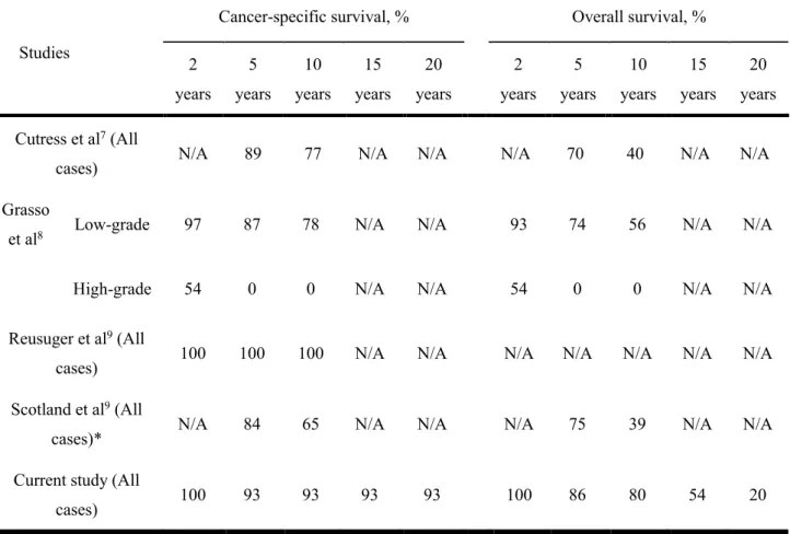
34 Table 5. Survival outcomes for ureteroscopic treatment of upper tract urothelial
carcinoma; long-term follow-up.
Studies
Cancer-specific survival, % Overall survival, % 2 years 5 years 10 years 15 years 20 years 2 years 5 years 10 years 15 years 20 years Cutress et al7 (All
cases) N/A 89 77 N/A N/A N/A 70 40 N/A N/A
Grasso
et al8 Low-grade 97 87 78 N/A N/A 93 74 56 N/A N/A
High-grade 54 0 0 N/A N/A 54 0 0 N/A N/A
Reusuger et al9 (All
cases) 100 100 100 N/A N/A N/A N/A N/A N/A N/A Scotland et al9 (All
cases)* N/A 84 65 N/A N/A N/A 75 39 N/A N/A
Current study (All
cases) 100 93 93 93 93 100 86 80 54 20