52:660 症例報告
ものわすれ外来でフォロー中に軽度認知障害から認知症へ移行した
嗜銀顆粒性認知症の 1 剖検例
岩崎
靖
1)2)*森
恵子
2)伊藤 益美
2)辰己 新水
1)三室 マヤ
1)吉田 眞理
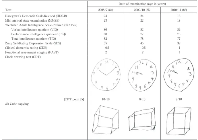
1) 要旨:症例は死亡時 87 歳の女性である.84 歳時に自ら記銘力の低下を訴えてものわすれ外来を受診し,約 3 年間外来フォローされた.初期には軽度認知障害と診断されたが自己中心的な行動がめだった.経過観察中に認知 症へ移行し,Alzheimer 病と臨床診断された.経時的な臨床検査所見では立方体図形模写課題が良好であり,画像所 見では左右差のある側頭葉内側面の萎縮,進行性の扁桃核と海馬の萎縮がみとめられた.病理学的検索により Al-zheimer 病変は軽度で,認知機能障害は嗜銀顆粒性認知症によると考えられた. (臨床神経 2012;52:660-665) Key words:嗜銀顆粒性認知症,Alzheimer病,認知症,精神症状,軽度認知障害 はじめに嗜銀顆粒性認知症(argyrophilic grain dementia)は Braak らにより記載された老年期の認知症性疾患である1)2).剖検症 例の検討から高齢者においては頻度的にまれでないことが明 らかにされているが2),診断には病理学的検索が必須であるた め臨床所見や経過,画像所見に関する 検 討 は 十 分 で は な い3)4).多くの症例は臨床的に Alzheimer 病や前頭側頭型認知 症,脳血管性認知症と誤診断されている3)4).われわれは自ら 記銘力の低下を訴えてものわすれ外来を受診し,約 3 年間の フォロー中に軽度認知障害から認知症へ移行した嗜銀顆粒性 認知症の高齢女性例を経験したので臨床経過,高次脳機能検 査所見,画像所見および病理所見を検討して報告する. 症 例 患者:死亡時 87 歳の女性 主訴:ものわすれ 家族歴・生活歴:特記すべきことなし.夫と死別し,2000 年に B 型軽費老人ホームに入所. 既往歴:2004 年に後頭部痛を訴えて小山田記念温泉病院 を受診し,頭部 CT にて左優位の軽度側脳室下角拡大をみと めた(Fig. 1a).2006 年に眩暈感を訴えて再診し,頭部 CT では側脳室下角の拡大が若干進行していた(Fig. 1b).他は特 記すべきことなし. 現病歴:2008 年(84 歳時)にものわすれを自覚して,もの わすれ外来を受診した. 初診時所見:意識は清明であった.発語は流暢で,構音障害 や明らかな失語はなかった.亀背だが歩行はスムーズで,失調 や小刻み歩行,すくみ足はみとめなかった.筋強剛や静止時振 戦はなく,パーキンソニズムは明らかでなかった.腱反射は上 肢はやや亢進,下肢は正常で左右差はなく,Babinski 徴候は 陰性だった.把握反射などの前頭葉徴候はみとめなかった.頭 部 MRI では脳萎縮と左優位の側脳室下角の拡大,扁桃核と海 馬の萎縮が前回 の 頭 部 CT と く ら べ て 若 干 進 行 し て い た (Fig. 1c).高次脳機能検査では Wechsler Adult Intelligence Scale-Revised(WAIS-R)にて言語性 IQ 86,動作性 IQ 80,全 IQ 82,改訂長谷川式簡易知能評価スケール(Hasegawa s De-mentia Scale-Revised;HDS-R)は 24 点,Mini mental state examination(MMSE)は 23 点,立方体模写は良好,時計描 画 試 験5)は 10!10 で,Zung の Self-rating Depression Scale (SDS)は 35 点だ っ た(Table 1).Clinical Dementia Rating (CDR)6)は 0.5,Functional Assessment Staging(FAST)7)は 2
と判定した.血清梅毒反応検査は陰性だった. 経過:初診時は軽度認知障害8)と診断し,ほぼ月に 1 回の通 院を継続した.ものわすれに加えて難聴,不眠,膝痛の訴えが 常同的であったが深刻感はなく,抑うつ傾向は明らかでな かった.多弁で,自己中心的な行動(都合の良い日に来院する, 予約時間外に受診する,他患者の診察中に診察室に入ってく る,診察中も一方的に話すなど)がめだった.初診 1 年後の頭 部 MRI では脳萎縮と側脳室拡大がやや進行し,高次脳機能検 * Corresponding author: 愛知医科大学加齢医科学研究所〔〒480―1195 愛知県長久手市岩作雁又 1 番地 1〕 1) 愛知医科大学加齢医科学研究所神経病理部門 2) 小山田記念温泉病院神経内科 (受付日:2012 年 2 月 22 日)
Fig. 1 Head CT and MRI images of the patient.
a: CT obtained at 80 years of age, 4 years before the onset of mild cognitive impairment. The pa-tient consulted her physician because of headache, and no apparent cognitive impairment was rec-ognized. Although mild ventricular dilatation is observed, cerebral atrophy is not readily apparent. The inferior horn of the lateral ventricle shows left-side-dominant dilatation. b: CT obtained at 82 years of age, 2 years before the onset of mild cognitive impairment. The patient consulted her phy-sician because of dizziness, and again, no apparent cognitive impairment was recognized. Left-side-dominant dilatation of the inferior horn of the lateral ventricle has progressed slightly with atrophy of the amygdala. c: MRI: T1 weighted image (0.5 Tesla, TR 350 ms, TE 17 ms) obtained 3 years
be-fore death. Atrophy of the cerebrum, amygdala and hippocampus, and lateral ventricular dilatation have progressed slightly. d: MRI: T1 weighted FLAIR image (3.0 Tesla, TR 2,100 ms, TE 10 ms)
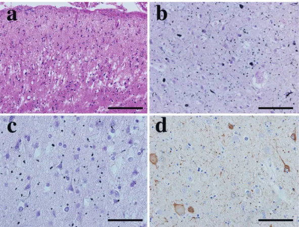
ob-tained 6 months before death. Cerebral atrophy and lateral ventricular dilatation have progressed. Upper images: At the level of the hippocampus. Lower images: At the level of the corona radiata. R: right side. 査 で は WAIS-R に て 言 語 性 IQ 82,動 作 性 IQ 77,全 IQ 78, HDS-R は 24 点,MMSE は 22 点,立方体模写は良好,時計描 画試験は 8!10 で,SDS は 45 点であった.CDR は 0.5,FAST は 2 と判定した.初診 2 年後(86 歳時)に自炊や服薬管理が 困難となり,日常生活に介助が必要となったため A 型軽費老 人ホームへ移動した.バスを利用し一人で通院することは可 能であったが,受診時に予約券や診察券を忘れるエピソード が続き,補聴器の使用が困難となったためコミュニケーショ ンが若干不自由となった.頭部 MRI では脳萎縮,側脳室拡大, 扁桃核と海馬の萎縮が進行し(Fig. 1d),高次脳機能検査では, WAIS-R にて言語性 IQ 82,動作性 IQ 75,全 IQ 77,HDS-R は 13 点,MMSE は 18 点,立方体模写は良好,時計描画試験は 8!10 で,SDS は 39 点であった.CDR は 1,FAST は 4 と判定 し,認知症へ移行したと臨床診断した.塩酸ドネペジルを投与 したが,臨床的に明らかな効果はなく,過剰反応などの副作用 もみとめられなかった.初診から約 3 年後に急性心筋梗塞の ため急死した.死亡直前まで疎通性は比較的保たれ,言語機能 の障害は明らかでなく,最終的な CDR は 1,FAST は 4 だっ た.臨床診断は Alzheimer 病だった. 病理所見:脳重は 1,250g で,肉眼的に限局性の脳萎縮はな かった.大脳冠状断では側頭葉の萎縮と側脳室の拡大,扁桃体 の萎縮がめだったが,海馬の萎縮は高度ではなかった(Fig. 2).黒質,青斑核の色調はよく保たれ,小脳,脳幹には著変を みとめなかった.組織学的には海馬傍回表層の神経細胞脱落 とグリオーシス,neuropil の粗鬆化,海綿状変化がめだち (Fig. 3a),海馬支脚では神経細胞脱落とグリオーシスをみと めた.扁桃核には神経細胞脱落をみとめ,ballooned neuron と肥胖性アストロサイトの増生がめだった.Gallyas-Braak 染色により明瞭に染色される嗜銀顆粒を迂回回(Fig. 3b),扁 桃核,海馬から移行嗅内野,後頭側頭回(Fig. 3c),下側頭回, 島葉,視床下部外側隆起まで広範にみとめ Saito stage III9)に 相当した.抗リン酸化タウ抗体(AT-8,Innogenetics)をもち いた免疫染色では,嗜銀顆粒に加えてタウ陽性の ballooned neuron,pretangle,coiled body,thread を み と め た(Fig.
臨床神経学 52巻9号(2012:9) 52:662
Fig. 2 Macroscopic image of a coronal section of the
cere-bral hemisphere at the level of the hippocampus. Although temporal lobe atrophy is present, atrophy of the hippocampus is not remarkable. Dilatation of the an-terior and inferior horns of the lateral ventriculus is ob-served. Scale bar, 10 mm. L: Left side.
Table 1 Progression in results of the cognitive function test battery.
Date of examination (age in years)
Test 2008/7 (84) 2009/10 (85) 2010/11 (86)
Hasegawa s Dementia Scale-Revised (HDS-R) 24 24 13
Mini mental state examination (MMSE) 23 22 18
Wechsler Adult Intelligence Scale-Revised (WAIS-R)
Verbal intelligence quotient (VIQ) 86 82 82
Performance intelligence quotient (PIQ) 80 77 75
Total intelligence quotient (TIQ) 82 78 77
Zung Self-Rating Depression Scale (SDS) 35 45 39
Clinical dementia rating (CDR) 0.5 0.5 1
Functional assessment staging (FAST) 2 2 4
Clock drawing test (CDT)
(CDT point [5]) 10/10 8/10 8/10
3D Cube-copying
3d).神経原線維変化は海馬支脚,海馬傍回に出現し,Braak stage III に相当した.側頭葉,後頭葉に CERAD stage B に相 当する,びまん性老人斑をみとめたが,アミロイドアンギオパ チーの所見はなかった.細動脈硬化はめだたず,梗塞巣や出血 巣はみとめられなかった.抗リン酸化 TDP-43 抗体をもちい た免疫染色では,海馬傍回の神経原線維変化や ghost tangle の出現部位に一致して少量の陽性所見をみとめた.海馬歯状 回顆粒細胞にもリン酸化 TDP-43 陽性の dot 状構造物をごく 少量みとめたが,脊髄の下位運動ニューロンに陽性所見はな かった.tuft-shaped astrocyte,astrocytic plaque,Lewy 小体, Pick 小体はみとめられなかった. 考 察 本症例は自覚的なものわすれを訴えてものわすれ外来を受 診し,経過中に軽度認知障害から認知症へ移行した高齢女性 例である.臨床的には Alzheimer 病と診断されたが,初期に は記銘力低下は高度ではなく,自己中心的な行動がめだった. 精神症状も高度ではなく,語義失語をふくめて明らかな失語 はなかった.病理学的には Alzheimer 病変は軽度で,嗜銀顆 粒が辺縁系を中心に広範に出現し,神経細胞脱落とグリオー シス,neuropil の粗鬆化を呈し,他の認知症を呈する神経変性 疾患や血管障害をみとめなかったことから,認知症の原因疾 患は嗜銀顆粒性認知症と考えられた.剖検多数例の検討から 嗜銀顆粒性認知症は神経変性症性の認知症疾患の中では Al-zheimer 病に次ぐ頻度であることが強調されており2)10)11),一
Fig. 3 Representative microscopic images of the limbic lesions.
a. Apparent spongiform degeneration and severe neuron loss with gliosis and tissue rarefaction are observed in the superficial layer of the transentorhinal cortex. b. Presence of numerous argyrophil-ic grains in the neuropil is apparent in the ambient gyrus. Many argyrophilargyrophil-ic threads are also visi-ble. c. Many argyrophilic grains with a few threads are visible in the occipitotemporal gyrus. d. Tau-positive ballooned neurons, pretangles, coiled bodies, and threads are visible in the amygdala. Numerous tau-positive grains are also recognized. a: Hematoxylin and eosin staining; b, c: Gallyas-Braak silver staining. d: AT-8 immunostaining. Scale bars, a: 200 μm; b, d: 100 μm; c: 50 μm.
般に高齢発症で臨床経過が長い2)3). 前頭葉徴候や歩行障害, パーキンソニズムをみとめる症例も報告されているが2)12),本 症例ではそれらの神経症候はみとめられなかった.また嗜銀 顆粒性認知症は Alzheimer 病に比して,記銘力,言語,注意 力や遂行能力の障害が軽いと報告されており4)13) ,retrospec-tive にみれば本症例の臨床経過は Alzheimer 病より嗜銀顆 粒性認知症を示していた可能性があり,臨床的な鑑別点とし て重要であると思われた.また本症例の立方体図形模写課題 が良好であった点も Alzheimer 病との臨床的鑑別点となる 可能性が推定された.本症例の画像所見では側脳室下角の拡 大,扁桃核の萎縮は左側に優位であったが,嗜銀顆粒性認知症 における側頭葉内側萎縮の左右差は Alzheimer 病との鑑別 上重要な所見と考えられている14). 嗜銀顆粒は Alzheimer 病,Lewy 小体病,進行性核上性麻 痺,皮質基底核変性症,Pick 病,多系統萎縮症など多彩な神 経変性疾患にしばしばみとめられるため2)3)12),嗜銀顆粒の出 現と認知機能障害や神経症候,精神症状との関連を検討する ことを困難にしている.また認知症のない高齢者においても しばしば観察され,その頻度は加齢と と も に 増 加 す る た め2)3),嗜銀顆粒の出現のみでは認知症は出現しないと考えら れている10).一方で,嗜銀顆粒が主な原因と考えられる認知症 は,全認知症患者の 5∼10% 程度を占めると推定され,高齢に なるほどその頻度が増加する9)10).嗜銀顆粒性認知症における 認知機能障害は,辺縁系における神経細胞脱落やグリオーシ ス,neuropil の粗鬆化と関連し3)4)11),本症例の病理所見もこれ に矛盾しない.迂回回を中心とする皮質基底核結合部位の萎 縮,変性が高度となり,嗜銀顆粒の出現が前方から後方へ拡大 すると認知機能障害を呈し,嗜銀顆粒の出現が限局するばあ いは認知機能が保たれることが嗜銀顆粒単独病変例の検討か ら示されている9)11).本症例では抗リン酸化 TDP-43 抗体をも ちいた免疫染色で少量の陽性所見をみとめたが,タウの沈着 領域に一致しており二次的な変性による所見と思われた. 本症例において立方体図形模写課題が良好であった点や, 左右差をともなう側頭葉内側面の萎縮,進行性の扁桃核と海 馬の萎縮をみとめた点は嗜銀顆粒性認知症の臨床診断におい て参考となる所見と思われた. 謝辞:本症例の病理解剖を施行していただいた小山田記念温泉 病院病理検査科・永岡昌光先生,抗リン酸化 TDP-43 抗体を供与し
臨床神経学 52巻9号(2012:9) 52:664 ていただいた東京都医学総合研究所・長谷川成人先生に深謝いた します. ※本論文に関連し,開示すべき COI 状態にある企業,組織,団体 はいずれも有りません. 文 献
1)Braak H, Braak E. Argyrophilic grains: characteristic pa-thology of cerebral cortex in cases of adult onset demen-tia without Altzheimer changes. Neurosci Lett 1987;76: 124-127.
2)Braak H, Braak E. Argyrophilic grain disease: frequency of occurrence in different age categories and neuropa-thological diagnostic criteria. J Neural Transm 1998;105: 801-819.
3)吉田眞理. Argyrophilic grain dementia の神経病理. 神経 進歩 2004;48:419-427.
4)岩崎 靖, 森 恵子, 伊藤益美ら. 嗜銀顆粒性認知症の 1 剖 検例における臨床所見の検討. 神経内科 2009;71:186-192. 5)Rouleau I, Salmon DP, Butters N, et al. Quantitative and
qualitative analyses of clock drawings in Alzheimer s and Huntington s disease. Brain Cogn 1992;18:70-87.
6)Morris JC. The clinical Dementia Rating (CDR); Current version and scoring rules. Neurology 1993;43:2412-2414.
7)Reisberg B. Dementia: a systematic approach to identify-ing reversible causes. Geriatrics 1986;41:30-46.
8)Petersen RC. Mild cognitive impairment as a diagnostic entity. J Intern Med 2004;256:183-194.
9)Saito Y, Ruberu NN, Sawabe M, et al. Staging of argyro-philic grains : an age-related tauopathy. J Neuropathol Exp Neurol 2004;63:911-918.
10)Tolnay M, Clavaguera F. Argyrophilic grain disease: A late-onset dementia with distinctive features among tauopathies. Neuropathology 2004;24:269-283.
11)Saito Y, Nakahara K, Yamanouchi H, et al. Severe in-volvement of ambient gyrus in dementia with grains. J Neuropathol Exp Neurol 2002;61:789-796.
12)Jellinger KA. Dementia with grains (Argyrophilic grain disease). Brain Pathol 1998;8:377-386.
13)Steuerwald GM, Baumann TP, Taylor KI, et al. Clinical characteristics of dementia associated with argyrophilic grain disease. Dement Geriatr Cogn Disord 2007;24:229-234.
14)Adachi T, Saito Y, Hatsuta H, et al. Neuropathological asymmetry in argyrophilic grain disease. J Neuropathol Exp Neurol 2010;69:737-744.
Abstract
Clinicopathologic findings of argyrophilic grain dementia in a case of mild cognitive impairment converting to dementia
Yasushi Iwasaki, M.D.1)2) , Keiko Mori, M.D.2) , Masumi Ito, M.D.2) , Shinsui Tatsumi, M.D.1) , Maya Mimuro, M.D.1)
and Mari Yoshida, M.D.1) 1)
Department of Neuropathology, Institute for Medical Science of Aging, Aichi Medical University 2)
Department of Neurology, Oyamada Memorial Spa Hospital
An 84-year-old Japanese woman with no family history of dementia visited our memory clinic complaining of memory disturbance. Neurological examination revealed no apparent motor abnormalities, focal cerebral signs, parkinsonism, or cerebellar dysfunction. Hasegawa s Dementia Scale-Revised (HDS-R) and Mini mental state ex-amination (MMSE) scores were 24 and 23 points, respectively. MRI revealed left-side-dominant dilatation of the in-ferior horn of the lateral ventricle. Although egocentric behavior was remarkable, no disturbance of intelligence was apparent at the first examination, and she was diagnosed as having mild cognitive impairment. Her memory disturbance and disorientation gradually worsened. Atrophy of the cerebrum and dilatation of the lateral ventri-cle advanced gradually on MRI. Two years later, she required care to perform activities of daily living. HDS-R and MMSE scores had dropped to 13 and 18 points, respectively, and conversion to dementia was diagnosed. Ability to perform 3D cube-copying was well preserved. The patient died due to acute myocardial infarction at the age of 87. The clinical diagnosis was Alzheimer disease.
At autopsy, the brain weighed 1,250 g, and argyrophilic grains were widely observed in the limbic system, corresponding to Saito s stage III. Neuron loss, gliosis, spongiform change, and tissue rarefaction were recognized in the superficial layer of the parahippocampal gyrus. Ballooned neurons, pretangles, oligodendroglial coiled bod-ies, and neuropil threads were also observed. Neurofibrillary tangles and senile plaques, mainly consisting of dif-fuse plaque, were recognized as corresponding to Braak stage III and CERAD stage B, respectively. Neither Lewy nor Pick bodies were observed. Although mild phosphorylated TDP-43 immunoreactivity was observed, it was suspected to be due to secondary degeneration of tau deposition. The patient was diagnosed pathologically as having argyrophilic grain dementia.
The clinical findings of the present patient reveal important observations that help to clinically discriminate between various dementias such as Alzheimer disease and argyrophilic grain dementia.
(Clin Neurol 2012;52:660-665)