Title
Oncocytic papillary renal cell carcinoma の2例
Author(s)
松岡, 崇志; 市川, 千宙; 福永, 有伸; 矢野, 敏史; 杉野, 善雄;
岡田, 卓也; 今井, 幸弘; 川喜田, 睦司
Citation
泌尿器科紀要 = Acta urologica Japonica (2016), 62(4): 187-
191
Issue Date
2016-04-30
URL
http://hdl.handle.net/2433/212520
Right
許諾条件により本文は2017/05/01に公開
Type
Departmental Bulletin Paper
Textversion
publisher
Oncocytic papillary renal cell carcinoma
の
2
例
松岡 崇志
1,市川 千宙
2,福永 有伸
1,矢野 敏史
1杉野 善雄
1,岡田 卓也
1,今井 幸弘
2,川喜田睦司
11神戸市立医療センター中央市民病院泌尿器科 2神戸市立医療センター中央市民病院病理検査科
TWO CASES OF ONCOCYTIC PAPILLARY RENAL CELL CARCINOMA
Takashi Matsuoka1, Chihiro Ichikawa2, Arinobu Fukunaga1, Toshihumi Yano1,Yoshio Sugino1, Takuya Okada1, Yukihiro Imai2and Mutsushi Kawakita1 1The Department of Urology, Kobe City Medical Center General Hospital 2The Department of Pathology, Kobe City Medical Center General Hospital
Oncocytic papillary renal cell carcinoma isa variant of papillary renal cell carcinoma (PRCC). We herein report two cases treated with retroperitoneoscopic partial nephrectomy. Histologically, tumor cells of both cases exhibit round and regular nuclei with CK7 positive areas in the cytoplasm typical of TYPE1 PRCC and eosinophilic granular cytoplasm with E-cadherin positive areas in the cytoplasmic membrane, which indicates TYPE2 PRCC. Out of 46 cases reported in the literature, only one died of disease, which revealsitslow malignant potential.
(Hinyokika Kiyo 62 : 187-191, 2016) Key words : Renal cell carcinoma, Papillary renal cell carcinoma, Oncocytic papillary renal cell carcinoma
諸 言
近年,病理学的に稀な腎腫瘍が新たに同定されてい る.通常papillary renal cell carcinoma(以下PRCC) は腎癌の10∼15%に見られるとされるサブタイプであ り,TYPE 1とTYPE 2の2つに分類される1).今回, そのどちらのTYPE にも分類されないPRCCの亜型 で あ る oncocytic papillary renal cell carcinoma(以 下
OPRCC)を2例経験したので報告する. 症 例 患者1 : 89歳,男性 主 訴: CTで偶発的に腎腫瘍を指摘 既往歴: 前立腺癌 現病歴 : PSA 85.8と高値を認め,前立腺生検の結 果,前立腺癌と診断.StagingのCTで偶発的に右腎 下極に18 mm の腫瘍を指摘された.右腎癌疑いとし て2012年4月後腹膜鏡下右腎部分切除術を施行され た. 画像所見 : CT では動脈相で造影効果乏しく,T1 強調像でhigh,T2強調像でlowに描出される腎腫瘍 を認めPRCCが第一に疑われ,造影効果弱く鑑別と して腎血管筋脂肪腫(以下AML)が挙げられた(Fig. 1A,B). 患者2 : 63歳,男性 主 訴: エコーで偶発的に腎腫瘍を指摘 既往歴:高血圧,高脂血症 現病歴:他院にて高血圧でフォロー中,スクリーニ ングのエコーで左腎腫瘍を指摘された.CT・MRIで 15 mm 大の左腎腫瘍認め,左腎癌疑いとして2013年 2月後腹膜鏡下腎部分切除術を施行された. 画像所見: CTでは動脈相で造影効果乏しく,MRI ではT1強調像でhigh,T2強調像でlowに描出され る腎腫瘍を認めPRCCが疑われた(Fig. 2A,B). 病理所見: 肉眼的には症例1,2ともに境界明瞭で あり,割面上褐色顆粒状の病変であった.腫瘍周囲に は被膜様構造を認め,症例2では内部に一部出血を 伴っていた.組織学的には血管性の軸を有する乳頭状 構造からなり,顆粒状好酸性の細胞質を持つ多角形の 細胞となっていた.泡沫細胞は認めず,核は類円形で 核小体は目立たず大小不同も目立たなかった(Fig. 3A,B). ここで,核は軽度の異型度でありPRCCのTYPE 1 の所見を示すにもかかわらず,細胞質は好酸性で泡沫 細胞を認めないTYPE 2の所見を示した. 免疫染色では,TYPE 1の多くで陽性となる CK7 が陽性となり,またTYPE 2で陽性となることが多い E-cadherinも陽性となった.以上の所見より,PRCC のTYPE 1,2の所見が混在するOPRCCと診断した. 症例1は22カ月,症例2は12カ月転移再発は認めてい ない.今後10年以上にわたり採血およびCTでフォ ローアップ予定である.
泌62,04,03-1A
A 泌62,04,03-1B
B
Fig. 1. Preoperative image findingsof case 1 : Contrast enhanced CT scan showed a less enhanced small renal mass in the venous phase (A : circle). MRI demonstrated a renal mass with low intensity in T2-weighted phase (B : circle).
泌62,04,03-2A
A 泌62,04,03-2B
B
Fig. 2. Preoperative image findingsof case 2 : Contrast enhanced CT scan showed a less enhanced small renal mass in the arterial phase (A : circle). MRI demonstrated a renal mass with low intensity in T2-weighted phase (B : circle).
考 察 PRCCのTYPE 1の病理組織学的所見としては乳頭 状パターンを示し,淡色の細胞質と小さい核異型の乏 しい楕円形の核の細胞からなるとされる.泡沫マクロ ファ―ジや砂粒体が乳頭の間質にあれば診断に有用で ある.TYPE 2は豊富な好酸性細胞質をもつ高い核異 型度をもつ細胞からなり,泡沫マクロファージの存在 は稀とされる2).OPRCCは近年同定されたPRCCの 亜型であるが3),OPRCCに対してTYPE 1,2とわけ て分類することに関しては否定的な見解もあり4),ま だ2004年の WHO分類には挙げられていない.しか しLefevreらはOPRCCはPRCC TYPE 1では通常見 られないoncocytic cellが存在することや17 トリソ ミーの欠失が見られること,TYPE 2と比べて予後良 好と考えられることなどから通常のPRCCの臨床病 理学的特徴と異なると報告している5).悪性度などま だ判明していないことも多く今後症例の蓄積が必要と 考え今回はわけて報告する. PRCCのTYPE 1の病理組織学的所見としては乳頭 状パターンを示し,淡色の細胞質と小さい核異型の乏 しい楕円形の核の細胞からなるとされる.泡沫マクロ ファ―ジや砂粒体が乳頭の間質にあれば診断に有用で ある.TYPE 2は豊富な好酸性細胞質をもつ高い核異 型度をもつ細胞からなり,泡沫マクロファージの存在 は稀とされる2). 今 回 わ れ わ れ が 経 験 し た PRCC の 亜 型 で あ る OPRCCを2005年にHesらが初めて報告3)して以来, 症例ごとの詳細が記載されている報告は本症例をいれ て46例あり,本邦では過去に2例報告されている6,7). その詳細をTable 1に示す6~13)が,男性37人,女性9 人と男性に多い.年齢の中央値は67歳でわれわれの症 例も含め小径腎癌の報告が多いが,15 cm 大や27 cm 大 の 巨 大 な OPRCC の 報 告 も あ る3,5).現 在, OPRCCは核異型が軽度であるPRCC TYPE 1の特徴 と,細胞質が好酸性で泡沫細胞を持たないTYPE 2の 所見を併せ持つhybridのタイプと考えられているが, 報告はまだ少なく発生頻度などは不明である.悪性度 泌尿紀要 62巻 4 号 2016年 188
泌62,04,03-3A A 泌62,04,03-3B B 泌62,04,03-3C C
Fig. 3. Pathological findingsof the tumor regions (A : gross appearance of case 1. B : case 1 HE stain×40, C : case 2×40) tumor cells had eosinophilic granular cytoplasm with round and regular nuclei.
Table 1. OPRCC in recent literature
No 46
Median age (years) 67
(Range) (40-89)
Male : Female (37 : 9) Median tumor size (mm) 28
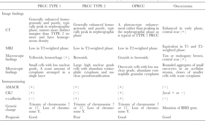
(Range) (6-270) Treatment Radical nephrectomy 33 Partial nephrectomy 13 Follow up (month) 78 (Range) (10-305) Recurrence 1 Metastasis 1 Clinical staging cT1aN0M0 32 cT1bN0M0 9 cT2aN0M0 2 cT2bN0M0 2 cT3aN0M0 1 Prognosis NED 45 DOD 1 は核の異型度に準ずるとされており,PRCC TYPE 1 と同等とされているが転移例や死亡例も1例報告され ていることから注意が必要である3,8).死亡例は術後 2年後に局所再発を来たし,さらに2年後に脳転移を 発症し死亡している3). OPRCCに関して免疫組織学的,形態学的に onco-cytomaとの相違点で議論されることが多い.形態学 的にはoncocytomaは乳頭状を取らない点が病理学的 に決定的に異なるとされている.また,免疫組織学的 に は 一 般 的 に PRCC は AMACR,CK7,CD10, E-cadherin,EMAや vimentinなどが陽性になることが 多 く,c-kit は 陰 性 と な る(Table 2).ま た 多 く の
oncocytoma で はc-kit,E-cadherin,EMA や vimentin
などが陽性となることが多く,AMACR や CD10, CK7やRCC,vimentinは陰性となる.PRCCにおい て AMACRは100%近い陽性率を示し2),oncocytoma で陽性になることが少ないことから両者の鑑別に有用 と さ れ て い る.PRCC に お い て は,TYPE 1 で は TYPE 2に比べCK7やMUC1が陽性になることが多 く,TYPE 2はTYPE 1に比べE-cadherinやCK20が 陽性のことが多いとされ,今回のわれわれの症例と一 致する. 画像所見としてはTseらが6例の患者を対象とし て造影CT所見を報告しており,OPRCCは皮髄相で 平均70.6 HU,腎実質相で69.9 HU,排泄相で58.7 HUでありPRCC TYPE 1と類似していると結論付け ている14).
また,oncocytoma では chromophobe cell carcinoma
との混在が時折見られるが11),PRCCと混在するこ とはきわめて稀とされ,OPRCCはPRCCと oncocy-tomaの混在とは別の病態と考えられている13).この ことは,OPRCCが染色体 7,17 のトリソミーやY 欠失といったPRCCに特徴的とされる遺伝子変異を 伴うことからも示されている3). 今回われわれは2症例を経験したが,比較的新しい 分類でもあることから現時点では報告が少なく,さら
Table 2. Variousfeaturesof PRCC TYPE 1, 2, OPRCC and Oncocytoma
PRCC TYPE 1 PRCC TYPE 2 OPRCC Oncocytoma Image findings
CT
Generally enhanced homo-genously and poorly, typi-cally peak in nephrographic phase, tumors more distinct marginsthan TYPE 2 tu-morsand have homoge-neousdensity
Generally enhanced homo-genously and poorly, typi-cally peak in nephrographic phase
A plateau-type enhance-ment rather than peaking in the nephrographic phase as istypical of TYPE 1 PRCC
Enhanced in early phase, central scar (+)
MRI Low in T2-weighted phase. Low in T2-weighted phase. Low in T2-weighted phase. Equivalent in T1 and T2-weighted phase. Macroscopic
findings Yellowish, hemorrhage (+). Brownish. Grayish to brownish. Tan or mahogany brown,central scar (+). Microscopic
findings
Small cellswith low nuclear grade, A scant amount of cytoplasm arranged in a single layer
Large high nuclear grade cellswith abundant eosino-philic cytoplasm and nu-clear pseudostratification
Oncocytic cellswith low nu-clear grade, abundant eosi-nophilic granular cytoplasm
Rounded aggregatesof small oncocytesin an acellular stroma, clones of smaller cells with scant cytoplasm Immunostaning AMACR (+) (+) (+) (−) CK7 (+) (−) (+) (local+or−) e-cadherin (−) (+) (+) (−) Genetic change Trisomy of chromosome 7 or 17. Loss of chromo-some Y. Trisomy of chromosome 7 or 17. Loss of chromo-some Y. Trisomy of chromosome 7 or 17. Loss of
chromo-some Y. Mutation of BHD gene.
Prognosis Good Poor Good Good
なる症例を蓄積し生物学的な特徴を明らかにする必要 があると思われる.
結 語
Oncocytic papillary renal cell carcinomaの2例を経験 した.画像ではPRCC TYPE 1の特徴を示し,病理所 見ではPRCC TYPE 1 とTYPE 2 両者の特徴を示し た.
文 献
1) Delahund B and Eble JN : Papillary renal cell carci-noma : a clinicopathologic and immunohistochemical study of 15 tumors. Mod Pathol 10 : 537-544, 1997 2) 大保亮一,吉田 修,荒井陽一 : 日常診療の疑問 に答える泌尿器科臨床病理学.96-99,インター メディカ,2008
3) HesO, Brunelli M, Michal M, et al : Oncocytic papillary renal cell carcinoma : a clinicopathologic study of 9 cases. Mod Pathol 18 : 145A, 2005 4) Srigley JR, Delahunt B, Eble JN, et al : The
international society of urological pathology (ISUP) Vancouver classification of renal neoplasia. Am J Surg Pathol 37 : 1469-1489, 2013
5) Lefevre M, Couturier J, Sibony M, et al. : Adult papil-lary renal cell tumour with oncocytic cells: clinico-pathologic, immunohistochemical, and cytogenetic features of 10 cases. Am J Surg Pathol 29 : 1576-1581, 2005
6) Masuzawa N, Kishimoto M, Nishimura A, et al. : Oncocytic renal cell carcinoma having papillotubular
groth : rare morphological variant of papillary renal cell carcinoma. Pathol Int 58 : 300-305, 2008 7) Okada A, Sasaki S, Fujiyoshi Y, et al. : A case of
oncocytic papillary renal cell carcinoma. Int J Urol
26 : 765-767, 2009
8) HesO, Brunelli M, Michal M, et al. : Oncocytic papillary renal cell carcinoma : a clinicopathologic, immunohistochemical, ultrastructural, and interphase cytogenetic study of 12 cases. Ann Diagn Pathol 10 : 133-139, 2006
9) Kunju LP, Wojno K, Wolf JS, et al. : Papillary renal cell carcinoma with oncocytic cell and nonoverlapping low grade nuclei : expanding the morphologic spect-rum with emphasis on clinicopathologic, immunohis-tochemical and molecular features. Hum Pathol 39 : 96-101, 2008
10) Park BH, Ro JY, Park WS, et al. : Oncocytic papillary renal cell carcinoma with inverted nuclear pattern : distinct subtype with an indolent clinical course. Pathol Int 59 : 137-146, 2009
11) Xia Q, Rao Q, Shen Q, et al. : Oncocytic papillary renal cell carcinoma : a clinicopathological study emphasizing distinct morphology, extended immuno-histochemical profile and cytogenetic features. Int J Clin Exp Pathol 6 : 1392-1399, 2013
12) Mai KT, Bicamumpaka C, Robertson SJ, et al. : Oncocytic renal cell carcinoma with immunohis-tochemical propertiesof renal oncocytoma. Pathol ResPract 205 : 119-244, 2009
13) Roesell C, Fleshner N, Marrano P, et al : Papillary renal cell carcinoma within a renal oncocytoma : case report of an incidental finding of a tumour within 泌尿紀要 62巻 4 号 2016年
tomour. J Clin Pathol 60 : 426-428, 2007
14) Tse G BS, Chow DS, Hsu M, et al : Multidetector computed tomographic featuresof oncocytic papillary renal cell carcinoma, a new subtype. J Comput Assist
Tomogr 34 : 380-384, 2010