Fukushima Medical University
This document is downloaded at: 2021-11-07T23:59:16Z
Title Integral role of receptor for advanced glycation end products (RAGE) in nondiabetic atherosclerosis
Author(s)
Uekita, Hironori; Ishibashi, Toshiyuki; Shiomi, Masashi;
Koyama, Hidenori; Ohtsuka, Shukuko; Yamamoto, Hiroshi;
Yamagishi, Shoichi; Inoue, Hiroyoshi; Itabe, Hiroyuki;
Sugimoto, Koichi; Kamioka, Masashi; Ohkawara, Hiroshi;
Wada, Ikuo; Yasuchika, Takeishi
Citation Fukushima Journal of Medical Science. 65(3): 109-121
Issue Date 2019
URL http://ir.fmu.ac.jp/dspace/handle/123456789/1056
Rights © 2019 The Fukushima Society of Medical Science. This article is licensed under a Creative Commons [Attribution- NonCommercial-ShareAlike 4.0 International] license.
DOI 10.5387/fms.2019-12
Text Version publisher
Vol. 65, No. 3, 2019
[Original Article]
Integral role of receptor for advanced glycation end products (RAGE) in nondiabetic atherosclerosis
Hironori Uekita
1), Toshiyuki Ishibashi
1,2), Masashi Shiomi
3), Hidenori Koyama
4,5), Shukuko Ohtsuka
4), Hiroshi Yamamoto
6), Shoichi Yamagishi
7), Hiroyoshi Inoue
7),
Hiroyuki Itabe
8), Koichi Sugimoto
1), Masashi Kamioka
1), Hiroshi Ohkawara
1), Ikuo Wada
9)and Yasuchika Takeishi
1)1)Department of Cardiology and Hematology, Fukushima Medical University, Fukushima, Japan, 2)De- partment of Internal Medicine, Ohara General Hospital, Ohara Memorial Foundation, Fukushima, Ja- pan, 3)Institute for Experimental Animals, Kobe University Graduate School of Medicine, Kobe, Japan,
4)Department of Metabolism, Endocrinology, and Molecular Medicine, Osaka City University Graduate School of Medicine, Osaka, Japan, 5)Department of Internal Medicine, Division of Diabetes, Endocrinol- ogy and Metabolism, Hyogo College of Medicine, Nishinomiya, Japan, 6)Department of Biochemistry and Molecular Vascular Biology, Kanazawa University Graduate School of Medical Sciences, Kanazawa, Ja- pan, 7)Department of Pathophysiology and Therapeutics of Diabetic Vascular Complications, Kurume University School of Medicine, Kurume, Japan, 8Division of Biological Chemistry, Department of Molec- ular Biology, Showa University School of Pharmacy, Tokyo, Japan, 9)Institute of Biomedical Sciences, Fu- kushima Medical University, Fukushima, Japan
(Received May 10, 2019, accepted November 7, 2019)
Abstract
An advanced glycation end products (AGE)/a receptor for AGE (RAGE) axis plays a central role in the pathogenesis of diabetic vascular remodeling. This study was conducted to clarify the role of RAGE in nondiabetic atherosclerosis. We used the aortic and coronary atherosclerotic lesions of Watanabe heritable hyperlipidemic (WHHL) rabbits prone to myocardial infarction (WHHLMI) at 1 to 14 months. Immunohistochemistry demonstrated the significant expression of RAGE as early as at 1 month with the stronger expression at 3 and 7 months, which was remarkably diminished at 14 months. RAGE expression was concordant with AGE accumulation. The major original sourc- es of RAGE expression were macrophages and smooth muscle cells in addition to endothelial cells, and RAGE expression was distributed in the areas of phospholipid products, a component of oxidized LDL and nitrotyrosine. The concentrations of serum AGE did not alter significantly with ag- ing. These findings suggested the expression of RAGE was induced by hyperlipidemia and oxida- tive stress independent of diabetes in WHHLMI rabbits. Additionally, our in vitro study showed that silencing of RAGE tended to attenuate oxidized-LDL-triggered PAI-1 expression in human cul- tured macrophages, as well as oxidized-LDL-induced tissue factor expression in peritoneal macro- phages, suggesting a possible role of RAGE in prothrombogenic molecular regulation. In conclu- sion, the present study provides in vivo evidence that RAGE plays an integral role in the initiation and progression of nondiabetic atherosclerosis, suggesting that RAGE may be a novel target for treating not only diabetic but also nondiabetic vascular complications.
Key words: Advanced glycation end products (AGE), RAGE, Atherosclerosis, WHHLMI rabbits, Oxidized LDL
Corresponding author : Toshiyuki Ishibashi E-mail : toshi5698@ohara-hp.or.jp https://www.jstage.jst.go.jp/browse/fms http://www.fmu.ac.jp/home/lib/F-igaku/
109
Introduction
Major risk factors such as diabetes, hyperlipid- emia and hypertension are recognized to increase atherothrombotic disorders. Especially, diabetes increases the morbidity and mortality of cardiovas- cular diseases
1,2). There is increasing evidence that inflammation, thrombogenicity and proteolysis are associated with the progression and instability of atherosclerotic plaques in diabetes
3-6). Several me- diators such as monocyte chemoattractant protein
-1 (MCP
-1), plasminogen activator inhibitor 1 (PAI
-1), tissue factor (TF) and matrix metalloproteinase
-9 (MMP
-9) have been shown to contribute to the pro- cess of diabetic vascular complications
3-6). Hyper- lipidemia is also a well
-known risk factor for cardio- vascular diseases, and lipid
-lowering therapy has been shown to prevent the risk of cardiovascular diseases
6,7). A hyperlipidemia
-dependent athero- sclerotic animal model, Watanabe heritable hyperlip- idemic (WHHL) rabbits prone to myocardial infarc- tion (WHHLMI), has been employed for the research of hyperlipidemic atherosclerosis
8).
Advanced glycation end products (AGE) are nonenzymatically glycated and oxidized modifiers of proteins and lipids
9). Activation of a major receptor of AGE (RAGE) enhances inflammatory cell infiltra- tion including macrophages and T cells and oxidant stress, and an AGE/RAGE axis plays a central role in the pathogenesis of diabetic vascular complica- tions
10,11). However, it remains to be elucidated whether RAGE mediates vascular responses in non- diabetic atherosclerosis associated with hyperlipid- emia
In this study, we attempted to clarify the role of RAGE in hyperlipidemia
-dependent atherosclerosis using WHHLMI rabbits. Furthermore, the effect of the knockdown of RAGE on atherosclerosis
-related molecular expression was investigated in human monocyte
-derived macrophages and RAGE−/−
mouse peritoneal macrophages.
Materials and Methods
Animals
Male and female WHHLMI rabbits with no sex
-related differences in atherosclerosis at 1 to 14 months were bred at the Institute for Experimental Animals, Kobe University Graduate School of Medi- cine
8). New Zealand White (NZW) rabbits were ob- tained from CLEA Japan Inc. (Tokyo, Japan). The animals were kept in rooms equipped with laminar
-flow filters at a temperature of ≈22°C and were fed a standard rabbit chow in the Experimental Animal Laboratory of Fukushima Medical University
8,12). The RAGE targeting construct and the generation of RAGE−/− mice were described previously
13,14). C57BL/6J wild
-type and RAGE−/− mice were pro- duced by mating C57BL/6J RAGE+/− mice, weaned at age 4
-5 weeks and maintained in a temperature
-controlled (22°C) facility in the Osaka City Universi- ty Graduate School of Medicine. Procedures in this study were approved by the animal care and use committee at the Osaka City University Graduate School of Medicine. The experiments of RAGE−/−
mice were also approved by the Animal Research Committee at Fukushima Medical University.
Study Protocol of WHHLMI Rabbits
One to 14 month
-old rabbits were selected on the basis of their serum cholesterol levels, which ranged from 13.0 to 18.1 mmol/l, to make the de- grees of atherosclerosis
8,15). Rabbits were anesthe- tized by an intravenous injection of 50 mg/kg sodium pentobarbital. The thoracic aortas and coronary ar- teries of the rabbits were removed and used for im- munohistochemical analysis of RAGE and its cellular origins in atheromatous plaques. These experi- ments were carried out under the control of the Ani- mal Research Committee in accordance with the Guidelines on Animal Experiments of Kobe Univer- sity Graduate School of Medicine and Fukushima Medical University, the Japanese Government Ani- mal Protection and Management Law (No. 105), as well as the Guide for the Care and Use of Laboratory Animals (NIH Publication No. 85
-23, revised 1996).
Immunohistochemistry
Immunohistochemical staining was performed as previously described
8,12). Briefly, rabbits were anesthetized and perfused with lactated Ringer’s solution and then Bouin’s fixative by use of a perfu- sion apparatus at a constant pressure of 100 mmHg.
After perfusion
-fixation, the atherosclerotic lesions
of the aortae and coronary arteries were excised and
then immersed in Bouin’s fixative for at least 24
hours. After immersion
-fixation, the atheroscle-
rotic lesions of descending thoracic aortae and coro-
nary arteries were embedded in paraffin and cut into
4
-μm-thick section. The sections were used for
immunohistochemical analysis. Sections were re-
acted at 4°C overnight with monoclonal antibodies
against rabbit macrophages (RAM11, Dako Cytoma-
tion, Carpinteria, CA) diluted 1 : 100, smooth muscle
α-actin (1A4, 1 : 100 dilution, Dako Cytomation) and
endothelial cells (CD31, 1 : 100 dilution, Dako Cyto- mation), phospholipid products, a component of oxi- dized LDL (ox
-LDL) (DLH3)
16)diluted 1 : 100, AGE (6D12, 1 : 100 dilution, Trans Genic Inc., Kumamo- to, Japan) and nitrotyrosine (Kamiya Biomedical Co., Seattle, WA) diluted 1 : 100, and with polyclonal an- tibody against RAGE (Millipore, Billerica, MA) di- luted 1 : 400. A streptavidin
-biotinylated horserad- ish peroxidase system (Nichirei, Tokyo, Japan) was used and antibody binding was visualized with 3, 3´
-diaminobenzidine and hydrogen peroxide (DAB SUBSTRATE KIT FOR PEROXIDASE, Vector Lab- oratories, Burlingame, CA).
RAGE Expression and Cellular Components of Ather- omatous Plaques
We defined the atheromatous lesion between endothelial cells and internal elastic laminae of ar- teries as plaque area under a light microscope at magnification ×100 as described previously
8,12,15). The percent area of RAGE expression was defined as the RAGE positive area to the total plaque area. In the same way, the percent areas of macro- phages and smooth muscle cells were defined as the RAM11
-positive and 1A4
-positive area to the total plaque area, respectively. Image analysis was per- formed to quantify the immunoreactive area using Image J 1.34 (National Institutes of Health, Bethes- da, MD).
Mesurement of Serum AGE Levels in WHHLMI Rab- bits
The level of serum AGE was measured using ELISA as described previously
17). Briefly, a 96
-well microtiter plate was coated by overnight incubation at 4°C with monoclonal antibody, which detects non
-carboxymethyllysine (CML) AGE. Then 100 μl of rabbit serum was added to each well and incubated for 2 hours at room temperature with gentle shaking on a horizontal rotary shaker. Immunoreactivity of each fraction was read from the calibration curve and was expressed as AGE unit (U) per ml, with one unit corresponding to the amount of antibody reac- tive material found in AGE
-bovine serum albumin at a protein concentration of 1 μg/ml.
Isolation of Peripheral Blood Monocytes and Culture of Monocyte
-Derived Macrophages
Human peripheral blood monocytes were iso- lated by density centrifugation and adherent method from normal healthy volunteers and the purity of isolated monocytes was greater than 95% as deter- mined by flow cytometry and cytohistochemistry as
described previously
18,19). Human monocytes were plated in collagen type I
-coated 6
-well plate (BD Biosciences, Bedford, MA) and incubated in a hu- midified incubator at 37°C in RPMI 1640 medium containing 10% fetal bovine serum (FBS) (Invitro- gen, Carlsbad, CA) for up to 30
-72 hours to differen- tiate into macrophages. Cells were more than 90
% viable as assessed with trypan blue exclusion.
Ethical approval was obtained from Fukushima Med- ical University for the study.
Preparation of Oxidized LDL and Lipoprotein Defi- cient Serum (LPDS)
Oxidized LDL and LPDS were prepared as de- scribed previously
20). Human LDL (density = 1.019
-1.063 g/ml) and LPDS (density > 1.21 g/ml) were isolated from serum of fasting normolipidemic volunteers by sequential ultracentrifugation. Oxi- dized LDL was prepared by incubating native LDL for 24 hours at 4°C in phosphate
-buffered saline (PBS) containing 5 μmol/l CuSO
4, then extensively dialyzed against PBS and sterilized by filtra- tion. LPDS was dialyzed against saline containing 20 mmol/l CaCl
2at room temperature and against saline to remove CaCl
2. Then, the sample was in- cubated with silicic acid at 37°C for 4 hours, adjusted to 50 mg/ml with saline and then sterilized by filtra- tion for use.
Fluorescent Immunohistochemistry
Monocytes were cultured in type I collagen
-coated chamber slides (BD BioCoat
TM, BD Biosci- ences) with RPMI 1640 medium containing 10%
FBS for 72 hours. Cytospin sample slides of mono- cytes were also prepared. Fluorescent immunohis- tochemistry of RAGE in monocytes and monocyte
-derived macrophages was performed as described previously
21). The slides were fixed in 4% formal- dehyde for 60 minutes and were not permeabilized, and incubated with anti
-RAGE antibody (Millipore) for 20 minutes. After three washes in PBS, Alexa
-Fluor 594
-conjugated secondary antibody (Molecular Probes, Eugene, OR) was reacted. Stained slides were stored in the dark until they were analyzed by a confocal microscope (Olympus, Tokyo, Japan) with argon (488 nm) lasers.
Knockdown of RAGE
RAGE expression was silenced by small inter-
fering RNA (siRNA) 5´
-CACUGCAGUCGGAGCU-
AAUGG
-3´ (sense strand)
22). Monocyte
-derived
macrophages were transfected with double
-strand
siRNA in serum
-free medium mixed with lipo-
fectamine (Invitrogen) according to the manufactur- er’s instructions. Four hours after transfection, monocyte
-derived macrophages were incubated in a medium containing 2 mg/ml LPDS for 24 hours and subsequently stimulated by 50 μg/ml oxidized LDL.
Alternatively, cells were treated with an irrelevant siRNA 5´
-GUACCGCACGUCAUUCGCAUC
-3´
(sense strand) as a negative control.
Western Blotting
The expression of RAGE, PAI
-1, TF and
β-actin was determined by Western blotting as de- scribed previously
23,24). Oxidized LDL (50 μg/ml) was added to nearly confluent human monocytes
-derived macrophages in collagen type I
-coated 6
-well plates and incubated for 18 hours with or without silencing of RAGE by siRNA. The whole cells were collected and solubilized with a hypotonic lysis buffer and the protein concentrations were measured by the Bradford method. Aliquots con- taining 20 μg of proteins were subjected to SDS/
polyacrylamide gel electrophoresis. For Western blotting, we used specific antibodies to RAGE (Santa Cruz Biotechnology Inc., CA) diluted 1 : 200, to PAI
-1 (Molecular Innovations, Southfield, MI) diluted 1 : 1,000, to TF (Santa Cruz Biotechnology) diluted 1 : 200, and to β
-actin (Santa Cruz Biotechnology) diluted 1 : 2,000. The signals from immunoreactive bands were visualized by an Amersham ECL system (Amersham Pharmacia Biotech UK Ltd., Bucking- hamshire, England).
Isolation of Mouse Peritoneal Macrophages
We used RAGE−/− mice and wild
-type litter- mate mice at the age of 7 weeks to isolate peritoneal macrophages. RAGE−/− and wild
-type mice were injected with 2 ml of thioglycollate medium (Becton, Dickinson and Company, Sparks, MD) into the peri- toneal cavities. After 3 days, the mice were sacri- ficed and injected with 5 ml PBS into peritoneal cav- ities to collect peritoneal macrophages. Cells were cultured with or without 5 μg/ml oxidized LDL for 18 hours in RPMI 1640 medium containing 2 mg/ml lipoprotein deficient bovine calf serum (Biomedical Technologies Inc., Stoughton, MA).
Densitometric Analysis
After scanning blots into a computer (CANON Canoscan 8400F, Canon Inc., Tokyo, Japan), the opti- cal densities of individual immunoblots were ana- lyzed using Image J 1.34 (National Institutes of Health, Bethesda, MD) as described previously
23,24).
Statistical Analysis
Statistical analyses were performed using ANOVA with Scheffé’s post hoc test and unpaired Student’s t
-test. A level of P<0.05 was considered significant. Data are represented as means±S.D.
Results
Atheromatous Plaques with Aging
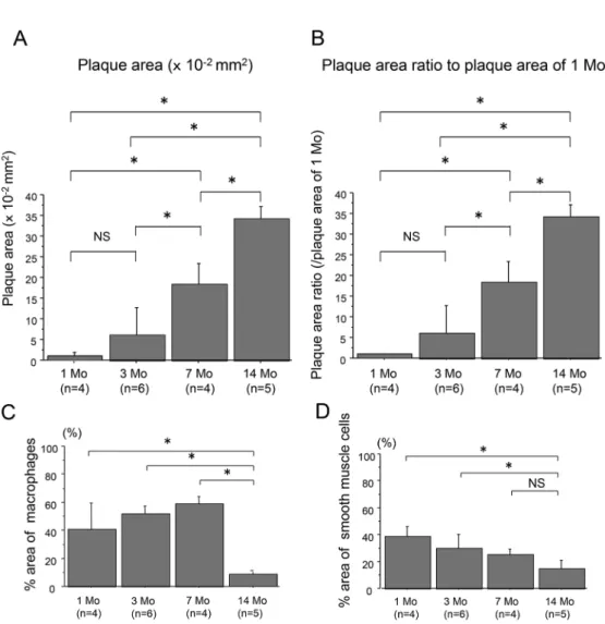
Atheromatous plaques of the aortae of WHHL- MI rabbits were significantly found as early as at 1 month and accelerated at 3 through 14 months (Figs.
1A and 1B). The data also show the infiltration of cellular components including macrophages and smooth muscle cells (Figs. 1C and 1D). However, at 14 months, the atheromatous plaques consisted of decreased cellular components and increased extra- cellular fibrous and lipid components compared to 7 months (Figs. 1C and 1D), which was consistent with the previous report
15).
RAGE expression
The expression of RAGE was noted as early as at 1 month in hyperlipidemia
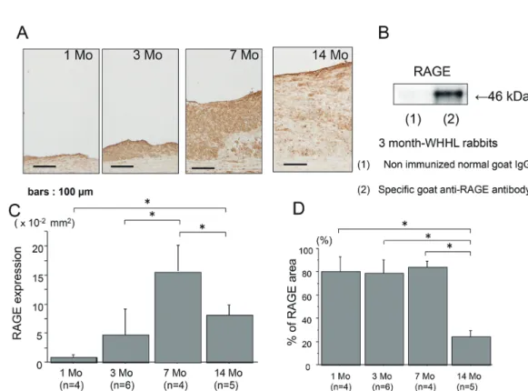
-dependent atheroscle- rotic lesions and progressively increased at 3 and 7 months (Fig. 2A), and the intensive expression of RAGE in the aortae of 3 months of WHHLMI rabbits as determined by Western blotting (Fig. 2B). How- ever, the atherosclerotic plaques at 14 months had the decreased expression of RAGE compared with 7 months (Figs. 2C and 2D).
RAGE, AGE and Cellular Component Expression Fig. 3 shows the cellular components and the expression of RAGE, AGE and phospholipid prod- ucts at 7 months in the atheromatous plaques by im- munohistochemistry. The expression of RAGE ap- peared to be distributed in the areas of cellular components including endothelial cells, smooth muscle cells and macrophages. In addition, RAGE and macrophage were distributed in phospholipid products expressing area. Interestingly, AGE and RAGE were accumulated in nondiabetic hyperlipid- emia
-dependent atheromatous plaques.
RAGE Expression in Coronary Atheromatous Plaques
The components of coronary atheromatous
plaques and the original sources of RAGE were sim-
ilar to those of aortic plaques at 7 months. The
abundant infiltration of macrophages and smooth
muscle cells were found in the coronary atheroscle-
rotic lesions. RAGE expression was identified as the cellular sources of macrophages and smooth muscle cells in addition to endothelial cells (Figs.
4A, 4B and 4C). The data also show the quantita- tive analysis of the area of coronary atheromatous plaques (Fig. 4D), and RAGE expression (Fig. 4E) and its cellular sources with aging (Figs. 4F and 4G). RAGE expression was progressively in- creased with aging up to 7 months and markedly de- creased at the age of 14 months, which was consis- tent with the extent of cellular infiltration.
Serum AGE Levels
Fig. 5A demonstrated the serum AGE levels at 2, 4, 8 and 15 months of WHHLMI rabbits, indicat- ing no significant difference in the serum AGE lev- els among the four groups. Fig. 5B shows the se- rum AGE levels of WHHLMI rabbits and control NZW rabbits at 4 months. There was no significant
difference between two groups. The results sug- gested that the expression of RAGE in hyperlipid- emia
-dependent atherosclerotic lesions was not re- lated to the serum AGE levels.
AGE Accumulation and Nitrotyrosine Expression with Aging
Fig. 6A shows that the deposition of AGE was increased with aging and that AGE was predomi- nantly recognized at the cellular components infil- trating into atheromatous plaques in WHHLMI rab- bit aortae. The deposition of AGE was noted at endothelial cells as early as at 1 month. The whole expression of AGE was increased with aging at 3 and 7 months, and diminished at the age of 14 months.
Moreover, immunohistochemistry demonstrated the expression of indirect oxidant marker nitrotyrosine with aging
25), which was similar to AGE deposition (Fig. 6B). The data suggested that AGE were pro-
Fig. 1. Quantitative analysis of the absolute plaque areas (A) and proportions (B) of aortic plaques of WHHLMI rab-bits with aging (1, 3, 7 and 14 months, n=4 to 6), as well as % area of macrophages (C) and smooth muscle cells (D). The proportions of plaques were determined by the control values at 1 month (B). Measured values are expressed as mean±S.D. *P<0.05. Mo, months and NS, not significant.
duced by oxidative stress and that the profiles of AGE accumulation and nitrotyrosine expression were similar to RAGE expression.
Role of RAGE in Atherosclerosis
-Related Molecular Expression
Fluorescent immunohistochemistry revealed the expression of RAGE on plasma membrane both of human monocytes and monocyte
-derived macro- phages (Figs. 7A and 7B). RAGE expression was strongly identified in macrophages cultured for 72 hours rather than for 24 hours by Western blotting (Fig. 7C), suggesting that RAGE expression was in- creased by the differentiation into macrophages.
Fig. 7D shows that oxidized LDL increased the ex- pression of RAGE and PAI
-1 in human monocyte
-derived macrophages. The silencing of RAGE in the macrophages tented to block PAI
-1 expression.
In addition, oxidized LDL promoted RAGE and TF expression significantly in peritoneal macro- phages from wild mice. The knockout of RAGE tented to decrease oxidized
-induced TF expression in peritoneal macrophages from RAGE−/− mice
(Fig. 8).
Discussion
We show here that RAGE expression was asso- ciated with the initiation and progression of athero- sclerosis in WHHLMI rabbits, as well as the RAGE
-mediated atherosclerosis
-related molecular expression.
The present study clearly shows that RAGE ex- pression is concordant with cellular infiltration of macrophages and smooth muscle cells in the athero- sclerotic lesions of nondiabetic WHHLMI rab- bits. This is consistent with the previous reports on nondiabetic human and animal atherosclerotic le- sions
11,26). AGE are shown to be generated by oxida- tive stress, and Calkin et al. reported that administra- tion of antioxidant attenuated the AGE accumulation in mice
9,27-29). Our data demonstrated that AGE was distributed in the areas of phospholipid products and nitrotyrosine in the atherosclerotic lesions. The present study suggested that hyperlipidemia trig- gered
-oxidative stress may accelerate AGE genera-
Fig. 2. The expression of RAGE in the aortic atherosclerotic plaques of WHHLMI rabbits with aging by immunohis-tochemistry (A) and Western blotting (B). A : RAGE expression at 1, 3, 7 and 14 months. B : Western blot- ting of RAGE in the aortae of 3-month-WHHLMI rabbits. Immunoblot was probed with a specific goat anti- RAGE antibody (lane 2), whereas nonimmunized normal goat IgG was used as a negative control antibody (lane 1). RAGE was identified at the band of 46 kD in the atherosclerotic lesions of 3-month-WHHLMI rabbit. C and D : Quantitative analysis of RAGE expression of area (C) and proportion (D) in the atherosclerotic plaques of WHHLMI rabbits at 1, 3, 7 and 14 months (n=4 to 6, each month). Measured values are represented as mean±S.D. *P<0.05.
tion and RAGE expression in cellular compo- nents
29-31), especially macrophages in the nondiabetic atherosclerosis.
It is well acknowledged that atheromatous RAGE expression is accelerated with high plasma glucose levels and stronger in diabetics than nondia- betics
5,26). Soro
-Paavonen et al. have shown that knockout of RAGE attenuates the development of atherosclerosis in diabetic mice
10). These suggested a central role of RAGE in the atherosclerosis compli- cated with diabetes. In addition, several studies have suggested that RAGE expression is involved in inflammatory cell infiltration, thrombogenicity and plaque destability through various signaling pathways in diabetic and nondiabetic atherosclerotic le- sions
32-36). Harja et al. have demonstrated that endo- thelial RAGE modulates vascular and inflammatory responses independent of diabetes in apoE−/− mice, suggesting the important role of RAGE in the patho- genesis of nondiabetic atherosclerosis
11).
We demonstrated that the significant expres- sion of RAGE was recognized in endothelial cells as
early as at 1 month in the aortic atherosclerotic le- sions of WHHLMI rabbits, as well as at 3 and 7 months. This finding concurred with the results reported by Roy et al. that RAGE is expressed in the endothelial cells of the aortic atherosclerotic lesions of diabetic 2
-month
-WHHL rabbits
37).
The glucose metabolism in WHHLMI rabbits is a critical issue in this study, which was extensively investigated in adult (10
-to 15
-month
-old) and mid- dle
-aged (17
-to 21
-month
-old) WHHLMI rab- bits
38). Briefly, changes in blood sugar levels were similar among the three groups, adult normal and WHHIMI rabbits and middle
-aged WHHIMI rab- bits. In terms of glucose tolerance, there were two groups in both adult and middle
-aged WHHLMI rab- bits. The rabbits with high fasting immunoreactive insulin (IRI) levels showed insulin resistance as de- termined by HOMA
-R and Matsuda
-insulin sensi- tivity index (ISI). However, overall, there was no significant difference in glucose tolerance among the three groups (normal adult rabbits, and adult and middle
-aged WHHIMI rabbits). Also, we have the
Fig. 3. Immunohistochemistry of the aortic atherosclerotic plaques of 7-month-WHHLMI rabbits. Results werefrom one representative out of four 7-month-WHHLMI rabbits. Immunostainings for endothelial cells (CD31), smooth muscle α-actin (1A4), AGE, RAGE, macrophages (RAM-11), phospholipid products (DLH3) and RAGE and AGE are shown.
data regarding the glucose metabolism including fasting blood sugar levels, IRI concentrations and HOMA
-R in 6
-month, 12
-month and 18
-month
-old WHHIMI rabbits, demonstrating that the changes in glucose metabolism are similar among the three groups (unpublished data).
Endothelial dysfunction accelerates the devel-
opment of atherosclerosis including monocyte che- motaxis, monocytes adhesion to endothelial cells and proteolysis
19,23,39). Moreover, MCP
-1, vascular cell adhesion molecule
-1 (VCAM
-1) and MMP
-2 are modulated by RAGE in endothelial cells under non- diabetic conditions
11). Taken together, our results regarding strong expression of RAGE in endothelial
Fig. 4. A, B, C : RAGE expression (A) and cellular components including macrophages (B) and smooth muscle cells(C) in coronary atherosclerotic plaques of 7-month-WHHLMI rabbits as determined by immunohistochemis- try. Results were the representative sections of the coronary atheromatous lesions from 5 rabbits of 7-month- WHHLMI. D, E, F, G : Quantitative analyses of the amounts of plaques (D), and the proportion of RAGE ex- pression (E) and cellular components including macrophages (F) and smooth muscle cells (G) in coronary plaques of WHHLMI rabbits with aging (1, 3, 7 and 14 months, n=5 to 6). Values are represented as mean±S.D.
*P<0.05.
cells suggested that endothelial RAGE may initiate and accelerate nondiabetic atherosclerosis indepen- dent of diabetes.
In the present study, we showed that RAGE ex- pression was accelerated with cellular infiltration
and that the atherosclerotic lesions with reduced cellular components at 14 months had marked de- crease in RAGE expression. However, we found no significant difference in the serum AGE levels with aging, as well as between WHHLMI rabbits and
Fig. 5. A : The serum AGE levels of WHHLMI rabbits with aging (2, 4, 8 and 15 months, n=4 to 5). B : The se-rum AGE levels of WHHLMI rabbits (n=4) and NZW rabbits (n=5) at 4 months. Data are expressed as mean±S.D.
Fig. 6. AGE accumulation (A) and nitrotyrosine expression (B) in aortic atherosclerotic plaques with aging as deter- mined by immunohistochemistry. Results were the representative sections of the aortic atheromatous lesions from 1, 3, 7 and 14 months of WHHLMI rabbits.
normal NZW rabbits. These results suggested that serum AGE levels were not a determinant of RAGE expression in atherosclerotic lesions.
We attempted to clarify the issue what regu- lates RAGE expression in nondiabetic atheromatous plaques. Since Oka et al. reported that the plasma cholesterol and oxidized LDL levels are markedly high as early as 1 month with the persistent high levels in WHHL rabbits
40), one possibility may be
hyperlipidemia which impairs endothelial function and accumulates macrophages in plaques
39). Al- though Roy et al. found RAGE expression in athero- sclerotic lesions of diabetic WHHL rabbits
37), we demonstrated the expression of RAGE in endothelial cells and macrophages of pure hyperlipidemia
-de- pendent atherosclerotic animal model independent of diabetes. Our in vitro study demonstrated that oxidized LDL increased RAGE expression in macro-
Fig. 7. A : Immunofluorescent detection of RAGE in human circulating monocytes (upper left panel, specific anti-RAGE antibody ; upper right panel, phase contrast ; lower left panel, non immunized normal goat IgG ; lower right panel, phase contrast). B : Immunofluorescent detection of RAGE in monocyte-derived macrophages cul- tured for 72 hours (upper left panel, specific anti-RAGE antibody ; upper right panel, phase contrast ; lower left panel, non immunized normal goat IgG ; lower right panel, phase contrast). C : The expression of RAGE was determined by Western blotting after 24 and 72 hours of culture. D : Effects of knockdown of RAGE on oxi- dized LDL-triggered PAI-1 expression in cultured macrophages. Isolated monocytes were incubated for 18 hours in the presence or absence of 50 µg/ml oxidized LDL after transfection of RAGE siRNA or scrambled siR- NA. PAI-1 expression was determined by Western blotting. Immunoblots are from one experiment represen- tative from three separate experiments.
phages. These findings suggest the regulation of RAGE expression by hyperlipidemia and oxidized LDL.
Another possibility of the mechanism of RAGE expression in nondiabetic athrosclerotic lesions may be oxidative stress
28), which nonenzymatically gen- erates AGE. It has been shown that statin sup- presses RAGE expression by decreasing oxidant stress
-dependent AGE generation in human diabetic atherosclerotic plaques
36). Statin has been also re- ported to attenuate RAGE expression probably via decreased oxidant stress in the absence of lipid
-low- ering effect
29,41). In the present study, RAGE ex- pression and AGE accumulation were associated with nitrotyrosine expression with cellular infiltra- tion in the atherosclerotic lesions. This finding suggested that the accumulation of peroxynitrate
-mediated protein oxidation reflected hyperlipidemia
-dependent oxidative stress, which may induce AGE accumulation and RAGE expression in plaques.
Burke et al. have suggested that RAGE expres- sion may be involved in the plaque instability in dia- betes since stronger RAGE expression in plaques was found in sudden death diabetic subjects than nondiabetics
26). Our study using WHHLMI rabbits suggested the possibility that the intensive expres- sion of RAGE in endothelial cells and macrophages of coronary atherosclerotic lesions may contribute to inflammation, thrombogenicity and extracellular ma- trix degradation in plaques, in turn, plaque destabili- zation. Further study is required for a better un- derstanding of the mechanism by which RAGE signaling leads to plaque instability.
In conclusion, we show that RAGE may play an integral role in the pathogenesis of atherosclerosis in WHHLMI rabbits independent of diabetes, sug- gesting that RAGE may be a good target to treat dia-
betic and nondiabetic vascular complications.
Acknowledgements
This work was supported in part by Grants
-in
-Aid for Scientific Research from Japan Society for the Promotion of Science (21590935 and 20790538), Fukushima Medical University Research Project, Takeda Science Foundation and The Uehara Memo- rial Foundation.
Conflict of Interest
The authors declare that they have no conflict of interest relevant to this article.
References
1. Haffner SM, Lehto S, Rönnemaa T, Pyörälä K, Laakso M. Mortality from coronary heart disease in subjects with type 2 diabetes and in nondiabetic subjects with and without prior myocardial infarc- tion. N Eng J Med, 339: 229-234, 1998.
2. Beckman JA, Creager MA, Libby P. Diabetes and atherosclerosis : epidemiology, pathophysiology, and management. JAMA, 287: 2570-2581, 2002.
3. Brownlee M. Biochemistry and molecular cell bi- ology of diabetic complications. Nature, 414: 813-820, 2001.
4. Sobel BE, Woodcock-Mitchell J, Schneider DJ, Holt RE, Marutsuka K, Gold H. Increased plas- minogen activator inhibitor type 1 in coronary ar- tery atherectomy speciments from type 2 diabetic compared with nondiabetic patients : a potential factor predisposing to thrombosis and its persis- tence. Circulation, 97: 2213-2221, 1998.
5. Cipollone F, Iezzi A, Fazia M, et al. The receptor RAGE as a progression factor amplifying arachido- Fig. 8. The role of RAGE in TF expression in the absence or presence of oxidized LDL in peritoneal macrophages of
RAGE−/− and wild-type mice. The macrophages were incubated for 18 hours with or without 5 µg/ml oxidized LDL. TF and RAGE expression was determined by Western blotting. In peritoneal macrophages from RAGE−/− mice, TF expression was very low at basal condition and the response to oxidized LDL was less com- pared to wild-type mice. Representative immunoblots from three separate experiments are shown.
nate-dependent inflammatory and proteolytic re- sponse in human atherosclerotic plaques : role of glycemic control. Circulation, 108: 1070-1077, 2003.
6. Howard BV, Roman MJ, Devereux RB, et al. Ef- fect of lower targets for blood pressure and LDL cholesterol on atherosclerosis in diabetes. The SANDS randomized trial. JAMA, 299: 1678- 1689, 2008.
7. Cholesterol Treatment Trialists’ (CTT) Collabora- tors. Efficacy and safety of cholesterol-lowering treatment : prospective meta-analysis of data from 90056 participants in 14 randomized trials of statins. Lancet, 366: 1267-1278, 2005.
8. Shiomi M
,Ito T
,Yamada S
,Kawashima S
,Fan J
. Development of an animal model for spontane- ous myocardial infarction (WHHLMI rabbit).Ar- terioscler Thromb Vasc Biol
, 23: 1239-1244, 2003.9. Goldin A, Beckman JA, Schmidt AM, Creager MA. Advanced glycation end products : sparking the development of diabetic vascular injury. Cir- culation, 114: 597-605, 2006.
10. Soro-Paavonen A, Watson AMD, Li J, et al. Re- ceptor for advanced glycation end products (RAGE) deficiency attenuates the development of athero- sclerosis in diabetes. Diabetes, 57: 2461-2469, 2008.
11. Harja E, Bu DX, Hudson BI, et al. Vascular and inflammatory stresses mediate atherosclerosis via RAGE and its ligands in apoE−/− mice. J Clin Invest, 118: 183-194, 2008.
12. Shindo J
,Ishibashi T
,Yokoyama K
, et al. Gran- ulocyte-macrophage colony-stimulating factor pre- vents the progression of atherosclerosis via chang- es in the cellular and extracellular composition of atherosclerotic lesions in Watanabe heritable hy- perlipidemic rabbits. Circulation, 99: 2150- 2156, 1999.13. Shoji T, Koyama H, Morioka T, et al. Receptor for advanced glycation end products is involved in im- paired angiogenic response in diabetes. Diabetes, 55: 2245-2255, 2006.
14. Yamamoto Y, Doi T, Kato I, et al. Receptor for ad- vanced glycation end products is a promising target of diabetic nephropathy. Ann N Y Acad Sci, 1043: 562-566, 2005.
15. Shiomi M, Ito T. The Watanabe heritable hyper- lipidemic (WHHL) rabbit, its characteristics and history of development : a tribute to the late Dr.
Yoshio Watanabe. Atherosclerosis, 207: 1-7, 2009.
16. Itabe H, Takeshima E, Iwasaki H, et al. A mono- clonal antibody against oxidized lipoprotein recog- nizes foam cells in atherosclerotic lesions. J Biol Chem, 269: 15274-15279, 1994.
17. Takeuchi M, Makita Z, Bucala R, et al. Immuno- logical evidence that non-carboxymethyllysine ad- vanced glycation end-products are produced from short chain sugars and dicarbonyl compounds in vivo. Mol Med, 6: 114-125, 2000.
18. Nagata K, Ishibashi T, Sakamoto T, et al. Effects of blockade of the renin-angiotensin system on tis- sue factor and plasminogen activator inhibitor-1 synthesis in human cultured monocytes. J Hy- pertens, 19: 775-783, 2001.
19. Ishibashi T, Sakamoto T, Ohkawara H, et al. Inte- gral role of RhoA activation in monocyte adhesion- triggered tissue factor expression in endothelial cells. Arterioscler Thromb Vasc Biol, 23: 681- 687, 2003.
20. Yokoyama K, Ishibashi T, Yi-qiang L, Nagayoshi A, Teramoto T, Maruyama Y. Interleukin-1 beta and interleukin-6 increase levels of apolipoprotein B mRNA and decrease accumulation of its protein in culture medium of HepG2 cells. J Lipid Res, 39: 103-113, 1998.
21. Okumura AJ, Hatsuzawa K, Tamura T, et al. In- volvement of a novel Q-SNARE, D12, in quality control of the endomembrane system. J Biol Chem, 281: 4495-4506, 2006.
22. Naito Y, Yamada T, Ui-Tei K, Morishita S, Saigo K. siDirect : highly effective, target-specific siR- NA design software for mammalian RNA interfer- ence. Nucleic Acids Res, 32(Web Server issue) : W124-W129, 2004.
23. Sakamoto T, Ishibashi T, Sakamoto N, et al. En- dogenous NO blockade enhances tissue factor ex- pression via increased Ca2+ influx through MCP-1 in endothelial cells by monocyte adhesion. Arte- rioscler Thromb Vasc Biol, 25: 2005-2011, 2005.
24. Sakamoto T, Ishibashi T, Sugimoto K, et al. RhoA- dependent PAI-1 gene expression induced in endo- thelial cells by monocyte adhesion mediates gera- nylgeranyl transferase I and Ca2+ signaling.
Atherosclerosis, 193: 44-54, 2007.
25. Peluffo G, Radi R. Biochemistry of protein tyro- sine nitration in cardiovascular pathology. Cardio- vasc Res, 75: 291-302, 2007.
26. Burke AP, Kolodgie FD, Zieske A, et al. Morpho- logic findings of coronary atherosclerotic plaques in diabetics. A postmortem study. Arterioscler Thromb Vasc Biol, 24: 1266-1271, 2004.
27. Baynes JW. Role of oxidative stress in develop- ment of complications in diabetes. Diabetes, 40: 405-412, 1991.
28. Anderson MM, Heinecke JW. Production of Nε- (carboxymethyl) lysine is impaired in mice defi- cient in NADPH oxidase. A role for phagocyte- derived oxidants in the formation of advanced gly- cation end products during inflammation.
Diabetes, 52: 2137-2143, 2003.
29. Calkin AC, Giunti S, Sheehy KJ, et al. The HMG- CoA reductase inhibitor rosuvastatin and the an- giotensin receptor antagonist candesartan attenu- ate atherosclerosis in an apolipoprotein E-deficient mouse model of diabetes via effects on advanced glycation, oxidative stress and inflammation. Dia- betologia, 51: 1731-1740, 2008.
30. Faria A, Persaud SJ. Cardiac oxidative stress in diabetes : Mechanisms and therapeutic poten- tial. Pharmacol Ther, 172: 50-62, 2017.
31. Matsui T, Higashimoto Y, Nishino Y, et al. RAGE- Aptamer Blocks the Development and Progression of Experimental Diabetic Nephropathy. Diabetes, 66: 1683-1695, 2017.
32. Yan SF, Ramasamy R, Naka Y, Schmidt AM. Gly- cation, inflammation, and RAGE. A Scaffold for the macrovascular complications of diabetes and beyond. Circ Res, 93: 1159-1169, 2003.
33. Schmidt AM, Yan SD, Wautier JL, Stern D. Activation of receptor for advanced glycation end products : a mechanism for chronic vascular dysfunction in diabetic vasculopathy and atherosclerosis. Circ Res, 84: 489-497, 1999.
34. Naka Y, Bucciarelli LG, Wendt T, et al. RAGE axis : animal models and novel insights into the vascular complications of diabetes. Arterioscler Thromb Vasc Biol, 24: 1342-1349, 2004.
35. Aikawa M, Rabkin E, Sugiyama S, et al. An HMG-CoA reductase inhibitor, cerivastatin, sup- presses growth of macrophages expressing matrix
metalloproteinases and tissue factor in vivo and in vitro. Circulation, 103: 276-283, 2001.
36. Cuccurullo C, Iezzi A, Fazia ML, et al. Suppres- sion of RAGE as a basis of simvastatin-dependent plaque stabilization in type 2 diabetes. Arteri- oscler Thromb Vasc Biol, 26: 2716-2723, 2006.
37. Roy H, Bhardwaj S, Babu M, et al. VEGF-A, VEGF-D, VEGF receptor-1, VEGF receptor-2, NF-κB, and RAGE in atherosclerotic lesions of dia- betic Watanabe heritable hyperlipidemic rab- bits. FASEB J, 20: 2159-2161, 2006.
38. Shiomi M, Kobayashi T, Kuniyoshi N, et al. Myo- cardial infarction-prone Watanabe heritable hyper- lipidemic rabbits with mesenteric fat accumulation are a novel animal model for metabolic syndrome.
Pathobiol, 79: 329-38, 2012.
39. Simionescu M. Implications of early structural- functional changes in the endothelium for vascular disease. Arterioscler Thromb Vasc Biol, 27: 266-274, 2007.
40. Oka K, Yasuhara M, Suzumura K, Tanaka K, Sawamura T. Antioxidants suppress plasma lev- els of lectinlike oxidized low-density lipoprotein receptor-ligands and reduce atherosclerosis in Watanabe heritable hyperlipidemic rabbits. J Car- diovasc Pharmacol, 48: 177-183, 2006.
41. Lim S, Barter P. Antioxidant effects of statins in the management of cardiometabolic disorders. J Atheroscler Thromb, 21: 997-1010, 2014.