Fukushima Medical University
福島県立医科大学 学術機関リポジトリ
This document is downloaded at: 2021-11-08T00:32:10Z
Title TBX19 is overexpressed in colorectal cancer and associated with lymph node metastasis
Author(s)
Ando, Jin; Saito, Motonobu; Imai, Jun-Ichi; Ito, Emi;
Yanagisawa, Yuka; Honma, Reiko; Saito, Katsuharu;
Tachibana, Kazunoshin; Momma, Tomoyuki; Ohki, Shinji;
Ohtake, Tohru; Watanabe, Shinya; Waguri, Satoshi; Kono, Koji; Takenoshita, Seiichi
Citation Fukushima Journal of Medical Science. 63(3): 141-151
Issue Date 2017
URL http://ir.fmu.ac.jp/dspace/handle/123456789/667
Rights © 2017 The Fukushima Society of Medical Science
DOI 10.5387/fms.2017-08
Text Version publisher
141 TBX19 associates colon cancer metastasis
Fukushima J. Med. Sci., Vol. 63, No. 3, 2017
[Original Article]
TBX19 is overexpressed in colorectal cancer and associated with lymph node metastasis
Jin Ando
1), Motonobu Saito
1), Jun
-ichi Imai
2), Emi Ito
2), Yuka Yanagisawa
3), Reiko Honma
3), Katsuharu Saito
1), Kazunoshin Tachibana
4), Tomoyuki Momma
1), Shinji Ohki
1), Tohru Ohtake
4),
Shinya Watanabe
2), Satoshi Waguri
5), Koji Kono
1)and Seiichi Takenoshita
1)1)
Department of Gastrointestinal Tract Surgery,
2)Medical
-Industrial Translational Research Center,
4)De- partment of Breast Surgery, and
5)Department of Anatomy and Histology, Fukushima Medical University School of Medicine, Fukushima 960
-1295, Japan,
3)Nippon Gene Co.,Ltd., Tokyo 104
-0054, Japan
(Received April 24, 2017, accepted October 31, 2017)
Abstract
The T
-box 19 (TBX19) gene encodes a transcription factor characterized by a highly conserved DNA
-binding motif (T
-box). Recent studies have revealed that TBX19 has been identified as one of the genes activated by KRAS mutations, and is upregulated in colon adenoma. These results in- dicate that TBX19 may work as an oncogene in colorectal cancer (CRC). However, the expression and role of TBX19 have yet to be investigated. Here, we investigated TBX19 mRNA and protein expressions in colon cancer cells or surgically resected CRC. We found that TBX19 mRNA expres- sion was significantly increased in tumorous tissues compared to that in non
-tumorous tissues, and increased TBX19 mRNA expression was associated with positive lymph node metastasis in our co- hort. The expression of TBX19 mRNA was not correlated with that of TBX19 protein in tissue sample taken from the CRC patients. Moreover, TBX19 showed positive staining even in the nor- mal colonic tissues and the adjacent non
-tumorous tissues. These results suggest that the expres- sion of TBX19 protein is not correlated with the expression of TBX19 mRNA. In addition, our re- sults promote further investigations into the impact of TBX19 upregulation on colorectal carcinogenesis, as well as the underlying mechanisms.
Key words :
TBX19, colorectal cancer, lymph node metastasis, diagnostic biomarker, cytoplasmic granular signal
Introduction
Colorectal cancer (CRC) is the third most com- mon type of cancer that occurs in both men and women all over the world
1,2). One of the most im- portant molecular pathogeneses of colorectal carci- nogenesis is the adenoma
-carcinoma sequence
1). The adenoma
-carcinoma sequence is stepwise ge- netic aberrations, including APC, KRAS and TP53 mutations, in CRC patients
3). APC mutations are an early event in this multistep process, followed by KRAS
-activating mutations and TP53
-inactivating mutations
3). While chemotherapy and molecular
targeting drugs for CRC treatment have progressed recently, genetic aberrations among APC, KRAS, and TP53 are not therapeutic targets
4,5). It is par- ticularly necessary to develop therapeutic agents for KRAS mutations as they are frequently detected in CRC patients, as well as other malignant tumors, to improve cancer mortality
6). Although there have been several studies on the development of targeted drugs for KRAS mutations
7-9), they have not yet been used in a clinical setting.
The T
-box (TBX) gene family encodes a large family of transcription factors and plays a fundamen- tal role in early embryogenesis during the develop- Corresponding author : Motonobu Saito E
-mail : [email protected]
https://www.jstage.jst.go.jp/browse/fms http://www.fmu.ac.jp/home/lib/F
-igaku/
141
142 J. Ando et al.
mental process
10). Several studies have revealed aberrations of TBX genes in inherited human disor- ders, such as TBX1 mutation in DiGeorge syn- drome, TBX3 mutation in Ulnar
-Mammary syn- drome, TBX5 mutation in Holt
-Oram syndrome, and TBX22 mutation in cleft palate with ankyloglossia
11). In addition, recent studies have also found that TBX genes may be associated with cancer development in various malignant tumors
12). TBX2 and TBX3, which are downstream targets of the Wnt/beta
-catenin pathway, are frequently mutated in ovarian cancer and are amplified in breast cancer. Further- more, they are associated with hepatocellular carci- noma, pancreatic cancer, and malignant melano- ma
13-18).
In the present study, we focused on the analysis of TBX19, which is expressed in the rostral ventral diencephalon and pituitary gland
19). TBX19 muta- tions lead to a lack of adrenocorticotrophin resulting in adrenal insufficiency
20). On the other hand, TBX19 has been identified as one of the genes acti- vated by KRAS mutation, and is upregulated in colon adenoma
21,22). These results indicate that TBX19 might work as an oncogene in CRC, but the expres- sion and role of TBX19 in CRC remain unknown.
Here, we investigated TBX19 mRNA and protein expressions in surgically resected CRC tissues, and examined the biological significance.
Materials and Methods
Clinical samples of patients
A total of 89 surgical specimens obtained from CRC patients who had undergone surgical resection at Fukushima Medical University Hospital between January 2008 and December 2010 were used for the experiments. All 89 cases are used for comprehen- sive gene expression analysis, 5 cases are used for protein expression analysis by western blotting, and 54 cases are used for immunohistochemical (IHC) staining. In addition, 3 cases of adenoma were used for IHC staining. Information regarding age, sex, TNM stage, and pathological diagnosis, including lymphatic and venous invasion, were retrospectively collected. The carcinomas at the time of primary tumor resection were staged according to the Union for International Cancer Control UICC classification (the 7th classification)
23,24). Written informed con- sent was obtained from all patients. This study was approved by the ethics committee of Fukushima Medical University.
Comprehensive gene expression analysis
TBX19 expression data were obtained using custom microarray analysis as previously de- scribed
25,26). In brief, the surgical specimen was ho- mogenized and mixed with ISOGEN reagent (NIP- PON GENE, Tokyo, Japan). Total RNA was subjected to purification of polyA(A)+RNA using MicroPoly(A) Purist Kit (Thermo Fisher Scientific, Waltham, MA, USA). The human common refer- ence RNA was prepared by mixing equal amounts of poly(A)+ RNA extracted from 22 human cancer cell lines (A431, A549, AKI, HBL
-100, HeLa, HepG2, HL60, IMR
-32, Jurkat, K562, KP4, MKN7, NK
-92, Raji, RD, Saos
-2, SK
-N
-MC, SW
-13, T24, U251, U937, and Y79).
Synthetic polynucleotides (80
-mers) represent- ing 31,797 human transcripts (MicroDiagnostic, To- kyo, Japan) were arrayed on aminosilane
-coated glass slides with a custom
-made arrayer. RNA (2 μg) was subjected to reverse transcription with Su- perScript II (Thermo Fisher Scientific). Sample RNA was labeled using Cyanine 5
-dUTP (Perkin
-Elmer, Boston, MA, USA) and reference RNA was labeled using Cyanine 3
-dUTP. Hybridization was performed with a labeling and hybridization kit (Mi- croDiagnostic). Signals were measured with a Ge- nePix 4000B scanner (Axon Instruments, Union City, CA, USA) and then processed into primary ex- pression ratios. The primary expression ratios were then converted into log2 values and compiled into a matrix. We assigned an expression ratio of 1 (log ratio of 0) for spots that exhibited fluorescence intensities under the detection limits, and we in- cluded these in the signal calculation of the mean averages. Data were processed by MDI gene ex- pression analysis software package (MicroDiagnos- tic).
Cell line culture
The colon cancer cell lines used in this study were originally obtained from the American Type Culture Collection (Rockville, MD, USA) and were cultured in the recommended media with 10% fetal bovine serum. These monolayer cells were main- tained in a 37°C incubator with 5% CO
2. Cells were checked regularly under a light microscope and sub- cultured once they had reached 80% to 90% conflu- ence.
Reverse transcription
-quantitative polymerase chain reaction (RT
-qPCR)
Total RNA was extracted from cells using
143 TBX19 associates colon cancer metastasis
TRIzol reagent (Thermo Fisher Scientific) according to the manufacturer’s instructions as previously de- scribed
27). Complementary DNA (cDNA) was syn- thesized from 5 μg of total RNA with a random hex- amer using the SuperScript III First
-Strand Synthesis System (Thermo Fisher Scientific).
These cDNAs were used for the measurement of gene expression with a 7500 Real
-time PCR system (Thermo Fisher Scientific) using TaqMan probes.
The assessors were blinded to patient information and performed experiments in triplicate. Taqman expression assays were purchased from Thermo Fisher Scientific ; TBX19 (Hs01113611_m1) and β
-actin (Hs99999903_m1). β
-actin was used as an internal control. Relative TBX19 gene expression was calculated using the 2
-ΔΔCT method, according to the supplier’s protocol (Thermo Fisher Scientif- ic)
28).
Western blotting
Cancer cell lines and surgical specimens were homogenized in a 100 mM Tris
-HCl (pH 7.6) buffer containing 0.15 M NaCl, 5 mM EDTA, 1% Triton X
-100, 5% glycerol by Polytron PT3100 homogeniz- er (Kinematica AG, Luzern, Switzerland). After centrifugation at 17,400 xg for 15 min at 4°C, the su- pernatants were collected. Then, 20 ug of each protein sample was run on SDS
-polyacrylamide gels (5
-15% gradient ; Thermo Fisher Scientific) and blotted onto Immun
-Blot PVDF membranes (Bio
-Rad Laboratories, Hercules, CA, USA). The blot- ted membranes were incubated with the indicated primary antibodies overnight at 4°C. Rabbit mono- clonal anti
-TBX19 (HPA005800, Sigma
-Aldrich, St.
Louis, MO, USA) was used at 1 : 500 dilution, while mouse monoclonal anti
-β
-actin antibody (sc
-69879, Santa Cruz Biotechnology, USA) was used as a load- ing control at 1 : 2,500 dilution. The blotted mem- branes were then incubated with the appropriate horseradish peroxidase (HRP)
-conjugated goat
-anti
-mouse IgG (sc
-2005, Santa Cruz Biotechnology) secondary antibody at a dilution of 1 : 5,000. Sig- nals were detected by ImageQuant LAS4000 (GE Healthcare Bio
-sciences, Pittsburgh, PA, USA) us- ing SuperSignal West Pico Chemiluminescent Sub- strate (Thermo Fisher Scientific).
Immunohistochemical staining and evaluation IHC staining was carried out on paraffin
-em- bedded histological sections (4 μm thick) using a polymer peroxidase method. Briefly, after deparaf- finization and rehydration, the sections were treated with 0.3% hydrogen peroxide in methanol for 30 min
to block endogenous peroxidase activity. Following rinsing in phosphate
-buffered saline (PBS) (Thermo Fisher Scientific), the sections were incubated with anti
-TBX19 antibody (HPA005800, 1 : 2,000 dilu- tion ; Sigma
-Aldrich) at 4°C overnight. Three fur- ther washes (5 min per wash) in PBS was followed by treatment with a peroxidase
-labeled polymer, conjugated to goat anti
-rabbit immunoglobulins (Dako EnVision+System
-HRP Labelled Polymer ; ready
-to
-use ; #K4003 ; Dako ; Agilent Technolo- gies), as the secondary antibody for 30 min at room temperature. The staining was visualized with di- aminobenzidine, followed by counterstaining with hematoxylin. Expression of these proteins was evaluated as positive when the nucleus of tumorous tissues and the total field of view were observed at 400×magnification. We evaluated the staining of each specimen. The rate of positively stained cells was counted among three randomly selected fields (200 μm×200 μm) in the tumorous and non
-tumor- ous tissues. Cytoplasmic granular signals were counted in the tumorous tissues.
Statistical Analysis
Data were presented as the mean±SD. Sta- tistical analyses were performed using GraphPad Prism 6 software (GraphPad Software, La Jolla, CA, USA). The Mann
-Whitney U test and Wilcoxon matched pairs test were used for comparison of the means of the two groups and Kruskal
-Wallis test was used for comparison of more than two groups.
Log
-rank test was used for survival comparisons.
P<0.05 was considered to indicate a statistically sig- nificant difference.
Results
Expression analysis of TBX19 from the comprehensive analysis data
Firstly, the mRNA level of TBX19 in the CRC
specimen was determined by using comprehensive
gene expression analysis data. The expression ra-
tios of TBX19 were compared between 89 tumorous
tissue samples and 60 non
-tumorous tissue samples,
which revealed that TBX19 had a significantly high-
er expression in tumorous tissues compared to non
-tumorous tissues (P<0.0001, Mann
-Whitney’s U
-test) (Fig. 1A). Of note, among available 40 pairs of
non
-tumorous and tumorous tissues, upregulation of
TBX19 was also observed in the tumorous tissues
compared to the non
-tumorous tissues (P<0.0001,
Wilcoxon matched pairs test) (Fig. 1B).
144 J. Ando et al.
Next, we analyzed TBX19 expression levels with clinicopathological factors in the CRC speci- mens (Table 1). The case with positive lymph node metastasis showed significantly higher expression of TBX19 (P=0.012). However, TBX19 expression level was not found to be associated with age, gen-
der, TNM stage, histology, tumor depth, lymphatic invasion, venous invasion, or distant metastasis.
Kaplan
-Meier analysis demonstrated no association between increased TBX19 levels and relapse
-free survival (P=0.9473, log
-rank test) (Fig. 1C).
Fig 1.
A
B
C
Disease-free survival0 10 20 30 40 50
70 80 90
100 TBX19 Low
TBX19 High
Month
Percent survival
P = 0.947 0 2 4
Expression ratio (log2)
Non-tumor Tumor P < 0.0001
0 2 4
Expression ratio (log2)
Non-tumor Tumor P < 0.0001
Fig. 1. The mRNA expression of TBX19 in CRC specimens. (A) Expression differences of TBX19 between 89 tu- morous and 60 non-tumorous tissues from our CRC cohort. Dot plot represents TBX19 expression from micro- array analysis. The expression level on the log2 scale is shown. Horizontal bars indicate mean expression val- ues. P<0.0001, the Mann-Whitney U-test. (B) TBX19 expression differences in each of 40 pairs tumorous and non-tumorous tissues. Dot plot represents TBX19 expression from microarray analysis. The expression level on the log2 scale is shown. P<0.0001, Wilcoxon matched pairs test. (C) Kaplan-Meier survival of 89 cases in our cohort stratified by TBX19 tumor expression. P=0.947, log-rank test.
145 TBX19 associates colon cancer metastasis
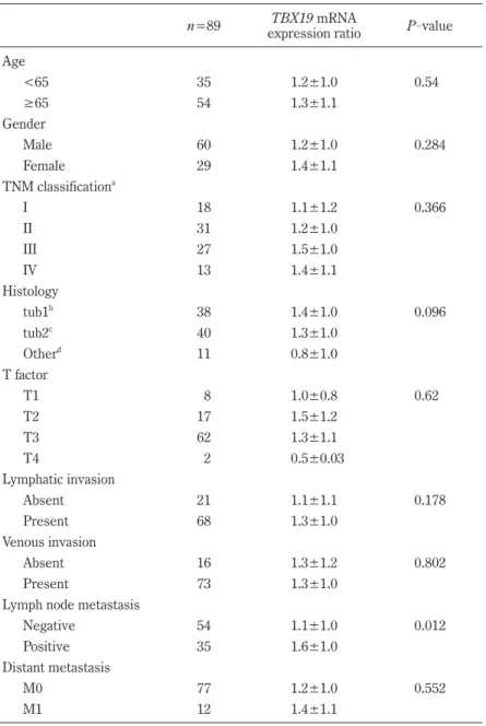
TBX19 mRNA and protein expression
To further confirm that TBX19 mRNA expres- sion is upregulated in CRC, we investigated TBX19 expression in 7 colon cancer cell lines. TBX19 mRNA expression was investigated by real
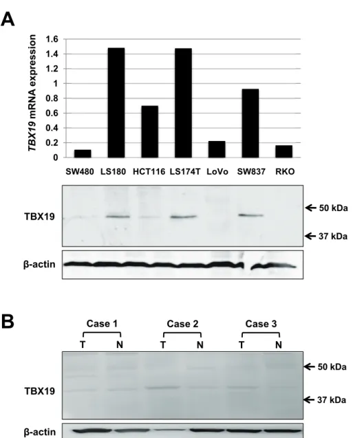
-time PCR, and TBX19 protein expression was investigat- ed by western blotting for the 7 colon cancer cells (Fig. 2A). Consistent with TBX19 mRNA expres- sion, TBX19 protein expression was upregulated in the LS180, LS174T, and SW837 cells. Then, we
analyzed TBX19 protein expression by western blot- ting in three representative non
-tumorous/tumorous CRC tissues that showed high TBX19 mRNA ex- pression (Fig. 2B). However, TBX19 protein was not highly expressed in the tumorous tissues com- pared to the non
-tumorous tissues. These results indicate that the upregulation of TBX19 mRNA did not result in upregulation of TBX19 protein expres- sion.
Table 1. Clinicopathological factors and TBX19 mRNA expression
n=89 TBX19 mRNA
expression ratio P-value Age
<65 35 1.2±1.0 0.54
≥65 54 1.3±1.1
Gender
Male 60 1.2±1.0 0.284
Female 29 1.4±1.1
TNM classificationa
I 18 1.1±1.2 0.366
II 31 1.2±1.0
III 27 1.5±1.0
IV 13 1.4±1.1
Histology
tub1b 38 1.4±1.0 0.096
tub2c 40 1.3±1.0
Otherd 11 0.8±1.0
T factor
T1 8 1.0±0.8 0.62
T2 17 1.5±1.2
T3 62 1.3±1.1
T4 2 0.5±0.03
Lymphatic invasion
Absent 21 1.1±1.1 0.178
Present 68 1.3±1.0
Venous invasion
Absent 16 1.3±1.2 0.802
Present 73 1.3±1.0
Lymph node metastasis
Negative 54 1.1±1.0 0.012
Positive 35 1.6±1.0
Distant metastasis
M0 77 1.2±1.0 0.552
M1 12 1.4±1.1
Values are expressed as the mean±standard deviation. aUICC TNM 7th classification23,24), bWell differentiated tubular adenocarcinoma. cModer- ately differentiated tubular adenocarcinoma. dSolid-type pooly differenti- ated adenocarcinoma, mucinous adenocarcinoma, papillary adenocarcino- ma or adenosquamous carcinoma. P-values were calculated using Mann- Whitney test or a Kruskal-Wallis test, where appropriate.
146 J. Ando et al.
IHC staining for TBX19
Next, we performed IHC staining for TBX19 in 3 adenoma and 54 CRC specimens. While normal colonic mucosa exhibited weak TBX19 staining (Fig. 3A), all 3 adenoma showed positive TBX19 staining (Fig. 3B). In the CRC specimens, TBX19 expression was observed in the nucleus of tumorous and adjacent non
-tumorous cells (Fig. 3C). When we assessed TBX19 staining intensity, the tumorous tissue samples showed a higher percentage of posi- tive TBX19 cells compared to the non
-tumorous tis- sue samples (Fig. 3D). However, this staining in- tensity was not associated with any clinicopathological
factors in our cohort (Table 2). We additionally performed TBX19 staining for metastatic liver can- cer from CRC. The metastatic liver tumor showed positive staining for TBX19, but normal liver tissue showed negative TBX19 staining (Fig. 3E). These results suggested that TBX19 was specifically up- regulated in CRC cells.
To further understand TBX19 staining, we fo- cused on the cytoplasmic granular signals in CRC cells (Fig. 3F). While the cytoplasmic granular sig- nals in cytoplasmic tumor cells were not observed in the normal tissue, they were observed in 15 of the 54 (27.8%) CRC tissues. The signals tended to be observed in undifferentiated histological types, such
00.2 0.4 0.6 0.8 1 1.2 1.4 1.6
SW480 LS180 HCT116 LS174T LoVo SW837 RKO
50 kDa
A
B
Fig 2.
TBX19 β-actin
Case 1 T N TBX19
β-actin
TBX 19 m R N A exp ressi on
37 kDa
50 kDa 37 kDa
Case 2
T N
Case 3 T N
Fig. 2. The expression of TBX19 in colon cancer cell lines and CRC specimens. (A) TBX19 expression analyses of mRNA by real-time PCR and protein by western blot in 7 colon cancer cells. Relative TBX19 mRNA expres- sion levels are shown in the upper panel (normalized to β-actin). TBX19 protein expressions are shown in the lower panel. β-actin was used as a loading control. (B) TBX19 protein expression analysis by western blot in 3 CRC patients. β-actin was used as a loading control.
147 TBX19 associates colon cancer metastasis
as poorly differentiated or mucinous adenocarcinoma (Table 3). These results further suggest that TBX19 mRNA was upregulated in CRC and associ-
ated with worse CRC outcomes.
Non-tumor Adenoma
A B
C
Fig 3.
Tumor
Cytoplasmic granular signal in tumor
E
D
Metastasis
F
Normal liver
Metastatic liver cancer from CRC
0 25 50 75 100
Positive cells (%)
Non-tumor Tumor
P < 0.0001
Fig. 3. Immunohistochemical staining of TBX19. Representative images of TBX19 staining in non-tumorous tis- sue (A), adenoma (B), CRC tissue (C and F), and metastatic liver cancer from CRC (E). (A) Weak positive stain- ing for TBX19 in non-tumorous tissue. The boxed area is shown at higher magnification. Scale bars=100 μm (left) and 25 μm (right). (B) Positive staining for TBX19 in adenoma. Scale bar=50 μm. (C) Positive staining for TBX19 in tumorous cells (arrow) and weak positive staining for TBX19 in adjacent non-tumorous cells (arrow head). Scale bars=100 μm (upper) and 25 μm (lower). (D) Difference in percentage of TBX19-positive stain- ing cells between 89 tumorous and 60 non-tumorous tissues from our cohort. P<0.0001, the Mann-Whitney U- test. (E) Weak positive staining for TBX19 in normal liver tissue and positive staining for TBX19 in metastatic liver cancer from CRC. Scale bars=50 μm. (F) Cytoplasmic granular signal of TBX19 in CRC. Scale bars=100 μm (upper) and 25 μm (lower).
148 J. Ando et al.
Discussion
In the present study, we found that TBX19 mRNA was upregulated in CRC and increased ex- pression of TBX19 mRNA was associated with posi- tive lymph node metastasis in CRC patients. On the contrary, TBX19 protein expression was not up- regulated in CRC and was not associated with any clinicopathological factors in our cohort. There- fore, our study shows that, while upregulated TBX19 mRNA may have a pivotal role in colon tumorigene- sis, the role of TBX19 protein in colonic tumorigen- esis is still unknown and need a further consider- ation. Furthermore, our results also suggest that the expression of TBX19 protein was not correlated with TBX19 mRNA expression. This was further confirmed by the result that the CRC patients, which showed high TBX19 mRNA expression, did not ex- hibit higher TBX19 protein expression in the tumor- ous tissues than in the non
-tumorous tissues by Western blotting in our small number of cases.
Even in the normal colonic tissues and the adjacent non
-tumorous tissues showed weak positive stain- ing for TBX19, suggesting that TBX19 protein may have a role in keeping normal mucosal homeostasis and in affecting CRC tumor development. There- fore, the evaluation of TBX19 in CRC by IHC stain- ing requires further investigation. Of course, the evaluation of sensitivity and specificity for anti
-TBX19 antibody remains to be required. When ob- serving IHC staining, we were interested in the cy- toplasmic granular signals in the tumor cells and found that they tended to associate with tumor dif- ferentiation in our cohort. To date, because the mechanism for the formation of granular signals in tumor cells has not yet been fully understood, fur- ther morphological and functional studies are re- quired.
Consistent with previous reports, TBX19 was also upregulated in adenoma
21,22). Because adeno- ma is considered to be a type of pre
-cancerous tu- mor, we believe that TBX19 plays a role in the ade-
Table 2. Clinicopathological factors and TBX19 IHC expressionTBX19 IHC
n=54 % of positive cells P-value
Age
<65 22 90.0±5.0 0.699
≥65 32 87.4±9.2
Gender
Male 36 88.2±7.9 0.673
Female 18 89.0±7.7
TNM classificationa
I 15 87.6±10.3 0.805
II 17 87.8±9.1
III 15 88.7±4.8
IV 7 91.1±1.8
Histology
tub1b 19 89.3±7.1 0.283
tub2c 23 86.3±9.5
Otherd 12 91.2±3.5
Lymph node metastasis
Absent 33 87.8±9.4 0.478
Present 21 89.4±4.3
Distant metastasis
Absent 47 88.0±8.3 0.787
Present 7 91.1±1.8
Values are expressed as the mean±standard deviation. aUICC TNM 7th classification23,24), bWell differentiated tubular adenocarcinoma. cModer- ately differentiated tubular adenocarcinoma. dSolid-type pooly differenti- ated adenocarcinoma, mucinous adenocarcinoma, papillary adenocarcino- ma or adenosquamous carcinoma. P-values were calculated using Mann- Whitney test or a Kruskal-Wallis test, where appropriate.
149 TBX19 associates colon cancer metastasis
noma
-carcinoma sequence. A previous in vitro experiment revealed that TBX19 is one of the down- stream genes that are activated by KRAS muta- tions
21). In fact, TBX19 was highly expressed in most colorectal adenomas with activated KRAS mu- tations
22). KRAS mutations are one of the steps in the adenoma
-carcinoma sequence that accumulate several genetic aberrations in CRC. Therefore, be- cause KRAS mutations occur in both colorectal ade- noma and cancer, it is understandable that TBX19 expression is upregulated in CRC as well as in ade- noma cases. In CRC treatment, the mutational sta- tus of KRAS is only used as a predictive marker for the effectiveness of anti
-EGFR antibodies, cetux- imab, or panitumumab
29). This is due to KRAS be- ing a downstream effector of EGFR. In addition, and most importantly, KRAS mutations have not yet to be a direct druggable target. KRAS mutations are the most frequently occurring aberrations in hu- man cancer, including CRC
6,30). In spite of huge ef- forts to develop molecular targeting drugs for KRAS mutations
7), no useful agents have yet been included in clinical treatment strategies for CRC. With re-
cent progress in the understanding of KRAS biology, KRAS has been considered as druggable by either targeting the mutations directly or targeting the downstream pathways, such as RAF
-MAPK and PIK3K
31). Of note, because TBX19 is also down- stream of KRAS, further experimental or mice stud- ies investigating the functional role of TBX19 may provide a therapeutic opportunity for KRAS
-driven cancers.
In conclusion, the current study reports the TBX19 expression status in CRC. Our results sug- gest that TBX19 as a candidate therapeutic target for CRC.
Acknowledgements
This research was partially supported by grants for translational research programs from New Ener- gy and Industrial Technology Development Organi- zation (NEDO) (Tokyo, Japan).
Table 3. Clinicopathological factors and cytoplasmic granular signal Cytoplasmic granular signal
Total (n=54) Positive (n=15) Negative (n=39) P-value
Age
<65 22 5 (33.3%) 17 (43.6%) 0.551
≥65 32 10 (66.7%) 22 (56.4%)
Gender
Male 36 10 (66.7%) 26 (66.7%) 1.000
Female 18 5 (33.3%) 13 (33.3%)
TNM classificationa
I 15 2 (13.3%) 13 (33.3%) 0.086
II 17 6 (40.0%) 11 (28.2%)
III 15 5 (33.3%) 10 (25.6%)
IV 7 2 (13.3%) 5 (12.8%)
Histology
tub1b 19 3 (20.0%) 16 (41.0%) 0.013
tub2c 23 5 (33.3%) 18 (46.2%)
Otherd 12 7 (46.7%) 5 (12.8%)
Lymph node metastasis
Absent 33 9 (60.0%) 24 (61.5%) 1.000
Present 21 6 (40.0%) 15 (38.5%)
Distant metastasis
Absent 47 13 (86.7%) 34 (87.2%) 1.000
Present 7 2 (13.3%) 5 (12.8%)
aUICC TNM 7th classification23,24), bWell differentiated tubular adenocarcinoma. cModerately dif- ferentiated tubular adenocarcinoma. dSolid-type pooly differentiated adenocarcinoma, mucinous adenocarcinoma, papillary adenocarcinoma or adenosquamous carcinoma. P-values were calcu- lated using Mann-Whitney test or a Kruskal-Wallis test, where appropriate.
150 J. Ando et al.
Conflict of Interest
The authors declare that there is no conflict of interest.
References