Primary Pulmonary Synovial Sarcoma Showing a Prolonged
Survival with Multimodality Therapy
Hirokazu Ogino
1, Masaki Hanibuchi
1, Hiromitsu Takizawa
2, Shoji Sakiyama
2,
Hiroyuki Sumitomo
2, Seiji Iwamoto
3, Hitoshi Ikushima
3, Kohei Nakajima
4, Shinji Nagahiro
4,
Taito Yamago
1, Yuko Toyoda
1, Yoshimi Bando
5and Yasuhiko Nishioka
1Abstract
A 54-year-old man was referred to our hospital due to a mass shadow noted on a chest X-ray. Thora-coscopic lobectomy yielded a diagnosis of primary pulmonary synovial sarcoma according to the histology and SYT-SSX1 gene analyses. Five months after the thoracic surgery, he developed brain metastasis; therefore, we performed resection of the brain metastatic focus followed by radiotherapy. As a local recurrence in the thoracic cavity concurrently emerged, systemic chemotherapy was also administered. These observations indi-cated that a multidisciplinary approach may be useful against primary pulmonary synovial sarcoma, although there is presently no established therapeutic strategy due to its rarity and highly aggressive nature.
Key words: primary pulmonary synovial sarcoma, spontaneous regression, brain metastasis, multimodality
therapy
(Intern Med 55: 381-387, 2016)
(DOI: 10.2169/internalmedicine.55.5169)
Introduction
Synovial sarcoma is a distinct soft tissue neoplasm which occurs mainly in the extremities and limb girdle, and it rep-resents 7-10% of all human soft tissue sarcomas (1, 2). It often metastasizes to the lung; however, primary pulmonary synovial sarcoma is extremely rare and has been reported to comprise less than 0.5% of pulmonary neoplasms (3, 4). Primary pulmonary synovial sarcomas have been increas-ingly reported as a result of growing awareness and im-proved diagnostic capabilities (5-9). This tumor is thought to be more locally aggressive and associated with a poorer prognosis than soft tissue synovial sarcoma (9); however, precise clinical data, such as on the prevalence rate, progno-sis, and metastatic pattern, as well as on the therapeutic strategy, are still unclear due to its rarity.
We herein report a rare case of primary pulmonary synovial sarcoma successfully treated with multimodality therapy.
Case Report
A 54-year-old man was referred to our hospital for the further examination of an abnormality noted on a chest ra-diograph. The patient had a smoking history of 3 pack-years and was undergoing treatment for hypertension. He had in-termittent left chest pain; however, no abnormality was noted on a physical examination. Blood tests showed slight elevation of inflammatory reactions; however, tumor markers for lung cancer and markers for mycotic infection were negative. An interferon-gamma release assay (QuantiF-ERONⓇ) was positive (Table). A chest X-ray showed a mass shadow in the left lower lung field (Fig. 1), and computed
1Department of Respiratory Medicine and Rheumatology, The University of Tokushima Graduate School, Institute of Health Biosciences, Japan, 2Department of Thoracic, Endocrine Surgery and Oncology, The University of Tokushima Graduate School, Institute of Health Biosciences, Japan,3Department of Radiology, The University of Tokushima Graduate School, Institute of Health Biosciences, Japan,4Department of Neuro-surgery, The University of Tokushima Graduate School, Institute of Health Biosciences, Japan and5Division of Pathology, Tokushima University Hospital, Japan
Received for publication February 16, 2015; Accepted for publication May 10, 2015 Correspondence to Dr. Yasuhiko Nishioka, [email protected]
Figure 1. The chest X-ray on admission. A mass shadow was seen in the left lower lung field.
PLT 29.0 × 10 /ȝL Ȗ-GTP 33 IU/L Infection marker Na 140 mEq/L
Coagulation K 4.1 mEq/L ȕ-D glucan <4.8 pg/mL CL 104 mEq/L
Aspergillus antigen (-) PT-INR 0.89 CRP 0.71 mg/dL
APTT 30.8 sec Cryptococcus
antigen (-) QuantiFERON® (+)
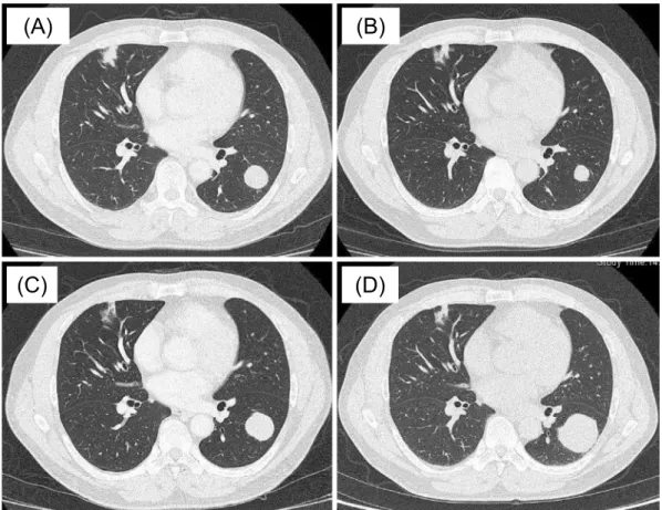
tomography (CT) showed a nodule which was 23 mm in di-ameter in the left lower lobe (S8) (Fig. 2A).
Positron emission tomography (PET)-CT showed a slightly increased fluorodeoxyglucose (FDG) uptake [stan-dardized uptake values (SUVs) of the nodule was 1.4] (Fig. 3A). No evidence of distant metastases was found on PET-CT or contrast-enhanced brain magnetic resonance im-aging (MRI) (data not shown). A transbronchial biopsy with bronchoscopy yielded no definitive diagnosis. No acid fast bacilli were detected in the sputum or bronchial lavage fluid, which suggested a latent tuberculosis infection (LTBI) that did not require treatment, rather than active tuberculo-sis. During the examination period, the nodule regressed spontaneously (from 23 to 18 mm in diameter) (Fig. 2B); therefore, we adopted a wait-and-see approach.
Two months later, the nodule showed regrowth (from 18 mm to 32 mm in diameter) (Fig. 2C, D), and the SUV-max of the nodule was elevated from 1.4 to 7.6 on follow-up PET-CT (Fig. 3B). Because a CT-guided percutaneous lung biopsy failed to establish a definitive diagnosis, we pro-ceeded with thoracoscopic left lower lobectomy. A
his-tologic examination of the specimens revealed the active proliferation of malignant tumor cells with a high nuclear cytoplasmic ratio (N/C ratio) and oval or short spindle nu-cleus (Fig. 4A). Immunohistochemistry (IHC) showed posi-tive reactivity for Bcl-2, AE1/3, CD56, and vimentin (Fig. 4B-E), indicating a strong possibility of monophasic synovial sarcoma. Fluorescence in situ hybridization (FISH) showed that the SYT split-signal was positive in 96% of the tumor cells (Fig. 5A), indicating the existence of a chromo-some translocation of the SYT gene (10). Finally, we per-formed reverse transcription-polymerase chain reaction (RT-PCR) and sequencing of the PCR product, and the results showed the existence of the SYT-SSX1 gene (Fig. 5B, C), which led to a diagnosis of primary pulmonary synovial sar-coma.
Five months after the operation, the patient showed the gradual development of visual field disturbance. A visual field test showed right homonymous hemianopia, and contrast-enhanced brain MRI showed a large ring-enhanced tumor with intratumoral hemorrhage and peripheral edema in the left occipital lobe (Fig. 6A). Because radiotherapy alone is thought to be insufficient to control brain metastasis of synovial sarcoma (11), we initially performed brain tumor resection followed by radiotherapy. A histologic examination of the brain tumor showed the proliferation of spindle-shaped malignant cells, which was similar to the findings in the primary lung tumor (Fig. 4F); therefore, we diagnosed it as a brain metastatic focus of primary pulmonary synovial sarcoma. After brain tumor resection, intensity-modulated radiation therapy (IMRT) was performed with 50, 40, and 30 Gy to the parietal region, occipital region, and whole brain, respectively. The combined therapy with craniotomy and radiation successfully eliminated the tumor (Fig. 6B) and no recurrence of the brain tumor has been observed up to this time. Although the results of objective tests, such as the visual field test, were not improved, his subjective symptoms, such as visual acuity, partially improved after the combined treatment.
Figure 2. Chest computed tomography findings. (A) At the initial visit. A nodule, which was 23 mm in diameter, was seen in the left lower lobe (S8). (B) One month after the initial visit, the nodule spon-taneously regressed (from 23 to 18 mm in diameter). (C) Two months later, the nodule showed re-growth (from 18 to 32 mm in diameter). (D) At the time of thoracic surgery, the nodule had further grown.
(A)
(B)
(C)
(D)
Figure 3. PET-CT findings. (A) A slight increase of FDG uptake (SUV: 1.4) was observed in the nodule in the left lower lobe. (B) The SUV-max (7.6) of the nodule in the left lower lobe was further elevated during the follow-up period.
(A)
(B)
At the same time of the appearance of brain metastasis, tumor recurrence in the left thoracic cavity was seen on chest CT (data not shown); therefore, we initiated chemo-therapy with doxorubicin (30 mg/m2
per day, days 1 to 2) plus ifosfamide (2 g/m2per day, days 1 to 5 with mesna and pegfilgrastim) every 21 days after IMRT for brain metasta-sis. Because grade 3 leukopenia emerged, these drugs were
administered every 35 days thereafter. After two cycles of chemotherapy, the nodule in the thoracic cavity slightly de-creased (from 33 to 30 mm in diameter). Although the best overall response was stable disease (SD) according to the Response Evaluation Criteria in Solid Tumors (RECIST) guideline (version 1.1), we judged it was clinically effective and continued therapy for a total of four cycles.
Figure 4. Histopathology and immunohistochemistry of the tumors (×40). Bar indicates 100 μm. (A) Hematoxylin and Eosin (H&E) staining of the thoracic tumor showed the proliferation of malig-nant tumor cells with a high nuclear cytoplasmic ratio (N/C ratio), showing an oval or short spindle nucleus. Immunohistochemistry revealed positive reactivity for (B) Bcl-2, (C) AE1/3, (D) CD56, and (E) vimentin. (F) H&E staining of the brain tumor showed the proliferation of spindle-shaped malig-nant cells similar to those in the primary lung tumor.
(D)
(E)
(F)
Discussion
Synovial sarcoma is a highly malignant tumor, and it eas-ily develops into local recurrence and/or distant metastasis, especially to the lung, which results in a poor prognosis. Several reports showed that the 5-year survival rates were approximately 60% despite aggressive treatment (12, 13). Primary pulmonary synovial sarcoma is thought to be more aggressive than that of soft tissue origin, and in a retrospec-tive study, 46% of pulmonary and mediastinal synovial sar-coma patients died within 5 years, and only 26% of them were alive with no evidence of disease after several treat-ments (9). In the present case, the tumor rapidly progressed and led to a large brain metastasis and local recurrence dur-ing the short follow-up period. This medical history with rapid tumor progression was consistent with the previous re-ports.
In the present case, small cell lung cancer was suspected after the first CT-guided percutaneous lung biopsy due to the observation of the aggregation of small oval cells with round bare nuclei. However, there was no evidence of me-tastasis to the lymph nodes and distant organs at that time, and IHC revealed that both chromogranin A and synapto-physin were negative (data not shown), which was incom-patible with small cell lung cancer. Therefore, we performed lobectomy, which successfully yielded a diagnosis of pri-mary pulmonary synovial sarcoma. Pripri-mary pulmonary synovial sarcoma is generally diagnosed in patients with un-usual clinical or histological features, such as lung can-cer (9), and thus we should aggressively proceed with
diag-nostic studies including a surgical approach, detailed IHC, or genomic testing.
Brain metastasis of primary pulmonary synovial sarcomas is extremely rare and has been reported in only a few cases (7, 14). Moreover, to the best of our knowledge, this is the first reported case of primary pulmonary synovial sar-coma with brain metastatic focus treated by brain tumor re-section. While there was no evidence of any brain metastasis before thoracic surgery, a large brain metastatic focus in the occipital lobe developed five months postoperatively. Given the aggressive nature of the disease, as observed in the pre-sent case, close follow-up should be conducted for the early detection of local recurrence and/or distant metastasis even after curative surgical resection.
In general, the combination of surgery and radiation ther-apy is more effective than radiation monotherther-apy for soft tissue sarcoma. Indeed, radiotherapy combined with surgery was reported to achieve better local control than either mo-dality alone for the majority of soft tissue sarco-mas (11, 15), and pre- and postoperative approaches could achieve acceptable local control (16). Therefore, we per-formed craniotomy for brain tumor resection followed by ra-diotherapy. As a result, we have not observed the recurrence of the brain tumor up to this time. Although the results of objective tests, such as visual field test, were not improved, the patient’s subjective symptoms, such as visual acuity, par-tially improved.
A phase 3 trial which assessed the efficacy of the first-line treatment of doxorubicin with ifosfamide for advanced or metastatic soft tissue sarcoma showed that the response rate, median progression-free survival and median overall
Figure 5. A chromosome translocation of the SYT gene was detected in the thoracic tumor. (A) Fluorescence in situ hybridization (FISH) showed that the SYT split-signal was positive in 96% of tumor cells, which indicated that the tumor cells had the chromosome translocation of the SYT gene. (B) Reverse transcription-polymerase chain reaction (RT-PCR) showed the existence of the
SYT-SSX1 gene in the tumor of the present case. The primers for SYT-SYT-SSX1 were as follows: F-primer:
5’-CAACAGCAAGATGCATACCA-3’ and R-primer: 5’-GGTGCAGTTGTTTCCCATCG-3’. The detection of a PCR product of 331 base pair size indicated the existence of the SYT-SSX1 gene. (C) The sequential analysis of SYT-SSX1. The resulting PCR product of SYT-SSX1 was purified, followed by direct sequencing. This revealed the break points of the SYT gene and SSX1 genes.
(A) (B)
(C)
331 bp
SYT SSX1
Figure 6. Contrast-enhanced brain MRI findings. (A) A large ring-enhanced tumor with intratu-moral hemorrhage and peripheral edema in the left occipital lobe was seen at the time of the develop-ment of visual field disturbance. (B) The brain tumor disappeared after brain tumor resection and radiotherapy.
showed that pazopanib, a multitargeted tyrosine kinase in-hibitor with activity against VEGF receptor-1, -2, and -3 and platelet-derived growth factor receptor-α, β, significantly in-creased the median progression-free survival (4.6 months with pazopanib versus 1.6 months with placebo) in patients who had at least one regimen containing anthracycline (22); as a result, it was approved for the treatment of soft tissue sarcoma in Japan and the U.S. Although the efficacy of pa-zopanib against primary pulmonary synovial sarcoma re-mains unclear, it may be a useful therapeutic option in the present case in the near future.
Interestingly, the primary tumor spontaneously regressed during the initial course of the disease in the present case. Spontaneous regression of a malignant tumor is defined as “the partial or complete disappearance of a malignant tumor in the absence of all treatment or in the presence of therapy which is considered inadequate to exert a significant influ-ence on neoplastic disease” (23). The incidinflu-ence of spontane-ous regression has been estimated to be no more than 1 in 60,000-100,000 cases (24), and there appear to be no reports of spontaneous regression of synovial sarcoma. Immune sys-tems and hormonal effects are thought to play important roles (25); however, the precise mechanisms are still un-known.
In summary, we encountered a rare case of primary pul-monary synovial sarcoma that developed a large brain me-tastatic focus with visual field disturbance. This case high-lights the importance of an intensive diagnostic approach and an awareness of the aggressive metastatic potential of this rare tumor. The combined modality treatment success-fully controlled the disease, indicating that a multidiscipli-nary approach may be a useful therapeutic strategy against primary pulmonary synovial sarcoma.
The authors state that they have no Conflict of Interest (COI).
Acknowledgement
The authors thank Dr. T. Hasegawa (Department of Surgical Pathology, Sapporo Medical University School of Medicine) for kindly performing the fluorescence in situ hybridization.
References
1. Thorson JA, Weigelin HC, Ruiz RE, Howard JK, Lucas DR. Iden-tification of SYT-SSX transcripts from synovial sarcomas using RT-multiplex PCR and capillary electrophoresis. Mod Pathol 19: 641-647, 2006.
2. He R, Patel RM, Alkan S, et al. Immunostaining for SYT protein
positive cases from the French Sarcoma Group and the Mesopath Group. Am J Surg Pathol 29: 339-346, 2005.
6. Essary LR, Vargas SO, Fletcher CD. Primary pleuropulmonary synovial sarcoma: reappraisal of a recently described anatomic subset. Cancer 94: 459-469, 2002.
7. Okamoto S, Hisaoka M, Daa T, Hatakeyama K, Iwamasa T, Hashimoto H. Primary pulmonary synovial sarcoma: a clinicopa-thologic, immunohistochemical, and molecular study of 11 cases. Hum Pathol 35: 850-856, 2004.
8. Suster S, Moran CA. Primary synovial sarcomas of the mediasti-num: a clinicopathologic, immunohistochemical, and ultrastruc-tural study of 15 cases. Am J Surg Pathol 29: 569-578, 2005. 9. Hartel PH, Fanburg-Smith JC, Frazier AA, et al. Primary
pulmo-nary and mediastinal synovial sarcoma: a clinicopathologic study of 60 cases and comparison with five prior series. Mod Pathol 20: 760-769, 2007.
10. Miura Y, Keira Y, Ogino J, et al. Detection of specific genetic ab-normalities by fluorescence in situ hybridization in soft tissue tu-mors. Pathol Int 62: 16-27, 2012.
11. Tepper JE, Suit HD. Radiation therapy alone for sarcoma of soft tissue. Cancer 56: 475-479, 1985.
12. Spillane AJ, A’Hern R, Judson IR, Fisher C, Thomas JM. Synovial sarcoma: a clinicopathologic, staging, and prognostic as-sessment. J Clin Oncol 18: 3794-3803, 2000.
13. Guillou L, Benhattar J, Bonichon F, et al. Histologic grade, but not SYT-SSX fusion type, is an important prognostic factor in pa-tients with synovial sarcoma: a multicenter, retrospective analysis. J Clin Oncol 22: 4040-4050, 2004.
14. Nuwal P, Dixit R, Shah NS, Samaria A. Primary monophasic synovial sarcoma lung with brain metastasis diagnosed on transthoracic FNAC: Report of a case with literature review. Lung India 29: 384-387, 2012.
15. Yang JC, Chang AE, Baker AR, et al. Randomized prospective study of the benefit of adjuvant radiation therapy in the treatment of soft tissue sarcomas of the extremity. J Clin Oncol 16: 197-203, 1998.
16. O’Sullivan B, Davis AM, Turcotte R, et al. Preoperative versus postoperative radiotherapy in soft-tissue sarcoma of the limbs: a randomised trial. Lancet 359: 2235-2241, 2002.
17. Judson I, Verweij J, Gelderblom H, et al. Doxorubicin alone ver-sus intensified doxorubicin plus ifosfamide for first-line treatment of advanced or metastatic soft-tissue sarcoma: a randomised con-trolled phase 3 trial. Lancet Oncol 15: 415-423, 2014.
18. Sleijfer S, Ouali M, van Glabbeke M, et al. Prognostic and predic-tive factors for outcome to first-line ifosfamide-containing chemo-therapy for adult patients with advanced soft tissue sarcomas: an exploratory, retrospective analysis on large series from the Euro-pean Organization for Research and Treatment of Cancer-Soft Tis-sue and Bone Sarcoma Group (EORTC-STBSG). Eur J Cancer 46: 72-83, 2010.
19. Yudoh K, Kanamori M, Ohmori K, Yasuda T, Aoki M, Kimura T. Concentration of vascular endothelial growth factor in the tumour tissue as a prognostic factor of soft tissue sarcomas. Br J Cancer 84: 1610-1615, 2001.
of HER-2/neu, c-kit (CD117) and vascular endothelial growth fac-tor (VEGF) overexpression in soft tissue sarcomas. Anticancer Res 24: 333-337, 2004.
21. Pakos EE, Goussia AC, Tsekeris PG, Papachristou DJ, Stefanou D, Agnantis NJ. Expression of vascular endothelial growth factor and its receptor, KDR/Flk-1, in soft tissue sarcomas. Anticancer Res 25: 3591-3596, 2005.
22. van der Graaf WT, Blay JY, Chawla SP, et al. Pazopanib for me-tastatic soft-tissue sarcoma (PALETTE): a randomised,
double-blind, placebo-controlled phase 3 trial. Lancet 379: 1879-1886, 2012.
23. Everson TC, Cole WH. Anonymous Spontaneous Regression of Cancer. Saunders, Philadelphia, 1966: 164-220.
24. Cole WH. Efforts to explain spontaneous regression of cancer. J Surg Oncol 17: 201-209, 1981.
25. Ota S, Shinagawa K, Ueoka H, et al. Spontaneous regression of metastatic endometrial stromal sarcoma. Jpn J Clin Oncol 32: 71-74, 2002.
Ⓒ 2016 The Japanese Society of Internal Medicine http://www.naika.or.jp/imonline/index.html