近畿大学学術情報リポジトリ
65
0
0
全文
(2) Doctoral Dissertation. Effects of prenatal and postnatal nutritional environment on the onset of cerebrovascular disorders in stroke-prone spontaneously hypertensive rats. Lila Otani. Graduate School, Kinki University Division of Advanced Life Science Major: Applied Bioscience.
(3) Effects of prenatal and postnatal nutritional environment on the onset of cerebrovascular disorders in stroke-prone spontaneously hypertensive rats. Lila Otani September, 2005 Graduate School, Kinki University Division of Advanced Life Science Major: Applied Bioscience (Advisor: Prof. Tetsuo Murakami). ijiS$ 4' ~JeiIE·I~~1fn.J± § r.-&JeiIE:7 '/ r CSHRSP) C:h' ~t 0 ijiS1fn.1ff1!! ~O)JeiIE c~t-t 0 ija!JCM:h' J: lf~;fLMO)*.0)'3~. J1I~*~*~~Ji;. .~~ff~f~. Jitm~JfIJ{~~:WJX. *ii-. CmlJ : l-tJ:. fJ~ g~~~). Submitted to the Graduate school, Kinki University, to fulfill the requirement for the Doctorate Degree..
(4) ABBREVIATIONS. SHRSP. Stroke-Prone Spontaneously Hypertensive Rats. SHR. Spontaneously Hypertensive Rats. WKY. Wistar Kyoto Rats. RAAS. Renin Angiotensin Aldosterone System. ATI. Angiotensin II Type 1 receptor. AT2. Angiotensin II Type 2 receptor. ACE. Angiotensin Converting Enzyme. ARB. Angiotensin II Receptor antagonist. FOAD. Fetal Origins of Adult Disease. 11. fJ -HSD2. 11 beta-hydroxysteroid dehydrogenase type 2.
(5) CONTENTS INTRODUCTAON INTRODUCTION . . . . . . . . . . . . . . . . . . . . . . . . . . 1. CHAPTERl. The Effects of Maternal Mild Protein Restriction on Stroke Incidence and Blood Pressure in Stroke-Prone Spontaneously Hypertensive Rats (SHRSP). CHAPTER 2. Introduction· . . .. ·5. Methods· .. ·6. Results· . .. · 8. Discussion· . . . . . .. · 14. Summary· . . . . . .. · 17. Prenatal low protein exposure enhanced salt sensitivity in stroke-prone spontaneously hypertensive rats (SHRSP). CHAPTER 3. Introduction· . . .. · 18. Methods· . .. · 19. Results· . . .. ·21. Discussion· . . . . .. ·26. Summary· . . . . . .. ·29. Reduction of Salt Sensitivity in Stroke-Prone Spontaneously Hypertensive Rats Administered an AT 1 Receptor Antagonist During Suckling Introduction· . . .. ·30. Methods· . . . .. · 31.
(6) Results· . . . . . . . . . .. ·33. Discussion· . . . . . . . .. . 38. Summary .. ·41. GENERAL CONCLUTION· . . . . . . . . . . . . . . . . . . . . . 43 REFERENCES· . . . . . . . . . . . . . . . . . . . . . . . . . . 48 AKNOWLEGEMENT· . . . . . . . . . . . . . . . . . . . . . 57 PUBLICATIONS· . . . . . . . . . . . . . . . . . . . . . . . . . 58.
(7) GENERAL INTRODUCTION Stroke was the first leading cause of death in Japan previously. Although the incidence of stroke decreased since the latter half of the nineteen-sixties, a stroke still remains to be a main cause of death even at present (Statistics and information department, Ministry of health, Labour and Welfar of Japan 2004). A main risk factor for stroke is hypertension. The occurrence of hypertension is influenced by genetic factors of individuals and their life style in adult life, such as low physical activity, smoking habit and excessive intake of salt. The idea has recently been accepted more widely that not only genetic factors of individuals and their environment in adult life but also their prenatal intrauterine environment influence the incidence of hypertension, diabetes, and obesity. The exchanges of gases, nutrients and wastes between mother and fetus occur through the placental membrane.. Since the growth of the fetus depends upon its. mother, its development is influenced by maternal health and nutritional states. Numerous studies have hitherto been devoted to examine the influence of maternal nutritional conditions on the growth of fetuses and postnatal development of children (mainly development with respect to the body weight)(Lederman and Rosso 1981). The body weight at birth reflects the environment in the embryonic stage and consequently an indicator giving an idea about maternal nutritional states during gestation. In recent epidemiological investigations in the United Kingdom, Sweden, Jamaica, India, and the United States of America, low body weight at birth was reported to be risk factors for hypertension, cardiac diseases, and cerebrovascular diseases in later life (Curhan et al. 1996; Launer et al. 1993; Law et al. 1993; Moore et al. 1996; Stein et al. 1996). Hence much attention was attracted to the relationship between nutritional conditions in fetal and lactation period and the incidence of cardiovascular diseases and diabetes after arriving at adulthood. Barker et al. found a close relationship between delayed intrauterine fetal growth and the occurrence of cardiovascular diseases, and put forward the "fetal origins of adult diseases hypothesis". This hypothesis proposes that alterations in fetal nutrition and endocrine status result in developmental adaptations that permanently change structure, physiology, and metabolism, thereby predisposing individuals to cardiovascular, metabolic, and endocrine diseases in later life. The process whereby a stimulus or insult at a sensitive or critical period of development has long-term effects is termed programming. In evolutionary terms, the phenomenon is likely to reflect the benefits. of plasticity during early development.. 1. Although adaptations of fetuses to.
(8) malnutrition are beneficial to short-term survival, they exert serious influences on the occurrence of cardiovascular diseases, diabetes, and endocrine diseases in adult life (Scheme 1)(Barker 1997). Poor maternal diet. Low maternal fat stores. Reduced placental transfer. Physiological and metabolic adaptatio7s in utero. T [ Lifelong metabolic thrift (postnatal nutrition. )1------.. . .. J. 1--------1. ,r. Genetic factor. '-. Cardiovascular and Metabolic disease. Scheme 1 Fetal undernutrition hormone and metabolic adoptions which are of benefit in the short term by making the fetus nutritionally thrifty but in the longer term predispose to disease After the "fetal origins of adult diseases hypothesis" was proposed, studies have been made with regard to effects of nutritional states during gestation and in infancy on lifestyle-related diseases like cardiovascular diseases and diabetes. Even in an animal study, it was reported that rats born from the dams that had been exposed to nutritional or protein restriction during gestation had hypertension in adulthood. The following programming mechanisms are proposed for this phenomenon: 1) an increase in sensitivity to glucocorticoid, 2) a decrease in the number of nephron in the kidney, and 3) a rise in the activity level of the renin-angiotensin-aldosterone system (RAAS ; Scheme. 2). 1) Glucocorticoid increase transluminal Na+ and Ca2+ transport and expression of angiotensin II type 1 receptors (AT l ) , both acting in concert to increase vascular tone and raise blood pressure. It was reported that maternal nutritional or protein restriction led to an increase in expression of glucocorticoid receptors (Gardner et al.. 2.
(9) 1997; Langley-Evans et al. 1996a). 2) The kidney plays a role in regulation of the body fluids homeostasis.. In response to a decrease in the number of nephron, the. blood pressure level rises to maintain the water and sodium homeostasis. Results of a large number of studies indicate that nutritional and protein restriction during gestation results in a decrease in the number of nephron in the offspring (Mackenzie and Brenner 1995; Merlet-Benichou et al. 1994). Woods et al. showed that in rats born from dams exposed to protein restriction during gestation, suppression of RAAS brought about a decrease in the number of nephron in the kidney (Woods et al. 200 1). 3) An increase in expression of AT I receptors and high reactivity to angiotensin II were reported to contribute to a rise in blood pressure in rats born from the darns exposed to protein restriction during gestation(Langley-Evans and Jackson 1995). That RAAS contributes to rises in blood pressure in rats exposed to low protein diet has been demonstrated in studies in which ACE inhibitor and ARB were used (Sherman and Langley-Evans 1998; Sherman and Langley-Evans 2000) .. Maternal undernutrition Altered Pattern of Gene Expression. Hypersensitivity of Glucocorticoids. Increased Expression of RAAS components. Alteration of Renal function. Scheme 2 Possible mechanisms of prenatal programming cardiovascular disease.. As described hitherto, numerous studies have been made with respect to rises in blood pressure in rats born from dams exposed to nutritional and protein restriction in order to clarify the mechanism whereby the rises in blood pressure are brought about (Gardner et al. 1997; Sherman and Langley-Evans 2000; Woods et al. 200 1). However, litter is known about the effect of protein and nutritional restriction during. 3.
(10) gestation on the occurrence of stroke and ischemic cardiac diseases. This study was consequently designed to examine the effect of the environment in fetal and lactation period on cerebrovascular impairment after arriving at adulthood using SHRSP. A study to be described in the first chapter was made to examine the effect of restriction of protein intake of SHRSP dams during gestation on the blood pressure and the incidence of stroke in rats born from such dams. A study to be described in the second chapter was performed to examine the relationship between the incidence of stroke and the renin-angiotensin-aldosterone system in rats born from mother rats treated identically. A study to be described in the third chapter was performed to investigate the relationship between suppression of the renin-angiotensin-aldosterone system during the lactation period and the level of blood pressure as well as the occurrence of stroke.. 4.
(11) CHAPTER 1 The Effects of Maternal Mild Protein Restriction on Stroke Incidence and Blood Pressure in Stroke-Prone Spontaneously Hypertensive Rats (SHRSP) Introduction Various studies have shown that the blood pressure is affected by hereditary predisposition and environmental factors such as dietary habits (Boden-Albala and Sacco 2000; Caulfield et al. 1998; Hermansen 2000). Studies in SHRSP have demonstrated that excess salt intake may affect blood pressure and cause cerebral stroke, leading to life-shortening (Suzuki 1978), while the improvement of nutritional factors (including protein nutrition) are effective for the preventing cerebral stroke and lengthening life span (Murakami et al. 1994; Okamoto et al. 1977; Okamoto 1979; Wang et al. 1985). These studies have revealed that individuals with genetic factors regarding hypertension or cerebral stroke can prevent the onset of stroke via the improvement of environmental factors after growth. On the other hand, although it is well known that maternal nutritional restriction during pregnancy delays fetal growth (Abel 1990; Lederman and Rosso 1981), its effect on the onset of diseases after birth is less understood.. Recently, an epidemiological survey conducted in Britain, Sweden,. Jamaica, India, and the U.S. has demonstrated that low birth weight can be a risk factor in hypertension, ischemic heart diseases, and type II diabetes mellitus after growth (Curhan et al. 1996; Launer et al. 1993; Law et al. 1993; Moore et al. 1996; Osmond et al. 1993; Stein et al. 1996), attracting attention to the relationship between maternal nutritional state during pregnancy and the onset of diseases after growth. It has been hypothesized that maternal nutritional restriction may affect the development, metabolism, and physiology of a fetus in its adaptation to undernutrition in utero, and that the effects can persist even after birth, and contribute to the onset of diseases (Barker 1994). Langley-Evans et al. confirmed in an animal study using normotensive rats that maternal mild protein restriction during pregnancy elevated blood pressure levels of offspring, proving this hypotheses (Langley-Evans and Jackson 1996a; Langley-Evans et al. 1996b; Langley-Evans et al. 1996c). In low-protein-exposed rats, the number of renal nephrons decreased (Langley-Evans et al. 1999b), and the reactivity to glucocorticoids (Gardner et al. 1997) and angiotensin II increased(Langley-Evans et al. 1996d), evidently showing the relationship between protein restriction in pregnancy and the onset of hypertension after growth. However, the effect of maternal nutritional restriction during pregnancy on the offspring of animals having genetic factors is unknown. In this study, the author investigated the. 5.
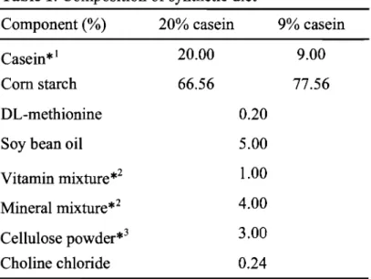
(12) effect of protein restriction during pregnancy on the offspring's blood pressure, using SHRSP that have a hereditary predisposition to hypertension and cerebral stroke. Furthermore, The author examined the effect of post-growth environmental factors on blood pressure, the incidence of cerebral stroke, and life span, using salt loading in low-protein-exposed SHRSP. Table 1. Composition of synthetic diet. Component (%). 20% casein. 9% casein. Casein*!. 20.00. 9.00. Com starch. 66.56. 77.56. DL-methionine. 0.20. Soy bean oil. 5.00. Vitamin mixture*2. 1.00. Mineral mixture*2. 4.00. Cellulose powder*3. 3.00. Choline chloride. 0.24. *lCrude protein 85.0%. *2AIN-76 prescription (Nippon Clea). *3Avicel (Nippon Clea).. Methods Animals. This study was done according to the guidelines of the animal experimental care committee of the Kinki University Faculty of Agriculture, and the rats were managed in conformation with the guidelines for Care and Use of Laboratory Animals. These guidelines are based on the Standards Relating to the Care and Management, etc. of Experimental Animals (Notification No.6, March 27,1980 of the Prime Minister's Office) and the Method for Sacrificing Laboratory Animals (Notification No.211, Decembers 1, 2000, of the Prime Minister's Office). Inbred SHRSP, which had been raised in our laboratory, were used for experiments. They were raised at a room temperature of 23 ± 1°C, humidity of 55 ± 5%, and under a 12-hour light-dark cycle (lighting from 7:00h to 19:00h). Mating. The animals were bred with sibling SHRSP. The SHRSP used in this study. were of a strain in which blood pressure elevation is relatively mild and whose life span is long. Virgin female SHRSP weighing 180g to 210g were mated to the same. 6.
(13) strain male (weighing 210 g or more, blood pressure 190 to 230 mmHg). Animals were paired with one male to one female and were placed together for one night. The date of pregnancy was determined with the confirmation of a vaginal plug and was set as pregnant day. o.. Animals were provided pair feeding and given a 20% and a 9%. casein diet in the control and low-protein groups, respectively, from pregnant day 0 to the confirmation of delivery (Table 1). During this period, the body weights and feed consumption of mother rats were measured every day. The feed was substituted by a commercial diet (Funabashi SP, Funabashi Farm Co., Ltd., Chiba, Japan) after the confirmation of delivery.. Observation ofdams andfetuses on pregnancy day 21. Some pregnant SHRSP (n=3 per group) were killed under ether anesthesia on pregnancy day 21. After abdominal section, the fetuses were isolated and weighed. Fetal organs also were dissected and weighed.. Breeding conditions for offspring rats. The day of delivery confirmation was set at postnatal day. o.. Birth weight was measured on postnatal day 1. Pups were culled on. postnatal day 4, resulting in a uniform litter size of 6. Offspring rats were breast weaned at the age of 4 weeks. All offspring were provided commercial diet and tap water ad libitum.. Offspring were weighed once weekly.. Blood pressure was. measured once weekly from 5 weeks of age.. Salt loading. The SHRSP in the control and low-protein groups were further divided into a tap water group or a 1% saline solution group, at 11 weeks old. All rats were given a commercial diet and drinking water ad libitum until their natural deaths. The number of rats per cage was set to four or less. Dead rats were autopsied. The removed brains were fixed with 10% formalin and then divided into five parts in order to confirm the presence or absence of stroke lesions (cerebral hemorrhage, cerebral softening, etc.).. Blood pressure measurement. After preliminary heating at 37°C, blood pressure was measured using the tail-cuff method (BP-98A; Softron Inc. Tokyo, Japan).. Statistical analysis. Experimental data were expressed as mean ± standard deviation. Data were tested using the one-way layout analysis of variance (ANOVA). When a difference was seen, a test of homogeneity of variance was done for the values, and. 7.
(14) Student's t-test or Welch's t-test (for values showing no homogeneity of variance) was done in order to determine a significant difference.. Results Observations ofpregnant rats and fetuses Figure 1 shows the body weight gain of dams during pregnancy. In the lowprotein group, although weight gain was suppressed during early gestation, weight increased smoothly on and after pregnant day 4.. The weight gain of this group,. however, did not reach the level of the control group. The numbers of corpus lutea, implantation sites, and resorption on pregnant day 21 were similar in the control group to those in the low-protein group (Data was not s shown).. 80 -.. -'ro Ow. 0). c. 60. 0). .c. 0). 40. :;:. 20. o. 2 4 6 8 10 12 14 16 18 20 22 Gestational days. Fig. 1. Effects of low protein diet during pregnancy on maternal weight gain. The rats were time-mated and then given either a 20% casein (0:n=9) or 9% casein (. :n=8) diet throughout pregnancy. The dams were provided pair feeding . The body weight gain was measured daily. Data are mean±S.D. *Significant difference from the control (p<0.05) .. Mild protein restriction scarcely affected maternal weight gain and the outcome of pregnancy in litter sizeso No differences were found in fetal weight on the pregnant day 21 between the control and the low-protein exposed group, whereas placental weight were lower than the control group in the low-protein exposed group. Fetus / placenta weight ratio were higher than the control group in the low-protein exposed. 8.
(15) group. Kidney weight to fetus weight to body weight were lower in the low-protein exposed group than in the control group (Table 2). Table 2. Effects of low protein diet during pregnancy on fetal weight, placental weight, and fetal organ size at day 21 gestation. 20% casein. 9% casein. (l5/3). (l5/3). Fetus weight (mg). 4.48±0.39. 4.38±O.4l. Placenta weight (mg). 0.4±O.06. 0.34±O.03 **. Fetus/litter. Fetus / placenta. 12.62±1.52 13.47±1.45 *. Kidney weight (mg). 54.95±8.00 47.33±6.04**. Kidney wt./body wt.. 1.22±0.13. 1. 118±O. 11 **. Dams fed a 20% or 9% casein diets were killed at day 21 and fetuses and placenta were obtained. The male and female fetuses were obtained from 3 dams per group. To avoid possible gender differences, only male pups were studied. Number of examined were 5 fetuses per dams. Significant difference from the 20% casein (*p<0.05, * * p<0.05).The values are expressed as mean ± S.D.. Changes in body weight and blood pressure during the growth of offspring rats. The birth weight of male pups was significantly lower in the low-protein group than in the control group (p<0.05) (Table 3). In female pups, however, no significant differences were observed between these groups.. Body weight was similar until. weaning but was subsequently slightly higher in the low-protein group than in the control group after weaning. Body weight at the age of 10 weeks was almost the same in the control group ( male, n= 10 ; 201 ± 19 g , female, n=6 ; 168 ± 8 g) as in the low-protein group ( male, n=8; 200 ± 7 g, female, n=6; 166 ± 6 g). Blood pressure reached similar levels in the control group ( male, n= 10 ; 189 ± 5 mmHg, female, n=6 ; 172 ± 6 mmHg ) and in the low-protein group ( male, n=8 ; 190 ± 7 mmHg, female, n=6 ; 168 ± 9 mmHg). No differences in blood pressure were seen in the control and low-protein-exposed SHRSP ever since. Life span and stroke incidence were observed on male offspring rats. The average life span (control group; 270 ± 89 days, low-protein group; 293 ± 104 days) and incidence of stroke (control and lowprotein group; 100%) were similar in both groups.. 9.
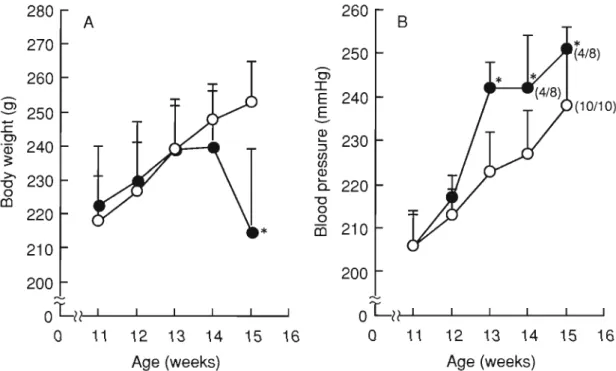
(16) Table 3. Birth weight of SHRSP exposed to low protein diet in utero.. Average per litter. Mean. 5.3 ± 0.3 (n=6) Male. 5.0 ± 0.5 (n=4). 5.4 ± 0.4. 5.9 ± 0.2 (n=5) 5.5 ± 0.2 (n=4). 20% casein. 4.9 ± 0.3 (n=6) 5.0 ± 0.5 (n=5) Female. 5.2 ± 0.2 5.4 ± 0.2 (n=3) 5.3±0.1 (n=4) 4.5 ± 0.8 (n=6). Male. 4.7 ± 0.6 (n=4). 4.7 ±0.2*. 4.8 ± 0.4 (n=8). 9% casein. 4.5 ± 0.6 (n=6) Female. 4.8 ± 0.3 (n=8). 4.7 ± 0.4. 4.9 ± 0.2 (n=4) Pups weighted 1 day after birth. The values are expressed as mean ± S.D. Significant difference from the 20% casein (*p <0.05).. Changes in blood pressure, life span, and the incidence of stroke by salt loading on low-protein-exposed SHRSP. Salt was administered to male offspring rats at the age of 11 weeks. At the start of the experiment, blood pressure levels of low-protein-exposed SHRSP were 206 ± 7 mmHg and those of the control rats were 206 ± 8 mmHg. After 2 weeks of salt loading, blood pressure was 223 ± 9 mmHg in the control group and 242 ± 6 mmHg in the low-protein group, showing a marked elevation of the pressure in the lowprotein group (Fig. 2B). Subsequently in the low-protein group, some rats showed weakness as well as symptoms commonly observed with the onset of stroke, such as convulsions and paralysis of the lower limbs. Feed consumption decreased (data not shown), and body weight decreased markedly (Fig. 2A). measurement was impossible in some rats 4 weeks after salt loading.. 10. Blood pressure.
(17) 280. 260. A. 270. -. -. :r:. E 240 E. >. "0 0. 230. ;:. co. --Q). 240. (4/8). 0>. 260. ·03. *. 250. . 9 250. . !: OJ. B. ~. ~. en en. (10/10). 230. Q) ~. a. 220 "0 0 0. 220. co 210. 210 200. 200 '". oLl?. OLl?. 0. 11. 12. 13. 14. 15. 16. Age (weeks). 0. 11. 12. 13. 14. 15. Age (weeks). Fig. 2. Effects of salt loading from 11 weeks of age on body weight (A) and blood pressure (B) of SHRSP ex posed to low protein diet in utero. Eleven week-old offspring born to dams fed a 20% casein (O: n= 10) or 9% casein (e :n=9) were given 1% saline fro m 11 weeks old. Offspring were obtai ned from 4 dams fed a 20% casein diet and from 3 dams fed a 9% casein diet. Offspring were g iven commercial diets (Funabashi SP diet, Funabashi Farm Co., Ltd. , Chiba, Japan). Body weight was meas ured once a week for 4 weeks, with a final measurement at 15 weeks. To avo id possible ge nder differences, only male pups were studied. Systolic blood pressure was measured using the tail-cuff method once weekly. (/): (No. of blood press ure measurable rats / No. of examined rats) . Data are mean±S.D. *Significant difference from the control (p<0.00 I).. 11. 16.
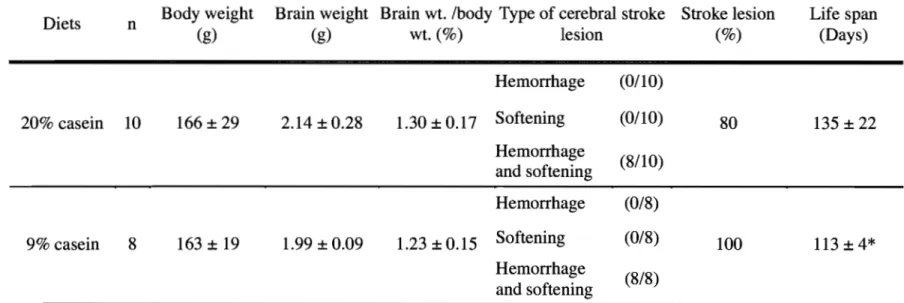
(18) Figure 3 shows the survival curves of these rats. Death began to occur at 105 days of age. The average life span was significantly lower in the low-protein group (113 ± 4 days) than in the control group (135 ± 22 days; p <0.05) (Table 4) . The incidence of stroke was 80% in the control group and 100% in the low-protein group.. The. brainlbody weight ratio was slightly lower in the low-protein group than in the control group, but the difference was not significant.. 100 .-.. '0:!2.. 75. ----. Q). C(l. ....... C(l. 50. > .:; ....... ::l (j). 25. o. o 14 15 16 17 18 19 20 21 22 23 Age (weeks). Fig. 3. Effects of salt-loading from 11 weeks of age on survival curve of SHRSP exposed to low protein diet in utero. Eleven week-old male offspring born to dams fed a 20% casein CO:n= lO) or 9% casein Ce :n=8) were given 1% saline. Offspring were obtained from 4 darns fed a 20% casein diet and from 3 dams fed a 9% casein diet. Offspring were given commercial diets CFunabashi SP diet, Funabashi Farm Co., Ltd., Chiba, Japan). To avoid possible gender differences, only male pups were studied. Data represents percentage survival.. 12.
(19) Table 4. Effects of salt-loading from 11 weeks of age on life span and stroke lesion of SHRSP exposed to low protein diet in utero Diets. 20% casein. n. 10. Body weight (g). 166 ± 29. Brain weight Brain wt. /body Type of cerebral stroke Stroke lesion (%) (g) wt. (%) lesion. 2.14 ± 0.28. 1.30 ± 0.17. Hemorrhage. (0/10). Softening. (0/10). 80. Life span (Days). 135 ± 22. M. 9% casein. 8. 163 ± 19. 1.99 ± 0.09. 1.23 ± 0.15. Hemorrhage and softening. (8/10). Hemorrhage. (0/8). Softening. (0/8). Hemorrhage and softening. (8/8). 100. 113 ± 4*. The male offspring were obtained from 4 dams fed a 20% casein diet and from 3 dams fed a 9% casein diet. The body and brain weight of male offspring were measured at the natural death. The values are expressed as mean ± S.D. *Significant difference from the 20% casein (p<0.05)..
(20) Discussion. Low birth weight due to maternal undernutrition has been considered a risk factor in hypertension and heart diseases, and studies have been done on the relationship between maternal nutrition and the onset of diseases in offspring (Curhan et al. 1996; Launer et al. 1993; Law et al. 1993; Moore et al. 1996; Osmond et al. 1993; Stein et al. 1996). It was reported that maternal undernutrition and protein restriction during pregnancy might cause hypertension in adult rat offspring (Langley-Evans and Jackson 1996a; Langley-Evans et al. 1996b; Langley-Evans et al. 1996c).. This. phenomenon, however, has not been examined in animals with hereditary factors regarding hypertension. The author investigated the effects of maternal protein restriction during pregnancy on offspring's blood pressure in SHRSP, which are the animal models for human essential hypertension and stroke. The minimum protein requirement is 9% for adult rats and 12% for pregnant rats (National Research Council 1978). Langley-Evans et al. advocated that the use of a 9% casein diet constituted mild protein restriction, because the breeding of normotensive rats on a 9% casein diet suppressed body weight gain in mothers but did not change the litter size and offspring's body weight (Langley and Jackson 1994; Langley-Evans 1994) after birth thus, the author adopted a 9% protein level for the low-protein diet in this study. It has been reported that a 9% casein diet during pregnancy in normotensive rats did not affect reproductive outcome but did increase placental weight, leading to a decrease in fetal weight (Langley-Evans et al. 1999b). Also, in SHRSP, it was shown that maternal weight gain and litter size were smaller, the placental weight was higher, and offspring's birth weight was lower than in the original strain, normotensive WKY(Yamada et al. 1981).. Since SHRSP are. programmed to develop hypertension due to genetic factors, we presumed that a 9% casein diet during pregnancy might significantly affect reproductive outcome and the offspring's growth. In the SHRSP genetic model of stroke, placental weights at day 21 were lower for dams fed a low protein diet than for control SHRSP. This finding was consistent with the report of Fernandez-Twinn et al. who found that placental weights of protein restricted Wistar rats were lower than the controls at both days 18 and 21 of gestation (Fernandez-Twinn et al. 2003). Low birth weight due to maternal undernutrition has been considered a risk factor for hypertension and heart disease (Barker 1997), and therefore studies have been carried out on the relationship between maternal nutrition and the onset of diseases in their offspring (Ozanne and Hales 1999) (Langley-Evans et al. 1996c). Maternal protein restriction reduced birth weight in SHRSP, the same as for Wi star rats. In SHRSP, fetus development was greatly. 14.
(21) influenced by mild maternal protein restriction during pregnancy, the same as for normotensive rats. In low-protein-exposed Wistar rats, blood pressure levels have been 20 to 30 mmHg higher even at 4 weeks of age than in control rats (Langley-Evans 1994), and the levels remain high throughout their lifetimes (Langley and Jackson 1994). In low-protein-exposed SHRSP, blood pressure levels were slightly higher than in the control group from 7 to 9 weeks of age but showed no differences between the two groups afterIO weeks of age. Likewise, the effect of maternal protein malnutrition does not alter the offspring's life span and incidence of stroke. Changes in blood pressure during the growth process vary fundamentally between SHRSP and normotensive rats. In WKY rats, blood pressure levels are 110 to 120 mmHg at 4 weeks of age and remain unchanged at 130 mmHg throughout the adult lifetime. In SHRSP, the levels are 120 to 130 mmHg at 5 weeks of age, then elevate by approximately 10 mmHg weekly up to 12 weeks of age, and increase to 230 mmHg or higher after growth. The possible mechanisms of blood pressure elevation in SHRSP are considered to be hyperactivity of the renin-angiotensin system (RAS) (Kim et al. 1992), the sympathetic nervous system,(Rascher et al. 1983; Tabei et al. 1995) or the endocrine system, or increased reactivity of peripheral resistance arteries, decreased number of renal nephrons (Nakamura et al. 1996; Skov et al. 1994), etc. Blood pressure elevation resulting from a low protein diets in normotensive Wistar rats, on the other hand, has been suggested to be related to a decreased number of renal nephrons (Langley-Evans et al. 1999b) and RAAS hyperactivity (Langley-Evans et al. 1996d; Sherman and Langley-Evans 1998; Sherman and Langley-Evans 2000). The mechanisms of the development of high blood pressure in low-protein Wistar rats and that in SHRSP share certain common characteristics. In SHRSP, it is the considered that the effects of intrauterine low-protein exposure were considered to have been concealed by the genetic mechanism of pressure elevation and to have not been revealed as an increase in blood pressure. Numerous studies have described a decrease in nephron numbers following maternal protein restriction (Langley-Evans 1999; Langley-Evans et al. 1999a; Langley-Evans et al. 1999b; Sherman et al. 1999) (Woods et al. 2001) or intrauterine growth restriction. Correlations of lower nephron number, or reduced kidney weight (Langley-Evans et al. 1999b; Merlet-Benichou et al. 1994), with lower weight at birth in humans (Mackenzie and Brenner 1995), have promoted the hypothesis, that intrauterine environmental effects upon nephron number may determine later blood pressure.. Knowing that nephron number is related to renal size, (Nyengaard and. 15.
(22) Bendtsen 1992) (Manalich et al. 2000), the results of the present study showed small kidney at day 21 of gestation in the low prqtein exposed SHRSP. However, blood pressure levels did not differ between the control and low protein exposed SHRSP at 10 weeks of age. Sajajpal and Aston demonstrated that elevated blood pressures in low protein exposed Wistar rats in utero were caused by RAAS upregulation but did not result in the development of fewer nephrons (Sahajpal and Ashton 2003; Sahajpal and Ashton 2004). Langley-Evans at al. indicated that renal programming, such as fewer nephron numbers, is not one of the primary mechanisms leading to hypertension following maternal protein restriction (Langley-Evans et al. 2003). In SHRSP, the effect of intrauterine low-protein exposure suggested that a reduction in kidney size may not underlay hypertension. In this context, we loaded low-protein-exposed SHRSP with salt from 11 weeks of age in order to investigate the effects of low-protein exposure in utero. The changes of blood pressure elevation and stoke incidence were not observed in low-proteinexposed SHRSP in water drinking. However, the low-protein-exposed SHRSP had a marked elevation in blood pressure as compared with the control group after salt loading, suggesting that low-protein exposure in the fetal period could continue to affect blood pressure even after growth. These results indicated that maternal mild protein restriction induces the incidence of stroke in salt-loading SHRSP. Moreover, the survival duration after salt loading was shorter in the low-protein-exposed SHRSP. In a report observing the morphologic changes of the brain in offspring rats exposed to low protein during gestation, anastomotic abnormality in the cerebral artery (Resnick et al. 1979) and decreased vascular density (Bennis-Taleb et al. 1999) were demonstrated. In the low-protein-exposed SHRSP group, it is unclear whether the shortened survival duration after salt loading might results from vascular disorders advanced by blood pressure elevation or might be associated with a morphologic developmental anomaly of the cerebral vessels. The incidence of stroke was almost identical between the control and low-protein groups, whereas the brain/body weight ratio was lower in the low-protein group than in the control group. This indicates that the period from the onset of stroke to death was short in the low-protein group. Generally, blood pressure elevation after salt loading is often caused by decreased Na excretion.. Recently, it has been shown that salt sensitivity may be related to an. enzyme, 11 [3 -HSD2 (Lovati et al. 1999; Takeda et al. 1998), which regulates the binding of aldosterone to mineralocorticoid receptors.. Maternal malnutrition and. glucocorticoid loading to mother rats during pregnancy have been reported to reduce renal 11 [3 - HSD2 activity in offspring rats (Saegusa et al. 1999; Whorwood et al.. 16.
(23) 2001). It is known that salt sensitivity is higher in SHRSP than in WKY rats. Salt loading in low-protein-exposed rats after growth did not elevate blood pressure in normotensive rats (Langley-Evans and Jackson 1996b) but increased blood pressure in SHRSP. In normotensive rats, upregulation of RAAS may play a role in prenatal programming of hypertension through maternal protein restriction during pregnancy (Langley-Evans et al. 1999b) (Woods et al. 2001).. Low-protein exposure might. abnormality of RAAS, resulting in further increased blood pressure and shortened life span. The author consider it necessary to investigate the mechanism governing the increase in salt sensitivity that in low-protein-exposed SHRSP in terms of the RAAS in offspring rats. Summary The effect of maternal protein restriction during pregnancy on the offspring's blood pressure was assessed in SHRSP which are genetically predisposed to hypertension and stroke. After the confirmation of pregnancy, the control group was given a 20% casein diet, and the low-protein group was fed a 9% casein diet. After the confirmation of delivery, commercial feed was given to both of the groups. No differences were seen between the control and low-protein offspring in regard to body weight, blood pressure elevation, or life span. One percent saline solution was put in the control and low-protein groups after the age of 11 weeks.. Blood pressure. increased markedly in the low-protein group, on the blood pressure level in the lowprotein group on week 2 after salt loading (242 ± 6 mmHg) was significantly higher than that in the control group (223 ± 9 mmHg; p<0.05). The survival duration was significantly shorter in the low-protein group (113 ± 4 days) than in the control group (135+22 days; p<0.05). These results suggest that maternal protein malnutrition in SHRSP exerted a high salt sensitivity and a malignant influence on stroke incidence on offspring.. 17.
(24) Chapter 2 Prenatal low protein exposure enhances salt sensitivity in SHRSP Introduction Stroke is a major cause of death in Japan, China and Europe and the most common risk factor for stroke is hypertension.. Recent epidemiologic evidence. indicates that the intrauterine nutritional environment is a strong determinant, not only of fetal growth, but also of hypertension and cardiovascular disease in later life. These epidemiological data are strongly supported by experimental animal studies. A number of dietary manipulations such as protein (Langley-Evans et al. 1999b; Woods et al. 2001) and calorie restriction (Merlet-Benichou et al. 1994; Woodall et al. 1996) during pregnancy in the rodent result in an increase in blood pressure in the offspring. Maternal protein restriction is the most commonly used rat model to investigate fetal programming of hypertension and it has been demonstrated clearly that maternal protein restriction results in elevated blood pressure levels in the offspring. Several mechanisms have been proposed to explain fetal programming of hypertension. In normotensive rats, upregulation of the renin angiotensin system may playa role in increased blood pressure in young rats exposed to low protein in utero.. Blood. pressure elevation in these rats was effectively prevented by early treatment with ACE inhibitor (Sherman and Langley-Evans 1998) or angiotensin II receptor antagonist (ARB) (Sherman and Langley-Evans 2000).. Treatment with the calcium channel. antagonist nifedipine was not effective, hence these results indicate that RAAS play an important role in programming the hypertensive effect of maternal protein restriction (Sherman and Langley-Evans 2000). Sahajpal and Ashton reported that rats exposed to maternal protein restriction in utero had increased expression of glomerular AT 1 receptor and reduced expression of AT 2 receptor (Sahajpal and Ashton 2004). Hypertension is not only a direct cause of death, but persistence of hypertension also is an important risk factor for stroke and ischemic heart disease. There are few studies that have investigated the influence of maternal protein or undernutrition on the onset of stroke or cardiac heart disease. The author is focusing on the influence of maternal protein restriction on the incidence of stroke in the offspring. The author have investigated previously the effect of maternal protein malnutrition during pregnancy on blood pressure, salt sensitivity and stroke incidence in SHRSP, as a genetic model of stroke (Otani et al. 2004).. 18. The results indicated that maternal.
(25) protein restriction during pregnancy induces salt sensItIvIty and promotes an increased incidence of stroke in SHRSP offspring. In this study, the author addressed the contribution of RAAS activation to high salt sensitivity in low protein exposed SHRSP. Methods Animals. This study was carried out according to the guidelines of the animal. experimental care committee of the Kinki University Faculty of Agriculture and the rats were managed in conformation with the guidelines for Care and Use of Laboratory Animals. Inbred SHRSP, which were raised in our laboratory, were used for the experiments. They were maintained at a room temperature of 23 ± 1°C, humidity of 55 ± 5%, and under a 12-hour light-dark cycle (lighting from 7:00h to 19:00h). Mating. The animals were bred with sibling SHRSP. The SHRSP used in this study. were of a strain in which blood pressure elevation is relatively mild and whose life span is long. Virgin female SHRSP weighing 150 g to 180 g were mated to males of the same strain (weighing 210 g or more, blood pressure 175 to 185 mmHg). Animals were paired and housed together for one night.. The date of pregnancy was. determined with the confirmation of a vaginal plug and was set as pregnancy day o. Animals were pair fed and given a 20% and 9% casein diet in the control and lowprotein groups, respectively, from pregnancy day 0 to the confirmation of delivery (Table 1). During this period, the body weights and feed consumption of mother rats were measured daily. The feed was substituted by a commercial diet (Funabashi SP, Funabashi Farm Co., Ltd., Chiba, Japan) after confirmation of delivery. Care ofoffspring rats. The day of confirmed delivery was set at postnatal day o. Birth. weight was measured on postnatal day. o.. Offspring rats were culled on postnatal day. 4, leaving a uniform litter size of 6. Offspring rats were weaned at the age of 4 weeks. All offspring were provided with a commercial diet (Funabashi SP, Funabashi Farm Co., Ltd., Chiba, Japan) and tap water ad libitum after weaning. weighed once weekly.. Offspring were. Salt loading. The SHRSP in the control and low-protein groups were further divided at. 10 weeks of age into a tap water group and a 1% saline solution group. All offspring were provided with a commercial diet ad libitum. Offspring were killed under ether. 19.
(26) anesthesia at 12 weeks of age. Blood pressure, after preliminary heating at 37°C, was measured using the tail-cuff method (BP-98A; Softron Inc. Tokyo, Japan). Spironolactone treatment. At 10 weeks of age, all SHRSP received a subcutaneous. injection of 10 mg/kg/day spironolactone, an aldosterone antagonist. The SHRSP in the control and low-protein groups were given a commercial diet and 1% saline ad libitum until their natural death. Dead rats were autopsied. The brains were removed. and fixed with 10% formalin and then divided into five parts in order to confirm the presence or absence of stroke lesions (cerebral hemorrhage, cerebral softening, etc.). Hormonal and biochemical assessment. Blood samples were collected into chilled 1.5. ml EDTA tubes at 8:00 to 10:00 a.m. Samples were collected from the tail veins of 12-week-old rats without anesthesia. The blood samples were centrifuged for 10 min at 1500 rpm to obtain plasma, which was then stored at -80°C. Plasma aldosterone concentrations were estimated using an EIA kit (Cayman Chemical Company, USA) after extraction three times with methylene chloride. Recovery of aldosterone was in the 97 to 100% range. ACE activity determination. Blood was collected from the abdominal aortas of rats. anesthetized with diethyl ether into heparinized test tubes. Tissue samples were prepared as follows: Aorta were gently flushed with saline containing heparin and removed immediately after sacrifice. Vessels were carefully stripped of surrounding fat and connective tissue. The kidneys were homogenized in 0.05 M Tris-HCI buffer (pH 7.8) with 0.5% Nonidet-P40 and centrifuged at 20,000 Xg for 20 min at 4°C. The supernatant was used as the enzyme preparation and for protein concentration and then stored at -80°C. The ACE activity was measured within three days of sacrifice. The plasma and aorta ACE activities were measured by a modified method, (Horiuchi et al. 1982) using a synthetic substrate Hip-His-Leu, specifically designed for ACE (Peptide Institute, Inc., Osaka, Japan). Fifty microliters of plasma or kidney extract were incubated for 30 min at 37°C with 5 mM Hip-His-Leu in 250 III of 100 mM phosphate buffer, pH 8.3, containing 400 mM NaCl. The reaction was terminated by addition of 750 III of 1 N HCl. The mixture was centrifuged for 5 min, and then filtered through 13 mm membrane filters (ADVANTEC, Toyo Roshi Kaisha, Ltd). The supernatants were subjected to analysis using reversed-phase HPLC. The HPLC equipment consist of a Shimadzu LC-6A pump, a Rheodyne injector fitted with a 20 III loop and a Shimadzu SPD-10A SPECTROPHOTOMETRIC detector set at. 20.
(27) 280nm. Analyses were performed in a Cosmosil 5CI8-AR-II column (4.6 x 1.0) 250mm, Nacalai Tesque, Kyoto, Japan) at ambient temperature with a mobile phase of methanol-lO mM KH2P04 (1:1) and adjusted to pH 3.0 with phosphoric acid at a flow rate 0.5 mUm!. One unit of activity is defined as the amount of enzyme catalyzing the release of 1 ~mol of hippuric acid from Hip-His-Leu per minute at 37°C. Statistical analysis. The data are presented as mean ± SD. Data with only a single grouping comparison were analyzed statistically by student's t test.. Data with. multiple group comparisons were analyzed statistically by one-way or two-way ANOVA.. Where appropriate, directed pairwise comparisons of individual groups. were conducted using Tukey's test and the Bonferroni method. Kaplan-Meier plots of survival were analyzed using a log-rank test. The analyses were performed using a commercially available statistical package (SPSS ver. 12.0 for windows; SPSS Japan Inc.). A probability value of P <0.05 was considered statistically significant. Results Growth The birth weights of male pups were significantly lower in the low-protein exposed (4.68 ± 0.49 g) group than the control group (5.00 ± 0.37 g).. However,. growth after birth was unaffected by the maternal diet. Body weight at the age of 10 weeks was almost the same in the control group (male, n=20; 222 ± 15 g) as in the low-protein exposed group (male, n=17; 219 ± 11 g). Blood pressure levels were the same in the control group (male, n=20; 200 ± 7 mmHg) and the low-protein exposed group (male, n= 17; 199 ± 7 mmHg) at 10 weeks of age. Both the control and low-protein exposed groups were divided into tap water and 1% saline groups at 10 weeks of the age. Figure 4 shows blood pressure and body weight changes in the control and low-protein exposed groups after salt loading. In the tap water drinking groups, no differences in blood pressure and body weight were seen between the control group and low-protein exposed group. After 2 weeks of salt loading, however, blood pressure levels increased significantly in the 1% saline drinking control group (215 ± 5 mmHg ) compared to the tap water drinking group (203 ± 5 mmHg). After two weeks of salt loading, blood pressure levels were 215 ± 5 mmHg in the control group and 230 ± 6 mmHg in the low-protein exposed group, showing marked elevation in the low-protein exposed group.. 21.
(28) 280. 280. Water. 260. 260. ~. ~. .9 E. .9 E 240 0>. 0>. .Ci). 240. .Ci). ::. ::. >-.. "0. 0. (I). 1% saline. >-.. "0. 220. 0. (I). 200. 220 200. 0 CON (n=6). •. 0 0. 10. 0 CON (n=7). •. LP (n=6). 0. 11. 12. 13. 0. 10. Age (weeks). 240. OJ. 11. 12. 13. Age (weeks). 240. Water. 230. 1% saline. 230. *. OJ. I. I. -. ~. -....E. ....0.. ..... E E 220 Q) .... (f) 210 (f). E 220. Q). ~ (f) (f). Q). "0 0 0. LP (n=7). 210. Q). 0.. "0 0 0. 200. iIi. 200. (I). 190. o. •. 0 0. 10. 190. CON (n=6). 11. o. CON (n=7). •. LP (n=6). LP (n=7). 0 12. 13. 0. Age (weeks). 10. 11. 12. Age (weeks). Fig. 4 Body weight and blood pressure in rats exposed to low protein diet in utero , drinking water or 1% saline. Animals were maintained on water or I % saline starting at 10 weeks of age. Offs pring were given commercial diets (Fun abashi SP diet, Funabashi Farm Co., Ltd., Chiba, Japan). To avoid possible gender di fferences, only male pups were studied. Systolic blood pressure was measured using the tail-cuff method once weekly. Data are mean±S .D. *Sign ificant difference from the control (p<O.OOI ).. 22. 13.
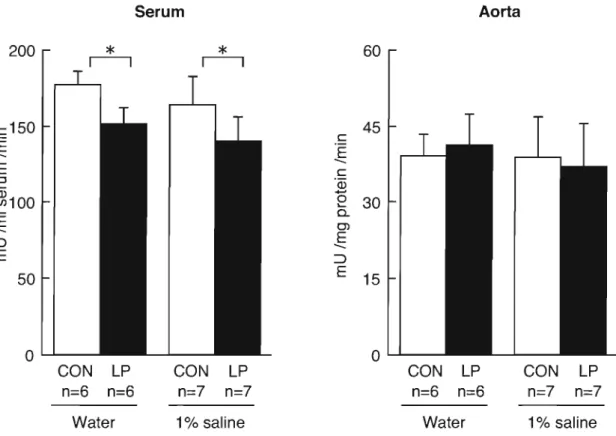
(29) A CE activity. Figure 5 shows the ACE activity in plasma and kidney at 12 weeks of age. There were no signific differences in the control group between the tap water and saline drinking rats. Maternal protein restriction significantly reduced plasma ACE activity in the offspring.. Plasma ACE activity of the low-protein exposed group was. unaffected by salt loading, as for the control group. The ACE activity in the aorta was unaffected by maternal protein restriction and salt loading. Serum. 200. *. r--,. Aorta. 60. *. r--,. c150 E E. c. 45. 'E. --. --. C. ::l .... ~100. 'Q5. 0 .... 30 c... --EE. C'l. --EE. :::>. :::>. 50. 15. 0 ..........- -. 0 ..........- CON LP. CON. LP. CON. LP. CON. LP. n=6. n=7. n=7. n=6. n=6. n=7. n=7. n=6. Water. 1% saline. Water. 1% saline. Fig. 5 Plasma and aorta ACE activity in tats exposed to low protein diets in utero drinking water or 1% saline. The plasma and kidney ACE activities were measured using a synthetic substrate Rip-Ris-Leu at 12 weeks of age. Offs pring were given commercial diets (Funabashl SP diet, Funabashi Farm Co., Ltd. , Chiba, Japan). To avoid possible gender differences, only male pups were studied. Data are mean±SD. Data are significantly different by two-way ANOVA, *p<O.05.. 23.
(30) Plasma aldosterone concentration Plasma aldosterone levels at 12 weeks of age are shown in Figure 6. In tap water drinking rats, plasma aldosterone levels in the low-protein exposed group were similar to the control group. Salt loading was associated with a marked suppression of plasma aldosterone levels in the control group. In contrast, the aldosterone levels after salt loading were not changed in the low-protein exposed group.. 160. **. 120. .€. C). 80. 0.. 40. OL...J...._CON. n=6. LP. CON. LP. n=6. n=7. n=7. Water. 1% saline. Fig. 6 Plasma aldosterone concentration in rats exposed to low protein diets in utero drinking water or 1% saline. Plasma aldosterone concentration were measured at 12 weeks of age. Data are mean±SD. Offspring were given commercial diets (Funabashi SP diet, Funabashi Farm Co. , Ltd. , Chiba, Japan) . To avoid possible gender differences, only male pups were studied. Data are significantly different by two-way ANOVA, *p<O.05 ,. * * p<O.Ol.. Administration of spironolactone Figure 7 shows blood pressure and body weight changes in the control and low protein exposed groups after spironolactone treatment.. The body weights of both. groups of rats were unaffected by spironolactone treatment. All animals had similar blood pressure levels at the start of spironolactone treatment (10 weeks). The systolic blood pressure of the control SHRSP was unaffected by spironolactone treatment. Over the 14 days of the experiment, differences in blood pressure between the control SHRSP and low protein exposed SHRSP were not seen and, thereafter, blood pressure. 24.
(31) was similar in both groups. There was no significant difference in the average life span of the control SHRSP and low protein exposed SHRSP (Fig. 8). The incidence of stroke was 100% in both groups. The life spans of the control SHRSP group (330 ± 105 days) and low protein exposed SHRSP group (292 ± 79 days) were not influenced by exposure to low protein during gestation (Table 5). 300. 250. 280. 240 ~. I. OJ. 260. E E. OJ 'w 240. ::J. 9. ...... .!:. ....Q). ~. Ul Ul. >. "0 0. CIl. 220. ....Q). 210. "0 0 0. 200. c.. 220. 230. CIl. 200. 0 CON (n=6). •. 0 0. 10. 12. 14. o. 190. LP (n=?). •. 0 16. 18. 0. Age (weeks). 10. 12. 14. CON (n=6) LP (n=?). 16. 18. Age (weeks). Fig. 7 Effect of spironolactone treatment from 10 weeks on body weight (A) and systolic blood pressure (B) of salt loaded SHRSP. The control and low protein exposed group were kepl under the same condilions from 10 weeks of age. Animals were maintained on I % saline starting al 10 weeks of age. Offs pring were given commercial diels (Funabashi SP diet, Funabashi Farm Co. , Ltd., Chiba, Japan). To avoid possible gender differences, only male pups were studied. Systolic blood pressure was measured using the tail-cuff method once weekly. Data are mean±S.D.. 25.
(32) 100. --. 75. ro. 50. ~ 0. 0. •. CON (n=6). LP (n=7). Q). Cti ">. .~. :J (f). 25. o. L...-_ _- ' - -_. o. 50. _. -L.._ _-~--__{ } - - - - - - '. 100. 150. 200. 250. Days after salt loading F ig. 8 Effect of spironolactone treatment from 10 weeks on cumulative percentage survival in salt loaded SHRSP. The control and low protein exposed group were kept under the same conditions from 10 weeks of age. Animals were maintained on I % saline starting at 10 weeks of age. Offspring were given commercial diets (Funabashi SP diet, Funabashi Farm Co. , Ltd., Chiba, Japan). To avoid possible gender differences, only male pups were studied. The difference in percent survival between the control and low protein exposed SHRSP was statistically significant (Kaplan-Meier analysis followed by log rank test).. Table 5 Effect of spironolactone treatment from 10 weeks on body weight, brain weight, stroke lesion and life span Body weight. Brain weight (g). Brain wt. /body wt. (%). Stroke lesion. (g). (%). Life span (Days). 20% casein 6. 236 ± 56. 2.43 ± 0.66. 1.02 ± 0.32. 100. 89 ± 63. 9% casein 7. 209 ± 46. 2.12±0.12. 1.06 ± 0.25. 100. 72±27. Diets. n. Data mean±S.D.. Discussion The birth weights of low protein exposed SHRSP were decreased by mild maternal protein restriction during pregnancy, but there was no noticeable difference in blood pressure elevation between SHRSP exposed to a low protein diet in utero and those rats exposed to a control diet in utero .. On the other hand, a remarkable. elevation of blood pressure in response to salt loading was seen in SHRSP exposed to a low protein diet during gestation. These results were consistent with our data published previously.. The main findings of this study were that there was no. 26.
(33) reduction of plasma aldosterone levels in SHRSP exposed to low protein in utero, whereas reduction of plasma aldosterone levels in response to salt loading was seen in control rats. Treatment with spironolactone of SHRSP exposed to low protein during gestation suppressed the increase in blood pressure in response to salt loading and reversed the shortened life span. The author loaded low protein exposed SHRSP with salt from 10 weeks of age. In agreement with our previous findings, the low protein exposed SHRSP had a marked elevation in blood pressure after salt loading, compared to control rats. This indicates that the effect of mild maternal protein restriction was evident in later life and promoted salt sensitivity.. In normotensive rats, mild maternal protein restriction. induced hypertension in the offspring.. Salt sensitivity in the offspring of protein. restricted dams is produced by severe maternal protein restriction, (Manning and Vehaskari 2005; Rasch et al. 2004; Woods et al. 2004) but not demonstrated in mild protein restriction(Langley-Evans and Jackson 1996b; Zimanyi et al. 2004). In normotensive rats, salt sensitivity in offspring also is less susceptible to maternal protein restriction than blood pressure in the offspring of protein restricted dams (Langley-Evans 1994; Woods et al. 2001).. In SHRSP, mild maternal protein. restriction promoted high salt sensitivity in the offspring. These results suggested that salt sensitivity in SHRSP offspring was greatly influenced by maternal protein restriction, compared to normotensive rats. In normotensive rats, upregulation of RAAS may play a role in prenatal programming of hypertension through maternal protein restriction during pregnancy (Langley-Evans et al. 1999b; Woods et al. 2001). Maternal protein restriction during pregnancy has been reported to lead to elevated serum and pulmonary ACE activity (Langley-Evans and Jackson 1995).. Unlike those previous studies, in this study. plasma ACE activity was low in the low protein exposed group compared to control rats and aorta ACE activity did not differ between control rats and low protein exposed SHRSP, and was not affected by salt loading. Maternal protein restriction had no effect upon plasma aldosterone levels in SHRSP offspring given tap water. This result is consistent with that of Manning and Sahajipal et al., who examined plasma aldosterone levels and found no difference between their control group and low protein exposed group (Mackenzie and Brenner 1995; Sahajpal and Ashton 2004).. Hence, there was no evidence for a role for. aldosterone in determining the higher blood pressure of rats exposed to maternal low protein intake during fetal life. This study showed that plasma aldosterone levels in control rats were suppressed by high salt intake. However, in low protein exposed. 27.
(34) SHRSP, plasma aldosterone levels were not suppressed and were significantly higher than control rats. The author hypothesize that increased salt sensitivity in low protein exposed SHRSP is linked to high aldosterone levels under salt loading. Salt-sensitive hypertension is linked to end-organ damage and is a strong risk factor for cardiovascular disease (Weinberger et al. 2001). End-organ damage in saltloaded SHRSP is initiated by angiotensin II (Stier et al. 1989). Numerous studies with ACE (Stier et al. 1991) inhibitors and ARB (Stier et al. 1993) have clearly implicated angiotensin II as a major factor responsible for the development of endorgan damage in salt sensitive hypertensive disease. On the other hand, aldosterone is released in response to angiotensin II and is involved in the regulation of sodium and water homeostasis.. Recently, aldosterone also has been implicated in the. pathogenesis of vascular disease in genetically hypertensive rats and elevated plasma aldosterone is considered a risk factor for cerebrovascular disease (Rocha et al. 1998; Rocha et al. 1999) (Brilla and Weber 1992). It has been reported that SHRSP have abnormal aldosterone metabolism with high salt intake, which results in high levels of aldosterone biosynthesis and plasma aldosterone (Volpe et al. 1993) (Enea et al. 2000). The author reported previously that mild maternal protein restriction during pregnancy led to a shortened life span in salt loaded SHRSP (Otani et al. 2004). This study showed that plasma aldosterone levels were high in low protein exposed SHRSP given 1% saline. The autor predicted that the onset of stroke was caused by aldosterone.. Therefore we administered spironolactone, the aldosterone receptor. antagonist, to salt loaded, low protein exposed SHRSP and control rats. Treatment of low protein exposed SHRSP with spironolactone improved the exacerbation of blood pressure in response to salt loading, and prolonged the life span. The life span of the low protein exposed SHRSP administered spironolactone was not significantly different from control rats. It has been reported that aldosterone receptor antagonists reduce markedly the severity of renal damage and incidence of stroke in SHRSP (Rocha et al. 1998) (Chander et al. 2003).. Many observations suggest that. aldosterone plays a direct role in vascular injury and end organ damage in salt-loaded SHRSP (Endemann et al. 2004a; Endemann et al. 2004b) (Rocha et al. 1999). Lack of suppression of aldosterone in response to salt loading in low protein exposed SHRSP is one cause of early onset of stroke and a shortened life span. In summary, this study has shown that maternal protein restriction in SHRSP leads to salt sensitive hypertension and a shortened life span. Abnormality of aldosterone metabolism in response of salt loading was observed in low protein exposed SHRSP and this is considered to induce early onset of stroke and a shortened life span.. 28.
(35) Summary. The author is focusing on the influence of maternal protein restriction on the incidence of stroke in the offspring.. In this study, the author addressed the. contribution of RAAS activation to high salt sensitivity in low protein exposed SHRSP. The birth weights of low protein exposed SHRSP were decreased by mild maternal protein restriction during pregnancy, but there was no noticeable difference in blood pressure elevation between SHRSP exposed to a low protein diet in utero and those rats exposed to a control diet in utero.. On the other hand, a remarkable. elevation of blood pressure in response to salt loading was seen in SHRSP exposed to a low protein diet during gestation. These results were consistent with our data published previously. The main findings of this study were that there was no reduction of plasma aldosterone levels in SHRSP exposed to low protein in utero, whereas reduction of plasma aldosterone levels in response to salt loading was seen in control rats. Treatment with spironolactone of SHRSP exposed to low protein during gestation suppressed the increase in blood pressure in response to salt loading and reversed the shortened life span.. Therefore, this study has shown that maternal. protein restriction in SHRSP leads to salt sensitive hypertension and a shortened life span. Abnormality of aldosterone metabolism in response of salt loading was observed in low protein exposed SHRSP and this is considered to induce early onset of stroke and a shortened life span.. 29.
(36) CHAPTER 3 Reduction of Salt Sensitivity in SHRSP Administered an AT 1 Receptor Antagonist During Suckling Introduction. The onset of hypertension is caused by complex interactions between genetic predisposition(Okamoto et a1. 1974) and environmental factors (Murakami et a1. 1994). A high salt intake is one of the most important environmental risk factors for hypertension. There is evidence that salt sensitivity may be genetically determined, because normotensive and hypertensive salt-sensitive subjects tend to exhibit a familiar history of hypertension more frequently than do those who are salt resistant(Luft et a1. 1988) and, in addition, there is a familiar similarity in the blood pressure response to sodium restriction (Miller et a1. 1987). Recently, it was determined that capsaicin treatment in the neonatal period may impair the renal sodium and water excretion response to high sodium intake, resulting in increased salt sensitivity (Wang and Li 1999). Therefore, environmental factors, such as diet in very early life, are becoming seen as of great importance in preventing hypertension, stroke and cardiovascular disease in later life. Several studies have indicated that the intrauterine or suckling environments affect the onset of hypertension and cardiovascular disease in adult life (Barker 1993; Law et a1. 1993; Lucas 1998; Osmond et a1. 1993; Otani et a1. 2004). It is possible to suppress blood pressure elevation by changing the postnatal environment, even for those with a genetic predisposition to hypertension. Treatment during early life of SHRSP with agents targeting the RAAS leads to persistent reductions in blood pressure (Harrap et a1. 1990; Mizuno et a1. 2001; Morton et a1. 1992; Nakaya et a1. 2001). The RAAS is known to trigger or set in motion a mechanism which determines the ultimate pressure levels and end-organ damage in later life. Renin and angiotensin II levels in SHR and SHRSP are inappropriately high, suggesting RAAS overactivation. (Bunkenburg et a1. 1991; Kim et a1. 1992) The RAAS plays an important role in the control of blood pressure and pathophysiologic end-organ damage in these rats. Suppression of the RAAS in early life has an effect on blood pressure and the onset of cardiovascular disease in later life. The author hypothesized that suppression of the RAAS in early life induces possible moderate blood pressure elevation and salt sensitivity in later life, regardless of genetic background. To test this hypothesis, SHRSP were administered an AT 1 receptor antagonist during lactation, and their offspring was evaluated to determine. 30.
(37) whether suppression of the RAAS modified blood pressure and salt sensitivity in later life.. Methods Experimental Animals. This study was carried out according to the guidelines of the experimental animals care committee of the Kinki University Faculty of Agriculture, and the rats were managed in compliance with the guidelines for care and use of laboratory Animals. SHRSP and the parental strain, Wistar Kyoto rats (WKY), were bred in our laboratory for use in this study. Mothers and their pups were housed individually in plastic cages. All litters were culled to leave six pups 4 days after delivery. Offspring were weaned at 4 weeks of age and then housed in groups of four or less per wire cage. The rats were raised at a room temperature of 23 ± 1°C, humidity of 55 ± 5% and under a 12 hour light/dark cycle.. Experiment 1.. Male SHRSP were used at two weeks of age and divided into control. and treatment groups. From 2 to 4 weeks postpartum, SHRSP mothers and pups were treated with the AT 1 receptor antagonist losartan (Banyu Pharmaceutical Co., Ltd., Tokyo, Japan), administered in their drinking water (lOOmg/L). For the control group, the drinking water was tap water. Losartan has been confirmed to be transferred to maternal milk by Spence SG et a1. (Spence et a1. 1996) The pups first began to take small quantities of liquid from feeder bottles from 2 or 3 weeks of age, but most fluid came from suckling.. Thus, provision of drug to the pups would have occurred. through milk and drinking water. All rats received a commercial diet (Funabashi SP, Funabashi Farm, Chiba, Japan).. The administration of losartan was stopped at 4. weeks of age. After weaning, offspring in both groups were fed a commercial diet with tap water ad libitum. Body weights of the offspring were determined once a week. Blood pressure was measured from 6 weeks of age. After preliminary heating at 37°C, blood pressure was measured using the tail-cuff method (BP-98A; Softron Inc., Tokyo, Japan). The SHRSP in the control and losartan groups were divided again at 10 weeks of age into a tap water group (n=7, respectively) and a 1% saline solution group (n=lO, respectively). The survival of all rats entered into the study was monitored. Members of the water or 1% saline drinking group SHRSP that died spontaneously were autopsied. The explanted brains were fixed in 10% formalin and then divided into five parts in order to confirm the presence or absence of stroke lesions (cerebral hemorrhage, cerebral softening, etc.).. 31.
(38) Experiment 2. Male SHRSP and age matched WKY were used in this study. At 2. weeks of age, SHRSP rats were divided into control (tap water) and losartan (lOOmg/L in drinking water) groups. Treatment continued for 2 weeks, and WKY rats received tap water. At 4 weeks of age, losartan treatment was stopped and some pups were sacrificed (WKY, n=5; untreated SHRSP, n=7; losartan treated SHRSP, n=7). At the time, brain, heart, lung, liver and kidney was also dissected and weighted. Rats were weaned at 4 weeks of age and divided into tap water ( WKY; n=5, untreated SHRSP ; n=7 ) and a 1% saline solution ( WKY; n=5, untreated SHRSP ; n=9, losartan treated SHRSP ; n=9 ) groups at 10 weeks of age. Blood pressure was measured by the tail-cuff method once a week from 10 weeks of age. Blood samples were collected into chilled 1.5 ml EDTA tubes at 8:00 to 10:00 a.m. Samples were collected from the abdominal aortas of 4-week-old rats under anesthesia with ether and from the tail veins of 15-week-old rats without anesthesia. The samples were centrifuged for 10 min. at 1500 rpm to obtain plasma, which was then stored at -80°C. Just after the blood collection, at 15 weeks of age, rats were sacrificed and fixed in 10% formalin. After fixation, the explanted brains were examined macroscopically for the presence or absence of stroke lesions. Seven days before the animals were killed, they were placed in individual matabolice cage and urinary collection was performed on the last 3 days. Urinary albumin concentrations were determined using a commercial assay (Wako Pure Chemical Industries Ltd., Osaka, Japan). Plasma renin activity was measured by the enzyme linked immunosorbent assay (ELISA) of Suzuki et al. (Hayashi et al. 2000; Nakanishi et al. 2001; Suzuki et al. 1990) Plasma aldosterone concentrations were estimated using an EIA kit (Cayman chemical Company, USA) after three times extraction with methylene chloride. Recovery of aldosterone was in the 97 to 100% range. Statistical analysis. The data are presented as mean ± SD. Data with only a single. grouping comparison were analyzed statistically by student's t test.. Data with. multiple group comparisons were analyzed statistically by one-way or two-way ANOVA.. Where appropriate, directed pairwise comparisons of individual groups. were conducted using Tukey's test and Bonferroni method. Kaplan-Meier plots of survival were analyzed using a log-rank test. The analyses were performed using a commercially available statistical package (SPSS ver. 12.0 for windows; SPSS Japan Inc.). A probability value of P <0.05 was considered statistically significant.. 32.
(39) Results Experiment 1 In experiment 1, the author examined the effects on blood pressure and survival in losartan treated SHRSP from 2 to 4 weeks of age. Pups were treated with losartan from 2 to 4 weeks of age. The control and losartan treated groups had similar body weights at the end of treatment (contro l, 47 ± 7 g; losartan, 46 ± 3 g). However, at 10 weeks of age, body weights were lower in the losartan treated group than the control group (control, 225 ± Ilg; losartan, 216 ± 109, p <0.05).. Blood pressure was. measured from 6 weeks of age and there was no difference between the control group (148 ± 8 mm Hg) and losartan treated group (153 ± 8 mmHg). Blood pressure in the SHRSP increased with age and rose in the control group to 202 ± 4 mm Hg at 10 weeks of age, the point at which the greatest increase was attained. Blood pressure in the losartan treated group reached similar levels to the control group (199 ± 5 mmHg) . On the other hand, the blood pressure of the control and losartan treated groups remained at about 240 mm Hg at 18 weeks of age. The blood pressure level of each group was the same during the stable phase. Moreover, the life spans of the control (330 ± 105 days) and losartan treated (292 ± 79 days) groups were not influenced by antihypertensive treatment during suckling. The incidence of cerebral stroke lesion was 100% in both groups (data not shown). 300. 270. A 0>. I. 275 (8/10). B. 255. E E. -. (10/10). Q) ~. 240. :::l C/) C/). ~ c... 225. "0. o o. (5/10). .0 .~. -. 210. "0 C/). >.. 200. (f). o Control (n=10). L-.__L~~~sa_rt_a_n~~n_=_1_0~). ~,~(-L_ _ _ _. o. 10. 12. 14. 16. 195. o Control (n=10) • Losartan (n=10). ~,~,...J......----'--_.l....-----'-----'. o. 18. Age (weeks). 10. 12. 14. 16. 18. Age (weeks). Fig. 9 Effect of salt loading from 10 weeks on body weight (A) and systolic blood pressure (B) ofSHRSP. A course of temporary treatment ofSHRSP with losartan was given between 2 and 4 weeks. After treatment, control group and losartan treated group were kept under same conditions. Animals were maintained on 1% saline starting at 10 weeks of age. ( / ): (No. of blood pressure measurabl e rats / No. of examined rats) Data are mean±S.D. *Significant difference ITom the control (p<O.05).. 33.
(40) Some rats were switched to 1% saline at the age of 10 weeks. Figure 9 shows the body weight and blood pressure after salt loading.. At the start point, the blood. pressures were similar in both groups and there was then a rise in blood pressure. However, the elevation of blood pressure levels in losartan treated group was moderate compared to the control group, being significantly lower (Fig. 9B). After 4 weeks of salt loading, the control group showed weakness and body weight decreased markedly (Fig. 9A).. Blood pressure measurement was impossible in some rats 4. weeks after salt loading. Figure 10 shows survival curves after salt loading. In the control group, the rats began to die 16 days after salt loading, and their life span was 37 ± 17 days. On the other hand, losartan treated rats did not start dying until 37 days after salt loading and the life span of the losartan-treated group was III ± 79 days, a marked prolongation of the life span. 100. r-----r----,. o •. ~ ~. Control (n=10) Losartan (n= 10). 75. 50. 25. o. o. 25. 75. 50. 100. 125. Days after salt loading Fig. 10 Effect of salt loading from 10 weeks on cumulative percentage survival in SHRSP. A course of temporary treatment of SHRSP with losartan was given between 2 and 4 weeks. After treatment, control group and losartan treated group were kept under same conditions. Animals were maintained on 1% saline starting at 10 weeks of age. The difference in percent survival between the control group and losartantreated SHRSP was statistically significant (Kaplan-Meier analysis followed by log rank test ; p<O.O1). 34.
(41) Experiment 2. In experiment 2, the author examined the effects on RAAS development in losartan treated SHRSP from 2 to 4 weeks of age, in comparison to WKY. Table 6 shows organ weights of 4-week-old WKY, SHRSP and losartan treated SHRSP. Body weights were similar for WKY and SHRSP and no differences were found between the body weights of the SHRSP and losartan treated SHRSP groups. Heart weight and ratios of heart weight to body weight were greater for SHRSP than WKY at 4 weeks of age. On the other hand, losartan treated SHRSP had lower heart weights than untreated SHRSP. Brain weight was lower for SHRSP than WKY. Losartan treated SHRSP had lower the ratio of lung and liver weight to body than untreated SHRSP. Table 6 The organ weight ofWKY, SHRSP and losartan treated SHRSP at the 4-week-old. WKY. SHRSP. Losartan SHRSP. 5. 7. 7. Body wt. (g). 45.0 ± 6.1. 48.0 ± 8.1. 47.4 ± 8.4. Brain wt. (g). 1.50 ± 0.07. 1.38 ± 0.08. a. 1.37±0.10. a. Heart wt. (g). 0.22 ± 0.02. 0.31 ± 0.03. a. 0.26 ± 0.04. b. Lung wt.(g). 0.44 ± 0.07. 0.42 ± 0.06. 0.37 ± 0.06. Liver wt. (g). 2.14 ± 0.28. 2.16 ± 0.34. 1.82 ± 0.35. Kidney wt. (g). 0.24 ± 0.03. 0.26 ± 0.04. 0.25 ± 0.03. Brain / wt.(%). 3.36 ± 0.31. 2.93 ± 0.33. a. 2.94 ± 0.42. Heart / wt.(%). 0.47± 0.04. 0.66 ± 0.10. a. 0.55 ± 0.08. Lung / wt.(%). 0.96 ± 0.03. 0.89 ± 0.10. a. 0.78 ± 0.08. a. Liver / wt.(%). 4.76 ± 0.12. 4.53 ± 0.32. 3.85 ± 0.30. ab. Kidney / wt.(%). 0.53 ± 0.02. 0.55 ± 0.02. 0.53 ± 0.06. n. Data mean±S.D.. ap. < 0.05 vs. WKY. bP. 35. < 0.05 vs. SHRSP.
(42) 125. **. A. 24. *. B. c: 0. 20. 100. ~ ...... -. -. c:. >0:;- 16. Q) 0_. c:- 75. +=:E: 0-. o E 0--. ell. c:-S. -. 50. «. 25. 0 ...... 12. c: c:. Q)-. a:. Q) CIJ. 0 -0. E. c:-._ C>. Q)C>. 8. 4. 0 WKY. n=5. SHRSP n=7. 0. Losartan SHRSP n=7. WKY. n=5. SHRSP n=7. Losartan SHRSP n=7. Fig. 11 Plasma aldosterone concentration of 4-week-old WKY, SHRSP and losartan treated SHRSP. A course of temporary treatment of SHRSP with losartan was given between 2 and 4 weeks. Plasma aldosterone concentration were measured at 4 weeks of age at the end of treatment. Data are mean±S.D. Data are significantly different by two-way ANOVA, *p<O.05, * * p <O.Ol.. Plasma aldosterone concentrations were measured at 4 weeks of age, as shown in Figure llA. The plasma aldosterone concentrations at 4 weeks of age were increased in SHRSP in comparison to WKY. The plasma aldosterone concentrations of SHRSP treated with losartan were significantly lower than the untreated SHRSP. The renin activity in plasma was lower in SHRSP than WKY, but there was no significant difference (Fig. 11 B). No differences were observed in plasma renin activity between untreated SHRSP and losartan treated SHRSP. WKY and SHRSP were divided into tap water and saline groups at 10 weeks of age. Table 2 shows the body weight, blood pressure and incidence of stroke after salt loading. For WKY rats, no differences were found in blood pressure between the tap water and saline groups at 15 weeks of age.. On the other hand, blood pressures. increased in salt-loaded SHRSP. However, blood pressure was moderately elevated in the losartan treated SHRSP compared to the untreated SHRSP. There was no effect of salt loading on albumin excretion in urine in WKY (Fig. 12A). Moreover, albumin excretion in urine in the salt-loaded SHRSP increased compared to water-drinking SHRSP.. In losartan treated SHRSP, albumin excretion in urine and incidence of. stroke were comparable to SHRSP with an intake of tap water in spite of salt loading.. 36.
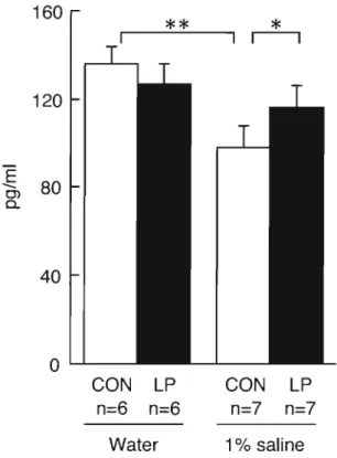
(43) 80. 0.>. c. .~. A. ::J. -. 60. -U--E. 40. 0.>. rc. .~~. o "0 ro ._ 0.>0l. x-. 0.> C. 20. E. ::J. .0. «. 0 30. B. *. 1*1 1*1. *. 1*1 1*1. 25. -- ->-. .:;.- r-. 20. u-. ro. E 15. . c-_ Ol. C C 0.>-. a:. 10 5 0 300 C. c. 0. -..:;. ro c. 250. *. ~. u_. 200. u--. 150. 0.>. co E. 0.>0l. II. cS 0 ~. 0.>. 100. ( J). 0 "0. «. 50 0. Salt Water Salt Losartan n=7 n=9 n=9 SHRSP. Water Salt n=5 n=5 WKY. Fig. 12 Urinary albumin excretion (A), plasma renin activity (B) and plasma aldosterone concentration (C) and of 15-week-old WKY, SHRSP and losartan treated SHRSP. Plasma aldosterone concentration and albumin excretion in the urine was measured at 15 weeks of age. A course of temporary treatment of SHRSP with losartan was given between 2 and 4 weeks. After treatment, all animals were kept under the same conditions. Animals were divided into tap water and I % saline groups at 10 weeks of age. Data are mean±S.D. Data are significantly different by two-way ANOVA, *p<O.OI.. 37.
図


+7


関連したドキュメント
The pharmacokinetic profiles of clenbuterol enantiomers following intravenous and intraduodenal administration of clenbuterol racemate (2 mg/kg) in rats were
東京大学 大学院情報理工学系研究科 数理情報学専攻. [email protected]
Effects of age on functional independence measure score gain in stroke patients in kaifukuki rehabilitation ward. Multivariate analysis of improvement and outcome
情報理工学研究科 情報・通信工学専攻. 2012/7/12
The issue of classifying non-affine R-matrices, solutions of DQYBE, when the (weak) Hecke condition is dropped, already appears in the literature [21], but in the very particular
Minimum rank, Symmetric matrix, Finite field, Projective geometry, Polarity graph, Bilinear symmetric form.. AMS
Protected Code Stroke: Hyperacute Stroke Management During the Coronavirus Disease 2019 (COVID-19) Pandemic. Temporary Emergency Guidance to US Stroke Centers During the
Amount of Remuneration, etc. The Company does not pay to Directors who concurrently serve as Executive Officer the remuneration paid to Directors. Therefore, “Number of Persons”
