ROLES OF AXIAL LIGANDS IN CATALYTIC
OXIDATIONS BY HEME ENZYMES
Akihiro Takahashi
DOCTOR OF PHILOSOPHY
Department of Functional Molecular Science School of Physical Sciences
The Graduate University for Advanced Studies
2009
CONTENTS
Part I. GENERAL INTRODUCTION 1
Part II. ROLES OF THE AXIAL LIGANDS IN OXIDATION REACTIONS BY COMPOUND I Chapter 1. Effect of Imidazole and Phenolate Axial Ligands on the Electronic Structure and Reactivity of Oxoiron(IV) Porphyrin !-Cation Radical Complexes: Drastic Increase in Oxo-Transfer and Hydrogen Abstraction Reactivities 12
Chapter 2. Thermodynamic Factor Determining the Reactivity of Oxoiron(IV) Porphyrin !-Cation Radical Complexes Bearing Various Anionic Axial Ligands: Linear Relationship between Reaction Rate and Free Energy of Reaction 57
Chapter 3. Functional Role of Neutral Axial Ligand on the Reactivity of Oxoiron(IV) Porphyrin !-Cation Radical Complexes: Comparison Between Anionic and Neutral Axial Ligand Effects 79
Part III. CHEMOSELECTIVITY AND ELECTRONIC STRUCTURE OF COMPOUND I Chapter 1. Activation Parameters for Cyclohexene Oxygenation by an Oxoiron(IV) Porphyrin !-Cation Radical Complex: Entropy Control of an Allylic Hydroxylation Reaction 94
Chapter 2. ENDOR Study of Oxoiron(IV) Porphyrin !-Cation Radical Complexes as Models for Compound I of Heme Enzymes 108
Part IV. SUMMARY AND CONCLUSIONS 118
LIST OF PUBLICATIONS 122
ACKNOWLEDGMENT 124
PART I.
GENERAL INTRODUCTION
Metal ions are common natural constituents of proteins and play crucial roles in life processes. Iron ions are the metal ions of choice for many biological processes because of their abundance in the geosphere, there inherent electronic properties, and their accessible redox potentials.1 The active site structures of iron proteins can be classified as either heme proteins or nonheme iron proteins. In heme proteins, the iron ion is bound to a porphyrin, which consists of a tetrapyrrole macrocycle, and is linked to a protein backbone via an amino acid residue (axial ligand).2 On the other hand, nonheme iron proteins generally form a mono-nuclear or multi-nuclear iron center with direct coordinations of amino acid residues.3 The functions of iron proteins are controlled by the structures of metal active sites which includes geometry and nature of the ligands attached to the iron ions, and the environment of the active site – the polarity and steric constraints of proteins in the immediate vicinity of the iron ions. The elucidation of the structural and functional relationships of iron proteins is a long-standing theme in bioinorganic chemistry.
Heme proteins display a diversity of biological functions. These functions include simple electron transfer reactions, such as those catalyzed by b- and c-type cytochromes, oxygen transport and storage via hemoglobin and myoglobin, oxygen reduction to the level of water by cytochrome oxidase, oxygenations of organic substrates as facilitated by cytochromes P450, and the reduction of peroxides by catalases and peroxidases.1 Heme proteins that perform a catalytic function are called heme enzymes: cytochromes P450, peroxidases, and catalases. The diversity of functions for the heme proteins results entirely from the influence of the axial ligand and protein environment around heme, since these heme proteins share a common the same iron protoporphyrin IX prosthetic group.
In heme enzymes, peroxidases are a ubiquitous class of enzymes that catalyze the oxidation of a wide variety of organic and inorganic substrate molecules with hydrogen peroxide.4 Most peroxidases contain an iron protoporphyrin IX (protoheme IX) as their prosthetic group and share common reaction intermediates, compound I and II, as described above.4,5 The initial step in the catalytic mechanism is heterolysis of the O-O bond of hydrogen peroxide, resulting in the two-electron deficient compound I (Fe4+=O, R•) and water, where R is the organic moiety of the heme or an amino acid residue. Then, compound I is reduced to the resting state by sequential one-electron oxidation of two substrates, SH, via the intermediate compound II (Fe4+=O). Interestingly, compound I is also believed to be the common intermediate in the catalytic cycles of cytochrome P450, chloroperoxidase (CPO), and catalase.2,5-8 Although these enzymes share a common reactive intermediate, the reactivity of compound I differs from enzyme to enzyme. Compound I in cytochrome P450 directly transfers a single oxygen atom to a variety of substrates, while those in peroxidase and catalase carry out one-electron oxidation of organic substrates, such as
Scheme 1.1. Schematic catalytic cycle of peroxidases.
FeIII ImH
FeIV ImH O
H2O2 H2O Compound I
FeIV ImH O
Compound II
Subs-H Subs + H+ Subs-H
Subs + H+ Resting State
N
N N
FeIIIN
HO2C CO
2H
Protoheme IX FeIII
=
amines and phenols, and oxidation of hydrogen peroxide to water and oxygen gas, respectively.2a,4-7 It is generally accepted that the diverse functions of compound I are controlled by heme environmental structures. One significant structural difference in these heme enzymes is the heme axial ligand.4-7,9 As shown in Figure 1, histidine and tyrosine residues (imidazole and phenolate) are coordinated to the heme iron as axial ligands in peroxidases other than CPO and catalases, respectively. In cytochrome P450 and CPO, a cysteine residue (thiolate) is the heme axial ligand. The amino acids that serve as the axial ligands are highly conserved in these heme enzymes, which suggestes a functional role.
Table 1.1. Reactions of heme-containing enzymes.
Enzyme Reaction
Horseradish peroxidase A-H2 + H2O2 ! A + 2H2O Cytochrome c peroxidase ROOH + 2H+ + 2e– ! ROH + H2O
Chloroperoxidase A-H2 + X– + 2H+ + H2O2! A-X + 2H2O
Catalase 2H2O2 ! 2H2O + O2
Cytochrome P450 R-H + O2 + 2H+ + 2e– ! ROH + H2O
Figure 1.1. Active site structures of peroxidase, catalase, and cytochrome P450.
Many synthetic model studies have been performed to investigate the functional role of the axial ligand in the reactivity of compound I.10-15 Catalytic oxygenation reactions with synthetic iron and manganese porphyrin complexes have demonstrated a significant axial ligand effect on their catalytic activity, yield, and selectivity.10-13 However, these studies have not directly shown the axial ligand effect on the electronic structure and reactivity of compound I because these catalytic reactions contain many simple reaction steps that are affected by the axial ligand.13a-d,16,17
The axial ligand effect on compound I has recently been studied on compound I model complexes and theoretical calculations based on density functional theory. 18-21 For example, Gross et al. reported a pronounced axial ligand effect on the reaction rate for the epoxidation of styrene with FeIVO(TMP+•)(L), where TMP = tetramesitylporphyrin and L = F–, Cl–, AcO–, CF3SO3–, ClO4–, and CH3OH (see Scheme 1.2).14 While the rates of styrene epoxidation by FeIVO(TMP+•)(L) complexes were increased in the order of F– >> CH3OH > Cl– > AcO– >> CF3SO3–, a cogent explanation could not be given. While,
Scheme 1.2.
N
N N
N FeIII
Ar Ar
Ar
Ar
N
N N
N FeIV
Ar Ar
Ar
Ar i) O3 gas O
ii) N2 gas
O
k2
– 78! L L
L = F–, Cl–, AcO–, CF3SO3–, ClO4–, MeOH
(TMP)FeIII(L) (TMP )FeIVO(L)
Styrene Oxide
numerous attempts have been directed at elucidating the axial ligand effect on the electronic structure and reactivity of compound I, the answer to that question has been remained open to further study.14d,15a,19 In particular, the following two questions are unresolved: (1) the difference between enzyme axial ligands, such as imidazole, phenolate, thiolate, and other inorganic anion ligands, such as F–, Cl–, and AcO–: and, (2) how the axial ligand regulates the reactivity of compound I.
The objective of this thesis is to answer the above-mentioned questions using synthetic compound I model complexes. Part II focuses on the effect of axial ligand on the reactivity of compound I. In chapter 1, the author attempted to synthesize compound I model complexes with imidazole and phenolate axial ligands, which were the first legitimate model complexes of compound I for peroxidases and catalases, respectively. In chapters 2 and 3, the author investigated the thermodynamic stability of compound I model complexes, as well as ferric heme complexes, to reveal how the axial ligand regulates the reactivity of compound I. Part III deals with the chemoselectivity and the electronic structure of compound I model complexes to reveal the axial ligand effect on the electronic structure and reactivity of compound I.
References
(1) See, for example: Lippard, S. J.; Berg. J. M. In Principles of Bioinorganic Chemistry; University Science Books: California, 1994.
(2) (a) Sono, M.; Roach, M. P.; Coulter, E. D.; Dawson, J. H. Chem. Rev. 1996, 96, 2841-2887. (b) Dawson, J. H.; Sono, M. Chem. Rev. 1987, 87, 1255-1276. (3) (a) Que, L., Jr.; Ho, R. Y. N. Chem. Rev. 1996, 96, 2607-2624. (b) Costas, M.;
Mehn, M, P.; Jensen, M, P.; Que, L., Jr. Chem. Rev. 2004, 104, 939-986.
(4) (a) Dunford, H. B. Heme Peroxidases, Wiley-VCH: New York, 1999. (b) Dunford, H. B.; Stillman, J. S. Coord. Chem. 1976, 19, 187-251.
(5) Schonbaum, G. R.; Chance, B. In The Enzymes; Boyer, P. D., Ed.; Academic Press: New York, 1976; Vol. 13, pp 363-408.
(6) Watanabe, Y.; Groves, J. T. In The Enzymes; Sigman, D. S., Ed.; Academic Press: New York, 1992; Vol. 20, pp 405-452.
(7) Oritz de Montellano, P. R. In Cytochrome P450: Structure, Mechanism, and Biochemistry, 2nd ed.; Plenum Publishing Corporation: New York, 1995.
(8) Dawson, J. H. Science 1988, 240, 433-438.
(9) Poulos, T. L. J. Biol. Inorg. Chem. 1996, 1, 356-359.
(10) (a) Robert, A.; Loock, B.; Momenteau, M.; Meunier, B. Inorg. Chem. 1991, 30, 706-711. (b) Meunier, B. Chem. Rev. 1992, 92, 1411-1456.
(11) Battioni, P.; Renaud, J. P.; Bartoli, J. F.; Reina-Artiles, M.; Fort, M.; Mansuy, D. J. Am. Chem. Soc. 1988, 110, 8462-8470.
(12) (a) Higuchi, T.; Uzu, S.; Hirobe, M. J. Am. Chem. Soc. 1990, 112, 7051-7052. (b) Suzuki, N.; Higuchi, T.; Urano, Y.; Kikuchi, K.; Uekusa, H. Ohashi, Y.; Uchida, T.; Kitagawa, T.; Nagano, T. J. Am. Chem. Soc. 1999, 121, 11571-11572. (c) Urano, Y.; Higuchi, T.; Hirobe, M.; Nagano, T. J. Am. Chem. Soc. 1997, 119,
12008-12009. (d) Ohno, T.; Suzuki, N.; Dokoh, T.; Urano, Y.; Kikuchi, K.; Hirobe, M.; Higuchi, T.; Nagano, T. J. Inorg. Biochem. 2000, 82, 123-125. (13) (a) Song, W. J.; Ryu, Y. O.; Song, R.; Nam, W. J. Biolog. Inorg. Chem. 2005, 10,
294-394. (b) Nam, W.; Lim, M. H.; Oh, S.-Y.; Lee, J. H.; Lee, H.J.; Woo, S.K.; Kim, C.; Shin, W. Angew. Chem., Int. Ed. 2000, 39, 3646-3649. (c) Nam, W.; Lim, M. H.; Oh, S.-Y. Inorg. Chem. 2000, 39, 5572-5575. (d) Nam, W.; Jin, S. K.; Lim, M. H.; Ryu, J. Y.; Kim, C. Inorg. Chem. 2002, 41, 3647-3652. (e) Nam, W.; Lim, M. H.; Moon, S. K.; Kim, C. J. Am. Chem. Soc. 2000, 122, 10805-10809.
(14) (a) Gross, Z.; Nimri, S. Inorg. Chem. 1994, 33, 1731-1732. (b) Gross, Z.; Nimri, S. J. Am. Chem. Soc. 1995, 117, 8021-8022. (c) Gross, Z. J. Biol. Inorg. Chem. 1996,1, 368-371. (d) Gross, Z.; Nimri, S.; Barzilay, C. M.; Simkhovich, L. J. Biol. Inorg. Chem. 1997, 2, 492-506.
(15) (a) Czarnecki, K.; Nimri, S.; Gross, Z.; Proniewicz, L. M.; Kincaid, J. R. J. Am. Chem. Soc. 1996, 182, 2929-2935. (b) Czarnecki, K.; Kincaid, J. R.; Fujii, H. J. Am. Chem. Soc. 1999, 121, 7953-7954.
(16) Collman, J. P.; Chien, A. S.; Eberspacher, T. A.; Brauman, J. I. J. Am. Chem. Soc. 2000, 122, 11098-11000.
(17) Vaz, A. D N.; Mcginnity, D. F.; Coon, A. M. Proc. Natl. Acad. Sci. USA. 1998, 95, 3555-3560.
(18) For a review, see: (a) Shaik, S.; Kumar, D.; de Visser, S. P.; Altun, A.; Thiel, W. Chem. Rev. 2005, 105, 2279-2328.
(19) Kamachi, T.; Kouno, T.; Nam, W.; Yoshizawa, K. J. Inorg. Biochem. 2006, 100, 751-754.
(20) Wang, R.; de Visser, S. P. J. Inorg. Biochem. 2007, 101, 1464-1472.
(21) (a) Green, M. T. J. Am. Chem. Soc. 1999, 121, 7939-7940. (b) Green, M. T. J. Am. Chem. Soc. 2000, 122 9495-9499.
PART II.
ROLES OF THE AXIAL LIGANDS IN OXIDATION
REACTIONS BY COMPOUND I
Chapter 1. Effect of Imidazole and Phenolate Axial Ligands on the Electronic Structure and Reactivity of Oxoiron(IV) Porphyrin !-Cation Radical Complexes: Drastic Increase in Oxo-Transfer and Hydrogen Abstraction Reactivities
Chapter 2. Thermodynamic Factor Determining the Reactivity of Oxoiron(IV) Porphyrin !-Cation Radical Complexes Bearing Various Anionic Axial Ligands: Linear Relationship between Reaction Rate and Free Energy of Reaction
Chapter 3. Functional Role of Neutral Axial Ligand on the Reactivity of Oxoiron(IV) Porphyrin !-Cation Radical Complexes: Comparison Between Anionic and Neutral Axial Ligand Effects
Chapter 1.
Effect of Imidazole and Phenolate Axial Ligands on the Electronic Structure and Reactivity of Oxoiron(IV) Porphyrin !-Cation Radical
Complexes: Drastic Increase in Oxo-Transfer and Hydrogen Abstraction Reactivities
Inorganic Chemistry in press
Akihiro Takahashi, Takuya Kurahashi, and Hiroshi Fujii
Abstract
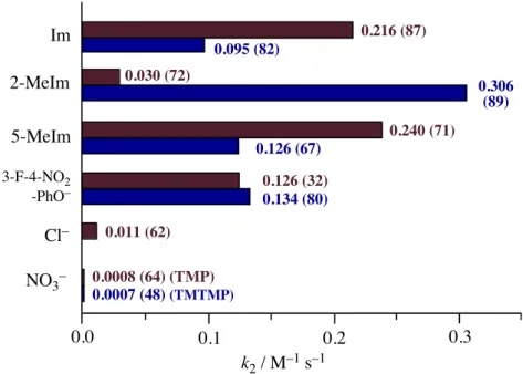
To study the effect of axial ligands on the electronic structure and reactivity of compound I of peroxidases and catalases, oxoiron(IV) porphyrin !-cation radical complexes with imidazole, 2-methylimidazole, 5-methylimidazole, and 3-fluoro-4-nitrophenolate as the axial ligands were prepared by ozone oxidation of iron(III) complexes of 5, 10, 15, 20-tetramesitylporphyrin (TMP) and 2, 7, 12, 17-tetra- methyl-3, 8, 13, 18-tetramesitylporphyrin (TMTMP). These complexes were fully characterized by absorption, 1H, 2H, and 19F NMR, EPR, and ESI-MS spectroscopy. The characteristic absorption peak of compound I at approximately 650 nm was found to be a good marker for estimation of the electron donor effect from the axial ligand. The axial ligand effect did not change the porphyrin !-cation radical state, the a2u state of the TMP complexes, or the a1u radical state of both the TMTMP complexes and compound I. The ferryl iron and porphyrin !-cation radical spins were effectively transferred into the axial ligands for the a2u complexes, but not for the a1u complexes. Most importantly, the reactivity of the oxoiron(IV) porphyrin !-cation radical complex was drastically increased by the imidazole and phenolate axial ligands. The reaction rate for cyclooctene epoxidation was increased 100 ~ 400-fold with axial coordination of imidazoles and phenolate. A similar increase was also observed for oxidation of 1,4-cyclohexadiene, N,N-dimethyl-p-nitroaniline, and hydrogen peroxide. These results suggest extreme enhancement of the reactivity of compound I by the axial ligand in heme enzymes. The functional role of axial ligands on the compound I in heme enzymes is discussed.
Abbreviation
TMP : 5, 10, 15, 20-tetramesitylporphyrin
TMTMP : 2, 7, 12, 17-tetramethyl-3, 8, 13, 18-tetramesitylporphyrin Im : imidazole
2-MeIm : 2-methylimidazole 5-MeIm : 5-methylimidazole
1,2-DiMeIm : 1,2-dimethylimidazole 1,5-DiMeIm : 1,5-dimethylimidazole 4-NO2-PhO : 4-nitrophenolate
3-F-4-NO2-PhO : 3-fluoro-4-nitrophenolate HRP : horseradish peroxidase
ASP: ascorbate peroxidase LiP : lignin peroxidase MnP : manganese peroxidase CIP : Coprinus cinereus peroxidase CPO : chloroperoxidase
BL-CAT : bovine liver catalase
Eo-CAT : Exiguobacterium oxidotolerans catalase Pm-CAT : Proteus mirabilis catalase
Ml-CAT : Micrococcus luteus catalase
compound I : oxoiron(IV) porphyrin !-cation radical
Introduction
Oxoiron(IV) porphyrin !-cation radical species in peroxidases and catalases, called compound I, is known as a reactive intermediate of these enzymes.1,2 It is also believed that compound I is formed as a reactive intermediate in the catalytic cycles of cytochromes P450.3 Although these enzymes share a common reactive intermediate, the reactivity of compound I differs from enzyme to enzyme. The compound I in cytochromes P450 directly transfers a single oxygen atom to a variety of substrates, while those in peroxidases and catalases carry out one-electron oxidation of organic substrates, such as amines and phenols, and oxidation of hydrogen peroxide to water and oxygen gas, respectively.1-3 It is generally accepted that the diverse functions of compound I are controlled by heme environmental structures, such as porphyrin peripheral structures, heme axial ligands, and protein structures around heme.
One significant structural difference in these heme enzymes is the heme axial ligand.1-3 As shown in Figure 2.1.1, histidine and tyrosine residues (imidazole and phenolate) are coordinated to the heme iron as axial ligands in peroxidases other than CPO and catalases, respectively. In cytochromes P450 and CPO, a cysteine residue Figure 2.1.1. Active site structures of (a) peroxidase (HRP, PDB file 1DZ9), (b) catalase (BL-CAT, PDB file 2A9E), and (c) cytochrome P450 (Cytochrome P450cam, PDB file 1HCH).
(thiolate) is the heme axial ligand. The amino acids that serve as the axial ligands are highly conserved in these heme enzymes, suggesting some functional roles in these enzyme functions. In the reaction cycles of these enzymes, the axial ligand can affect both the formation of compound I and the reaction of compound I with the substrate. The effect of the axial ligand on the rate of compound I formation has been studied by mutation of the aspartic acids that interact with the proximal histidine residues in peroxidases; the mutations weakened the electron donor effect of the proximal histidines, drastically decreasing in the rates of compound I formation.1,4 A similar axial ligand effect was also observed for synthetic heme enzyme model complexes. The rate of heterolytic cleavage of the O-O bond of iron bound m-chloroperbenzoate, which leads to formation of the compound I model complex, increased as the electron donor effect of the axial imidazole ligand increased.5 These mutation and synthetic enzyme model studies demonstrate that an increase in the electron donor effect of the axial ligand enhances the rate of compound I formation.
The axial ligand effect also affects the electronic structure and reactivity of compound I. The EPR spectra of compound I in peroxidases and catalases indicated a change in intramolecular spin coupling between the ferryl ion and radical spins, probably due to differences in the axial ligand effect.6-11 Catalytic oxidation reactions with synthetic iron porphyrin complexes having imidazoles, phenolates, and thiolates as axial ligands revealed significant effect of the axial ligand on product yield and selectivity.12-17 For example, the imidazole and chloride axial ligands had greater product yields than the thiolate axial ligand in catalytic competitive oxidation of cyclooctane/cyclooctene. However, the thiolate axial ligand favored hydroxylation, whereas the imidazole and chloride axial ligands preferentially yielded epoxidation.17 Moreover, the axial ligand effect on styrene epoxidation was reported using compound I
model complexes with chloride, fluoride, trifluoromethylsulfonate, methanol, and perchlorate axial ligands.18 More recently, the axial ligand effect was also studied in detail with other synthetic iron porphyrin systems and DFT calculations.19,20 Although these results clearly show the axial ligand effect on the electronic structure and reactivity of compound I, it is still unknown how the enzyme axial ligands (imidazole, phenolate, and thiolate) change the electronic state and reactivity of compound I. This is due to the difficulty of direct comparison of the reactivity of compound I in heme enzymes because of different protein structures, and due to the absence of stable compound I model complexes with imidazole, phenolate, and thiolate axial ligands because of rapid reduction of compound I model complexes by these axial ligands.
In previous studies, we reported the first preparation of oxoiron(IV) porphyrin
!-cation radical complexes with imidazole and 4-nitrophenolate as models for compound I of peroxidases and catalases.21,22 Although the imidazole complex could be characterized with absorption, 1H NMR, and resonance Raman spectroscopy, the
N
N N
N
FeIV
H3C O R
R
CH3 R
CH3 CH3 R
R = 2,4,6-trimethylphenyl
L
N
N N
N
FeIV O
L
R
R R
R
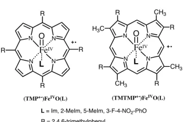
L = Im, 2-MeIm, 5-MeIm, 3-F-4-NO2-PhO (TMP+.)FeIVO(L) (TMTMP+.)FeIVO(L)
Figure 2.1.2. Structures of compound I model complexes prepared in this study.
4-nitrophenolate complex was too unstable to characterize, except for its absorption spectrum. To fully characterize and compare reactivities, we comprehensively prepared oxoiron(IV) porphyrin !-cation radical complexes with imidazole, 2-methylimidazole, 5-methylimidazole, and 3-fluoro-4-nitrophenolate as axial ligands by ozone oxidation of iron(III) complexes of 5, 10, 15, 20-tetramesitylporphyrin (TMP) and 2, 7, 12, 17-tetramethyl-3, 8, 13, 18-tetramesitylporphyrin (TMTMP), as shown in Figure 2.1.2. A stable catalase compound I model complex was synthesized with the 3-fluoro-4-nitrophenolate axial ligand. These imidazole and phenolate complexes were fully characterized using UV-visible absorption, 1H, 2H, and 19F NMR, EPR, and ESI-MS spectroscopy. Because of the structural similarity between TMTMP and protoporphyrin IX in heme enzymes, the TMTMP complexes more closely mimic the spectroscopic properties of compound I in peroxidases and catalases than the TMP complexes. The most important findings in this study were: 1) that the porphyrin
!-cation radical spin was significantly transferred in the axial imidazole (phenolate) and oxo ligands for the a2u radical complex, but not for the a1u radical complex, and 2) that the oxidation reactivity of oxoiron(IV) porphyrin !-cation radical complex was drastically increased, more than 400 times in the most significant case, with the coordination of imidazole and phenolate axial ligands, suggesting extremely reactive compound I in heme enzymes. Based on the present spectroscopic and kinetic studies, the functional roles of axial ligands on compound I in heme enzymes and catalytic oxidation reactions are discussed in the final section.
Experimental Section
Instrumentation. UV-visible absorption spectra were recorded on an Agilent 8453 (Agilent Technologies) equipped with a USP-203 low-temperature chamber (UNISOKU). 1H, 2H, 13C and 19F NMR were measured on a Lambda-500 spectrometer (JEOL). 1H, 2H, and 13C NMR Chemical shifts were reported versus tetramethylsilane (TMS) and referenced to residual solvent peaks (dichloromethane: 5.32 ppm). 19F chemical shift values were referenced to external hexafluorobenzene (C6F6) at 0 ppm. The concentrations of NMR samples were ~5 mM. EPR spectra were recorded at 4 K on Bruker E500 X-band spectrometer with an Oxford Instruments EPR910 helium-flow cryostat (Bruker). The concentrations of EPR samples were 1–3 mM. ESI-MS spectra were obtained with a LCT time-of–flight mass spectrometer equipped with an elecrospray ionization interface (Micromass). For oxoiron(IV) porphyrin !-cation radical complexes, desolvation and nebulizer gases were cooled with liquid nitrogen. The ESI-MS samples were prepared by oxidation with ozone in acetonitrile or dichloromethane at 233 K, followed by dilution with the same solvent, and injected into the ESI-MS with a gastight syringe cooled with dry ice. The sample solution was then transferred to the inlet of the mass spectrometer through pre-cooled capillary tube. Gas chromatography-mass spectroscopy (GC-MS) analysis was performed on a QP-5000 GC-MS system (Shimadzu) equipped with a capillary gas chromatograph (GC-17A, CBP5-M25-025 capillary column). Ozone gas was generated by UV irradiation of oxygen gas (99.9%) with an ozone generator PR-1300 (Clear Water) and used without further purification.
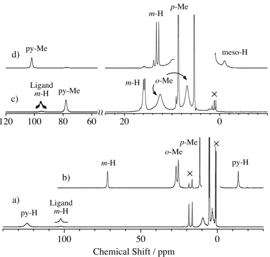
Materials. Imidazole-d4 (98 atom % D) was purchased from Aldrich. 2-Methylimidazole-d6 (98.1 atom % D) was purchased from CDN ISOTOPES (Canada). 4-Methylimidazole-d6 was prepared from 4-methylimidazole by activated 10% Pd/C in D2O, according to the literature.43 Deuterium incorporation into 4-methylimidazole, as analyzed by 1H and 13C NMR, was 2-H (95%), 5-H (95%) and 4-methyl group (50%). Dichloromethane was purchased from a commercial company as the anhydrous solvent and was stored in the presence of 4A molecular sieves. Other chemicals were purchased from commercial companies, and used without further purification. meso-Tetramesitylporphyrin (TMPH2) and 2,7,12,17-teramethyl-3,8,13,18-tetramesityl- porphyrin (TMTMPH2) were prepared according to the literature.44 (TMP)FeIIICl and (TMTMP)FeIIICl were prepared by the insertion of iron into TMPH2 and TMTMPH2 with FeCl2 and sodium acetate in acetic acid, respectively, and purified by silica gel column using CH2Cl2/CH3OH as an eluent. (TMP)FeIIIOH and (TMTMP)FeIIIOH were obtained from (TMP)FeIIICl and (TMTMP)FeIIICl, respectively, by passing through a basic alumina (10% water) column with dichloromethane as an eluent.45 (TMP)FeIIIClO4 and (TMTMP)FeIIIClO4 were prepared by the reaction of (TMP)FeIIICl and (TMTMP)FeIIICl, respectively, with silver(I) perchlorate in dichloromethane solution, and purified by re-crystallization from dichloromethane/n-hexane.46 (TMP)FeIIINO3 and (TMTMP)FeIIINO3 were obtained by mixing (TMP)FeIIIOH and (TMTMP)FeIIIOH in toluene with 1 M nitric acid, and purified by re-crystallization from ether/n-hexane.47 (TMP)FeIII(NO3) ; 1H NMR (CD2Cl2, 25"), 4.0 and 6.2 (ortho-Me), 15.3 and 16.5 (meta-H), 4.3 (para-Me), 74.0 (pyrrole-H). UV-vis. (CH2Cl2, 25"), 412, 514, 580, 647, and 691 nm. (TMTMP)FeIII(NO3) ; 1H NMR (CD2Cl2, 25"), –49.6 (meso-H), 5.1 and 8.4 (ortho-Me), 12.9 and 13.3 (meta-H), 6.3 (para-Me), 58.7 (pyrrole-Me). UV-vis. (CH2Cl2, 25"), 388, 506, 532, and 637 nm.
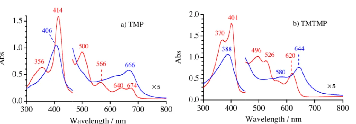
Synthesis. Iron(III) phenolate complexes. Iron(III) phenolate complexes were prepared by the published method.48 (TMP)FeIIIOH and (TMTMP)FeIIIOH were dissolved in toluene and 1 equiv of phenol derivative (4-nitrophenol, pentafluorophenol, 3,4,5-trifluorophenol, or 3-fluoro-4-nitrophenol) was added with stirring. The green or red solution changed to brown as the phenol dissolved. The solution was evaporated to dryness and the residue was purified by crystallization from ether/hexane. Microcrystals of the iron(III) phenol complex were collected by filtration. (TMP)FeIII(4-NO2-PhO) ; 1H NMR (CD2Cl2, 25"), 3.0 and 5.2 (ortho-Me), 12.5 and 13.5 (meta-H), 3.7 (para-Me), 79.1 (pyrrole-H), –91.4 (phenol ortho-H), 83.0 (phenol meta-H). UV-vis. (CH2Cl2, 25"), 420, 497, 559, 635 and 667 nm. (TMP)FeIII(F5-PhO) ; 1H NMR (CD2Cl2, 25"), 2.9 and 5.2 (ortho-Me), 12.7 and 13.6 (meta-H), 3.8 (para-Me), 78.7 (pyrrole-H). UV-vis. (CH2Cl2, 298K), 415, 500, 566, 642 and 673nm. (TMP)FeIII(3,4,5-F3-PhO) ; 1H NMR (CD2Cl2, 25"), 3.0 and 5.1 (ortho-Me), 12.3 and 13.3 (meta-H), 3.7 (para-Me), 79.8 (pyrrole-H), –101.0 (phenol ortho-H). UV-vis. (CH2Cl2, 25"), 420, 490, 557, 620 and 666 nm. (TMP)FeIII(3-F-4-NO2-PhO) ; 1H NMR (CD2Cl2, 25"), 3.1 and 5.2 (ortho-Me), 12.7 and 13.7 (meta-H), 3.8 (para-Me), 78.9 (pyrrole-H), –85.1 and –81.5 (phenol ortho-H), 72.4 (phenol meta-H). 19F NMR (CD2Cl2, 25"), 58.4 (meta-F). UV-vis. (CH2Cl2, 25"), 419, 500, 560, 640 and 668 nm. (TMTMP)FeIII(4-NO2-PhO) ; 1H NMR (CD2Cl2, 25"), –40.5 (meso-H), 4.8 and 7.4 (ortho-Me), 11.9 and 12.0 (meta-H), 6.0 (para-Me), 47.7 (pyrrole-Me), –83.1 and –79.9 (phenol ortho-H), 67.9 (phenol meta-H). UV-vis. (CH2Cl2, 25"), 371, 398, 498, 526 and 619 nm. (TMTMP)FeIII(3-F-4-NO2-PhO) ; 1H NMR (CD2Cl2, 25"), –47.0 (meso-H), 4.5 and 7.3 (ortho-Me), 11.9 and 12.0 (meta-H), 5.0 (para-Me), 47.0 (pyrrole-Me), –83.1 and –79.9 (phenol ortho-H), 67.9 (phenol meta-H). 19F NMR (CD2Cl2, 25"), 58.3 (meta-F).
UV-vis. (CH2Cl2, 25"), 370, 401, 496, 524 and 619 nm.
Iron(III) mono-imidazole complexes. Iron(III) porphyrin mono-imidazole complex was generated in situ by adding 1.0 equiv of imidazole, 2-methylimidazole, or 4-methylimidazole to perchlorate iron(III) porphyrin in dichloromethane.49 [(TMP)FeIII(Im)]ClO4 ; 1H NMR (CD2Cl2, 25"), 3.1 and 5.3 (ortho-Me), 12.6 and 13.4 (meta-H), 4.3 (para-Me), 19.9 (pyrrole-H), –15.5 (Im 2-H), 30.1 (Im 4-H), 85.1 (Im 5-H), 100.4 (Im N-H). UV-vis. (CH2Cl2, 25"), 412, 510, 575 and 690 nm. [(TMP)FeIII(2-MeIm)]ClO4 ; 1H NMR (CD2Cl2, 298K), 2.7 and 5.7 (ortho-Me), 12.7 and 13.1 (meta-H), 4.1 (para-Me), 34.9 (pyrrole-H), 16.6 (2-MeIm 2-Me), 31.8 (2-MeIm 4-H), 65.8 (2-MeIm 5-H), 109.0 (2-MeIm N-H). UV-vis. (CH2Cl2, 25"), 412, 511, 572 and 692 nm. [(TMP)FeIII(5-MeIm)]ClO4 ; 1H NMR (CD2Cl2, 25"), 13.0 and 13.9 (meta-H), 4.4 (para-Me), 31.7 (pyrrole-H), –18.6 (5-MeIm 2-Me), 31.7 (5-MeIm 4-H), 22.0 (5-MeIm 5-Me), 100.8 (5-MeIm N-H). UV-vis. (CH2Cl2, 25"), 412, 510, 572 and 692 nm. [(TMTMP)FeIII(Im)]ClO4 ; 1H NMR (CD2Cl2, 25"), –16.6 (meso-H), 4.8 and 8.0 (ortho-Me), 13.5 and 14.4 (meta-H), 5.5 (para-Me), 69.2 (pyrrole-Me), –16.1 (Im 2-H), 31.2 (Hm 4-H), 82.6 (Im 5-H), 98.5 (Im N-H). UV-vis. (CH2Cl2, 25"), 385, 503 and 626 nm. [(TMTMP)FeIII(2-MeIm)]ClO4 ; 1H NMR (CD2Cl2, 25"), –31.9 (meso-H), 5.1 and 8.8 (ortho-Me), 13.8 and 14.5 (meta-H), 6.2 (para-Me), 70.0 (pyrrole-Me), 17.5 (2-MeIm 2-Me), 35.9 (2-MeIm 4-H), 66.6 (2-MeIm 5-H), 110.0 (2-MeIm N-H). UV-vis. (CH2Cl2, 298K), 385, 503 and 626 nm. [(TMTMP)FeIII(5-MeIm)]ClO4 ; 1H NMR (CD2Cl2, 298K), -20.1 (meso-H), 5.0 and 8.2 (ortho-Me), 13.7 and 14.5 (meta-H), 5.8 (para-Me), 70.3 (pyrrole-Me), -15.4 (5-MeIm 2-H), 32.6 (5-MeIm 4-H), 20.7 (5-MeIm 5-H), 99.5 (5-MeIm N-H). UV-vis. (CH2Cl2, 25"), 385, 503 and 626 nm.
Oxoiron(IV) porphyrin complexes. Oxoiron(IV) porphyrin complexes were prepared as previously described.45 (TMP)FeIII(ClO4)2 or (TMTMP)FeIII(ClO4)2, which was prepared from the oxidation of (TMP)FeIIIOH or (TMTMP)FeIIIOH with solid ferric perchlorate, in dichloromethane-d2 was transferred through a short basic alumina (20% water) column (0.5 # 1 cm) at ambient temperature, directly into a NMR tube in dry ice-acetone bath. 1 equiv of 1-MeIm or 1,2-DiMeIm in dichloromethane-d2 was slowly added to the cooled solution in the NMR tube.
Ozone oxidation of iron(III) porphyrin. For NMR or EPR measurement, iron(III) porphyrin complexes were dissolved in dichloromethane and placed in NMR or EPR cell. The solution was cooled by a dry-ice acetone bath. Ozone gas was slowly bubbled in the solution with a gastight syringe. With the formation of oxoiron(IV) porphyrin !-cation radical complexes, the brown solution changed to green. The oxidations were monitored by 1H NMR or EPR spectra. For UV-visible absorption spectra, the oxidation was carried out in a 1 cm quartz cuvette in a low-temperature chamber set with a UV-visible absorption spectrometer and monitored by absorption spectral change. Finally, excess ozone gas was removed by bubbling argon gas with a gastight syringe.
Kinetics and product analysis. Oxoiron(IV) porphyrin !-cation radical (100 µM) in dichloromethane was prepared in a 1 cm quartz cuvette in a low-temperature chamber set with a UV-visible absorption spectrometer, as described above. An excess of cyclooctene (20–1000 equiv) was then added to the solution with vigorous stirring, and the reactions were monitored by the absorption spectral change at constant time intervals. After confirming the completion of the reactions, 10 equiv of
tetrabutylammonium iodide (nBu4N+I–) was added to the solution at the same temperature. After warming to room temperature, quantitative product analyses were subsequently performed with GC-MS using undecane as an internal standard. The reaction rate constants were determined by computer simulation of absorption versus time for the reactions.
FeIII X
FeIV X O3 O
FeIII L
O3
FeIV O
L L
L Route A
Route B X = ClO4–, NO3– Scheme 1
Results
Synthesis of Compound I Models. The oxoiron(IV) porphyrin !-cation radical complexes with imidazole and phenolate derivatives as axial ligands were prepared using two alternative routes, as shown in Scheme 2.1.1. [(TMP+•)FeIVO(L)](ClO4) and [(TMTMP+•)FeIVO(L)](ClO4), where L is Im, 2-MeIm, and 5-MeIm, were prepared from (TMP)FeIII(ClO4) and (TMTMP)FeIII(ClO4) using route B, respectively. [(TMTMP+•)- FeIVO(L)](ClO4) also was prepared from (TMTMP)FeIII(ClO4) using route A. However, pure [(TMP+•)FeIVO(L)](ClO4) could not be prepared using route A, because oxidation of (TMP)FeIII(ClO4) with ozone did not produce pure (TMP+•)FeIVO(ClO4). To explore the effects of counter anions, we examined the preparation from nitrate iron(III) porphyrin complexes. In contrast to the perchlorate complexes, [(TMP+•)FeIVO(L)]-
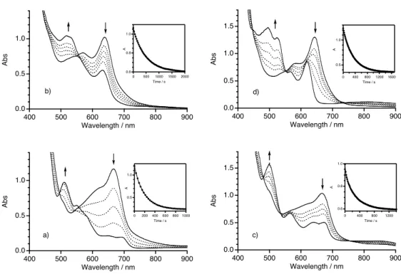
(NO3) and [(TMTMP+•)FeIVO(L)](NO3) were prepared from (TMP)FeIII(NO3) and (TMTMP)FeIII(NO3) using route A, respectively. On the other hand, route B was not effective, except for the case of TMTMPFeIII(NO3) and 2-methylimidazole, because the addition of 1 equiv of the imidazole derivative to the nitrate complex did not afford its mono-adduct complex, but rather a mixture of the parent and bis-adduct complexes. The oxoiron(IV) porphyrin !-cation radical complexes with imidazole derivatives, prepared from nitrate complexes using route A, were as stable as those prepared from perchlorate complexes at temperatures below 233 K in dichloromethane.
The preparation of oxoiron(IV) porphyrin !-cation radical complexes with phenolate derivatives (4-nitrophenolate, pentafluorophenolate, 3,4,5-trifluorophenolate, and 3-fluoro-4-nitrophenolate) as axial ligands was examined using route B for TMP and TMTMP complexes. The oxoiron(IV) porphyrin !-cation radical complexes with 4-nitrophenolate, 3,4,5-trifluorophenolate, and pentafluorophenolate were very unstable even at 183 K and quickly reduced to iron(III) porphyrin complexes with dissociation and decomposition of the axial phenol. However, the oxoiron(IV) porphyrin !-cation radical complex with 3-fluoro-4-nitrophenolate was stable enough for spectroscopic characterization at temperatures below 213 K in dichloromethane. In route A, the addition of 1 equiv of phenol derivatives to oxoiron(IV) porphyrin !-cation radical complex resulted in rapid reduction of the complex.
Characterization of Peroxidase Compound I Models. Figure 2.1.3, shows UV-visible absorption spectral changes for the formation of oxoiron(IV) porphyrin
!-cation radical complexes with imidazole from the route A in dichloromethane at –80˚C. Oxidation of (TMP)FeIII(NO3) with ozone at –80 ˚C yielded (TMP+•)FeIVO- (NO3), which showed a Soret band with decreased intensity at 401 nm and an
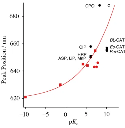
absorption peak near 666 nm, as shown in Figure 2.1.3 a). The careful addition of 1 equiv of imidazole to (TMP+•)FeIVO(NO3) showed a small but significant spectral change, indicating the formation of [(TMP+•)FeIVO(Im)](NO3) without reduction of the porphyrin !-cation radical. Similarly, Figure 2.1.3 b) shows the formation of [(TMTMP+•)FeIVO(Im)](NO3) using route A. With the coordination of imidazole, the intensity of the Soret band decreased and the absorption at 630 nm shifted to 643 nm. The absorption spectra of oxoiron(IV) porphyrin !-cation radical complexes having 2-methylimidazole and 5-methylimidazole were also measured, and the results are summarized in Table 2.1.1. The axial coordination of 2-methylimidazole and 5-methylimidazole did not change the peak positions for the TMP complexes, but did change them for the TMTMP complexes. The absorption spectral features of [(TMTMP+•)FeIVO(Im)](NO3) more closely resembled those of compound I of peroxidases than those of [(TMP+•)FeIVO(Im)](NO3) (Table 2.1.2).23-28 Similar complexes with perchlorate anions were also prepared, and the peak positions were the same as those observed with nitrate anions.
1
0
Abs
400
300 500 600 700 800
1
0
Abs
400
300 500 600 700 800
Wavelength / nm 410
512
576 658 693 a) TMP 404
401 666
!5 643 630 395
391 565
386
505
637 530 576
Wavelength / nm b) TMTMP
!5
Figure 2.1.3. UV-visible absorption spectra of oxoiron(IV) porphyrin !-cation radical complexes (10 mM) with imidazole in dichloromethane at –80 ". Red lines : (P)FeIII(NO3), black lines : (P+•)FeIVO(NO3), blue lines : [(P+•)FeIVO(Im)](NO3). (a) P = TMP, (b) P = TMTMP.
Figure 2.1.4 shows 1H NMR spectra of [(TMP+•)FeIVO(L)](ClO4) and [(TMTMP+•)FeIVO(L)](ClO4), where L is either Im, 2-MeIm, or 5-MeIm, in dichloromethane-d2. The 1H NMR signals were assigned by deuterium-labeled compounds, signal intensities, and comparison with those of previously characterized (TMP+•)FeIVO(ClO4).29 The 1H NMR shifts of [(TMP+•)FeIVO(L)](ClO4) are summarized in Table 2.1.3. [(TMP+•)FeIVO(Im)](ClO4) exhibits the pyrrole proton signal at –10.4 ppm, the m-protons of the meso mesityl group at 63.9 and 65.4 ppm, the o-methyl protons at 22.8 and 23.8 ppm, and the p-methyl protons at 10.3 ppm. The
separation of the o-methyl and m-proton signals was reasonable for the coordination of two different axial ligands, oxo and imidazole. The large downfield shifts of the meso mesityl protons indicate an unpaired electron in the a2u porphyrin !-orbital, a2u porphyrin !-cation radical state. As shown in Figure 2.1.4, the 1H NMR shifts of
100 80 60 40 20 0 -20
Chemical shift / ppm
py-H p-Me
m-H o-Me
d)
c)
a) b)
Figure 2.1.4. 1H NMR spectra of [(TMP+•)FeIVO(L)](ClO4) in dichloromethane-d2 at –60 ˚C. a) L = Im, b) L = 5-MeIm, c) L = 2-MeIm, d) L = 2-MeIm at –90 ˚C
[(TMP+•)FeIVO(5-MeIm)](ClO4) were almost identical to those of [(TMP+•)FeIVO- (Im)](ClO4). The 1H NMR shifts of [(TMP+•)FeIVO(2-MeIm)](ClO4) were similar to those of [(TMP+•)FeIVO(Im)](ClO4) and [(TMP+•)FeIVO(5-MeIm)](ClO4), but the 1H NMR signals of [(TMP+•)FeIVO(2-MeIm)](ClO4) were broader. When the temperature was lowered further, the 1H NMR signals of [(TMP+•)FeIVO(2-MeIm)](ClO4) became much broader and split at –90 ˚C as shown in Figure 2.1.4 d). This was because the rotation of the axially bound 2-methylimidazole along the Fe-N(2-MeIm) axis was hindered by a steric interaction between the 2-methyl group of 2-methylimidazole and the o-methyl group of the meso mesityl group. The 1H NMR spectra of [(TMP+•)FeIVO(L)](NO3) were identical to those of [(TMP+•)FeIVO(L)](ClO4).
Figure 2.1.5 shows the 1H NMR spectra of [(TMTMP+•)FeIVO(L)](ClO4), where L is either Im, 2-MeIm, or 5-MeIm, in dichloromethane-d2 at –80 ˚C. The 1H NMR signals of [(TMTMP+•)FeIVO(L)](ClO4) were also assigned using deuterium labeled
20 0 -20
140 120 100
meso-H 2-MeIm
py-Me
m-H o-Me
p-Me
a) b) c)
Chemical shift / ppm
4-H 2-H
Figure 2.1.5. 1H NMR spectra of [(TMTMP+•)FeIVO(L)](ClO4) in dichloromethane -d2 at –80 ". a) L = Im, b) L = 5-MeIm, c) L = 2-MeIm.
compounds, signal intensities, and comparison with those of previously characterized (TMTMP+•)FeIVO(ClO4).21,29 The 1H NMR shifts of [(TMTMP+•)FeIVO(L)](ClO4) are summarized in Table 2.1.4. As reported, [(TMTMP+•)FeIVO(Im)](ClO4) showed a large downfield shift of the pyrrole methyl signal at 113.2 ppm and small shift of the meso proton signal at 0.0 ppm.21 The 1H NMR spectra of [(TMTMP+•)FeIVO- (5-MeIm)](ClO4) and [(TMTMP+•)FeIVO(2-MeIm)](ClO4) were similar to that of [(TMTMP+•)FeIVO(Im)](ClO4), but paramagnetic shifts of [(TMTMP+•)FeIVO(2-MeIm)]- (ClO4) were larger than those of the other two. The small paramagnetic shifts of the meso proton signals for [(TMTMP+•)FeIVO(L)](ClO4) clearly indicate an unpaired electron in the a1u porphyrin !-orbital, a1u porphyrin !-cation radical state. Unlike [(TMP+•)FeIVO(2-MeIm)](ClO4), the rotation of the 2-MeIm in [(TMTMP+•)FeIVO- (2-MeIm)](ClO4) was not hindered. In fact, even at –90 ˚C, only a single pyrrole methyl signal was observed. This is because the o-methyl group of the pyrrole
!-mesityl group in TMTMP is farther from the iron center than that of the meso mesityl group in TMP, resulting in a smaller steric effect on the rotation of 2-methylimidazole in TMTMP. The 1H NMR spectra of [(TMTMP+•)FeIVO(L)](NO3) were similar to those of [(TMTMP+•)FeIVO(L)](ClO4). The temperature dependence of 1H NMR signals for [(TMP+•)FeIVO(L)](ClO4) and [(TMTMP+•)FeIVO(L)](ClO4) showed normal Curie’s law behavior.
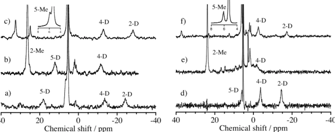
Direct evidence for the coordination of imidazole derivatives to oxoiron(IV) porphyrin !-cation radical complexes was obtained by 2H NMR spectra of iron-bound deuterium-labeled imidazole derivatives (Figure 2.1.6). The 2H NMR shifts are summarized in Table 2.1.5, together with the NMR shifts of iron-bound imidazole signals of oxoiron(IV) and iron(III) porphyrin complexes. The 2H NMR signals were assigned by their relative intensity and by substitution of each imidazole position with a
methyl group, which resulted in the disappearance of the signal for the substituted position and the appearance of a new signal for the introduced methyl group. The paramagnetic shifts of the 2H NMR signals of the deuterium-labeled imidazole derivatives clearly showed their coordination to the ferryl iron site as axial ligands. The temperature dependence of these 2H NMR signals showed normal Curie’s law behavior. The 2H NMR signals for the 2- and 4-positions of the iron-bound imidazole derivative showed upfield shifts, while those for the 5-position showed downfield shifts. With substitutions of the 2-positions with methyl groups, the 2H NMR signals of the methyl groups were observed downfield. The 2H NMR paramagnetic shifts of iron-bound imidazole signals for [(TMP+•)FeIVO(L)](ClO4) were larger than those of [(TMTMP+•)FeIVO(L)](ClO4).
40 20 0 -20 -40
Chemical shift / ppm
40 20 0 -20 -40
Chemical shift / ppm
8 6 4
8 6 4 2-D
5-D 2-Me 5-Me 4-D 2-D
5-Me
5-D 2-Me
a) b) c)
d) e) f)
4-D 4-D
5-D
4-D 2-D 4-D
4-D
2-D
Figure 2.1.6. 2H NMR spectra of a) [(TMP+•)FeIVO(Im-d4)](ClO4), b) [(TMP+•)FeIVO(2-MeIm-d6)](ClO4), c) [(TMP+•)FeIVO(5-MeIm-d6)](ClO4), d) [(TMTMP+•)FeIVO(Im-d4)](ClO4), e) [(TMTMP+•)FeIVO(2-MeIm-d6)](ClO4), and f) [(TMTMP+•)FeIVO(5-MeIm-d6)](ClO4) in dichloromethane. Temperature; a) – c) at –60 ˚C and d) – f) at –80 ˚C.
Figure 2.1.7 shows the EPR spectra of [(TMP+•)FeIVO(2-MeIm)](ClO4) and [(TMTMP+•)FeIVO(2-MeIm)](ClO4) at 4 K. [(TMP+•)FeIVO(2-MeIm)](ClO4) showed the EPR signals at g = 4.47, 3.45, and 1.97, which were similar to those of (TMP+•)FeIVO(ClO4).30 The EPR spectra of [(TMP+•)FeIVO(2-MeIm)](ClO4) were typical of those of S = 3/2 systems, indicating strong ferromagnetic interactions between ferryl iron (S = 1) and the porphyrin !-cation radical (S = 1/2). The EPR spectra of [(TMP+•)FeIVO(Im)](ClO4) and [(TMP+•)FeIVO(5-MeIm)](ClO4) were very close to that of [(TMP+•)FeIVO(2-MeIm)](ClO4), suggesting similar spin interactions (Table 2.1.3). The g values for the TMP complexes were hardly changed by the axial ligand effect. As shown in Figure 2.1.7 b), the EPR spectrum of [(TMTMP+•)FeIVO(2-MeIm)](ClO4) differed from that of [(TMP+•)FeIVO(2-MeIm)]- (ClO4), but was similar to that of [(TMTMP+•)FeIVO(MeOH)](ClO4) reported previously.30 As observed for [(TMTMP+•)FeIVO(MeOH)](ClO4),30 the EPR spectrum of [(TMTMP+•)FeIVO(2-MeIm)](ClO4) was interpreted as a weak ferromagnetic interaction between ferryl iron (S = 1) and the porphyrin p-cation radical (S = 1/2).
500 400
300 200
100 0
Field / mT
*
*
4.47
3.45 3.67
2.01
1.97
a) b)
Figure 2.1.7. EPR spectra of (a) [(P+•)FeIVO(2-MeIm)](ClO4) at 4K. (a) P = TMP, in dichloromethane-toluene (5:1), (b) P = TMTMP, in dichloromethane-propionitrile (5:1).! Signals denoted by asterisks are due to [(TMP)FeIII(2-MeIm)](ClO4) and [(TMTMP)FeIII(2-MeIm)](ClO4).
The EPR spectra of [(TMTMP+•)FeIVO(Im)](ClO4) and [(TMTMP+•)FeIVO(5-MeIm)]- (ClO4) were similar to that of [(TMTMP+•)FeIVO(2-MeIm)](ClO4), but the g" values were changed by the axial imidazole ligand (Table 2.1.4). This result suggests that the spin interaction was altered by the axial imidazole for the TMTMP complex. The EPR spectra of [(TMP+•)FeIVO(L)](NO3) and [(TMTMP+•)FeIVO(L)](NO3) were almost identical to those of [(TMP+•)FeIVO(L)](ClO4) and [(TMTMP+•)FeIVO(L)](ClO4), respectively.
The structural characterization of oxoiron(IV) porphyrin !-cation radical complexes having imidazole derivatives was examined using ESI-MS spectroscopy. ESI-MS peaks corresponding to [(TMP+•)FeIVO(L)]+ and [(TMTMP+•)FeIVO(L)]+ cations, where L = Im, 2-MeIm, and 5-MeIm, were detected (Table 2.1.6). The isotope distribution patterns of the observed ESI-MS peaks were almost identical to those calculated from [(TMP+•)FeIVO(L)]+ and [(TMTMP+•)FeIVO(L)]+ structures. All of the ESI-MS data supported the binding of imidazole derivatives as an axial ligand.
2.0 1.5 1.0 0.5 0.0
Abs
400
300 500 600 700 800
1.5
1.0
0.5
0.0
Abs
400
300 500 600 700 800
!5
Wavelength / nm 406
414
356
500 566
640 674 666 a) TMP
388 644
370 401
496526 620
580
Wavelength / nm
!5
b) TMTMP
Figure 2.1.8. UV-visible absorption spectra of (P)FeIII(3-F-4-NO2-PhO) and (P+¥)FeIVO(3-F-4-NO2-PhO) in dichloromethane (concentration 1.0 # 10-5 M, 1cm path length cell) at –80": a) P = TMP, b) P = TMTMP.

 in dichloromethane-d 2](https://thumb-ap.123doks.com/thumbv2/123deta/6164266.104591/31.892.176.691.165.528/figure-nmr-spectra-tmp-fe-iv-clo-dichloromethane.webp)
 in dichloromethane](https://thumb-ap.123doks.com/thumbv2/123deta/6164266.104591/32.892.173.702.581.877/figure-nmr-spectra-tmtmp-fe-iv-clo-dichloromethane.webp)