A Morphological Study of Lichen-Symbionts,
Ascomycetous Fungus Myelochroa leucotyliza and
Green Alga Trebouxia sp., with Special Reference to
the Mechanism of Lipid (Atranorin) Secretions
Satoko Arakawa-Kobayashi
DOCTOR OF PHILOSOPHY
Department of Molecular Biomechanics
School of Life Science
The Graduate University for Advanced Studies
Table of Contents
Page
Contents 1
Abbreviations List 2
Summary 3-5
General Introduction 6-8
Chapter I. Establishment of the Culture Line of the Lichen Symbiont 9
Introduction 10
Materials and Methods 11
Results & Discussion 12
Chapter II. Identification of crystalline material found in the lichen thallus, Myelochroa
leucotyliza 13
Abstract 14
Introduction 15
Methods 16
Results 17-18
Discussion 19
Chapter III. A Morphological Study of Lichen-Symbionts, Ascomycetous Fungus Myelochroa leucotyliza and Green Alga Trebouxia sp., with Special Reference to the
Mechanism of Lipid (Atranorin) Secretions 20
Abstract 21
Introduction 22
Materials and Methods 23-25
Results 26-30
Discussion 31-35
General Discussion 36
Tables 37-40
Figures and Legends 41-72
Appendices 73-74
Acknowledgements 75
References 76-80
Abbreviations list
EPC: Egg phosphatidylcholine ITS: internal transcribed spacer LC: liquid chromatography PC: phosphatidylcholine PKS: polyketide synthase
SAED: selected area and electron diffraction TLC: thin layer chromatography
Summary
Lichens are symbionts of fungi and algae (or cyanobacteria). Lichen forms a unique complex called thallus and produces large amount of lipids of various kinds. These lipids were secreted from the fungal cells and deposited in the thallus to form tremendous amount of crystalline materials. The crystalline materials are considered to have roles to protect the lichen from excess light, bacterial infection, freezing, and etc. This should be the reason that lichens can exhibit a wide ability of adaptations to extreme habitats even as those of alpine, desert, and polar region. From this aspect, we considered the significance of symbiosis of lichen must depend on the unusual lipid production and secretion.
As mentioned, the fungi produce lipids and secrete them out of cells. On the other hand, the algae were considered to provide precursor sugars for the fungi to produce lipids (Mosbach, 1969, Yamazaki et al., 1965.). Although fungi grown in the wild secrete large amount of lipids to form crystals, those grown in the culture both alone or with algae produce only small amounts and are found unable to form crystals in the culturing medium (Honegger, 1996).
This study is focused on the mode and the mechanism of the lipid secretion as we considered that the existence of large amount of crystals is signifying the symbiosis of lichens.
To begin the study we first respectively isolated the fungi and the algae from the thallus of Myelochroa leucotyliza. The isolated samples were cultured both alone or with each other. The cultured samples and the lichen in the wild were examined by means of quick freezing and replicated or substituted electron microscopy. Next, it seemed essential to identify the nature of crystals. X-ray and electron microscope diffraction methods and TLC analysis on the re-crystallized lipid extracted from the wild lichen did the identification. The major component of the re-crystallized material was atranorin.
According to the observations on the morphology of wild and cultured samples with electron microscopy the following results were obtained; 1) atranorin is the predominant component of the crystalline materials in the thallus, 2) the dynamic morphological change of the plasmamembrane of the fungi occurred in parallel to the activities of lipid secretion, 3) the zymogen granules contained in the fungal cytoplasm exocytose during the lipid secretion, 4) the amount of lipid bodies contained in the fungi inversely changed with those of the crystal deposition outside of the cell.
It was considered that if we could promote the cultured fungi into actively lipid-secreting cells, and if we could find crystals around the cells, we would have an opportunity to understand the entire scheme of the way the cell secretes lipid and the significance of symbiosis of lichens. Along with this consideration, we cultured fungi in the medium with higher concentration of sugar than usual. According to Hamada’s report (1993) that in these condition fungi synthesize large amount of lipid. We detected neither crystal by EM or existence of atranorin by TLC. It was observed, however, that in the fungal cytoplasm cultured in this sugar fortified medium existence of multiple lipid bodies together with numbers of zymogen granules. The appearance of these accumulations of multiple organelles suggested the cellular inhibition of secretions of both lipids and proteins. In order to release the inhibition, we added into the culturing medium various materials that had been reported to promote lipid secretion. Finally, we soaked the fungi growing in this sugar fortified culture with a few drops of the medium in which the algae had been cultured alone. The soaking looked the fungi to release the
inhibition and made them actively exocytose zymogen granules. The plasmamembrane changed the shape with many invaginations of omega appearances. The crystals were found outside of the cells.
The amount and the size of lipid bodies decrease during the active secretion of zymogen granules. However, the lipid-bodies may consist of reservoirs of sterol- derivatives and triacylglycerols. It is considered that they should represent the indirect source of atranorin. Atranorin belongs to polyketides. It has to be produced via acetate- malonate pathway by the interaction of polyketide synthase, in which two phenolic units derived from acetate join to become atranorin. The cytosolic location of polyketide synthase has been reported by showing fluorescent of GFP of the GFP-tagged enzyme. Further, we observed using a fluorescent microscope the possible cytosolic location of atranorin under UV excitation. As atranorin is extremely hydrophobic, the lipid must have being bound with some associating protein in the cytoplasm. However, it is not known how atranorin reaches the inner side of the plasmamembrane.
The modes of lipid secretion so far survived in the long course of histology are as follows. They are 1) the simple diffusion, 2) the exocytic secretion, and 3) the endoplasmocrine secretion. The simple diffusion is the hypothesis in which lipid in the cytoplasm diffuses freely through the lipidic plasmamembrane. The secretion by exocytosis represents the hypothesis that lipids bound by the biological membrane secrete themselves as the same mechanism to that of protein secretion. Rhodin (1971) has interpreted the endoplasmocrine secretion of lipids. The lipid-body encircled by the tubules of smooth ER makes contact with plasmamembrane by some confusing manner and in the consequence intact lipid-body is exocytosed outside.
We chose two out of above mentioned three in regard to the mechanisms of lipid secretion of our fungi for the following reasons. 1) Upon incubation with ethanol, Kabakibi et al. (1998) demonstrated the release of fatty acid ethyl ester of Hep-G2 cell into the culturing medium. The secretion stopped when BFA was added to the medium. However, the secretion resumed when lipoprotein or albumin was introduced into the medium that contained BFA. They concluded that the release of the lipid had occurred via an independent pathway of vesicular transport. That is, regardless of whether or not the protein secretion was inhibited by BFA, the release of lipid actually occurred due to the added protein operating outside of the cell. 2) We also did the experiment along the line shown by these authors. To determine whether albumin can pull atranorin out of lipid membrane we prepared atranorin-containing phosphatidyl choline (PC) liposome. The obtained atranorin-PC-liposome was added with BSA-albumin and after extensive shaking the mixture was centrifuged. The pellet consisted of lipid-bileaflet membrane. The supernatant was examined using a spectrofluorometer to detect whether it contains atranorin. Added albumin actually pulled atranorin out of the lipid membrane. In order to try to isolate “effective” protein/or proteins that pull atranorin out of fungal cells we employed anionic columns. The columns separated plenty of proteins from the fungal colony. A few of them showed the extraction ability of atranorin from the atranorin-PC- liposome. However, many of them had no ability to pull atranorin out. The elution of the homogenate of the fungal colony by the “albumin column” showed the most
zymogen granules once secreted elicit atranorin locating on the other side of the membrane. Atranorin goes through the plasmamembrane in the mode of simple diffusion providing if there exists the force of proteins outside.
General Introduction
Lichen forms a unique complex called thallus and produces a large amount of lipids of various kinds. More than 700 kinds were so far reported (Purvis, 2000). Some of them have been exploited as medicines, antibiotics, anti-tumor agents, dyes, and perfumes (Gilbert, 2000; Brodo et al., 2001). Lichen consists of at least two organisms in symbiotic associations: one is fungus, which belongs mostly to the Ascomycete, and the other consists of photosynthetic partner, a green alga or a cyanobacterium. Fungus and its photosynthetic partner had specifically evolved to make lichen by accommodating symbiotically in the wild life. This photosynthetic fungal partner is not usually found independently in free-living state in the wild life (Ahmadjian, 1993; Tschermak-Woess, 1988; Hawksworth et al., 1995; Honegger, 1996). For instance the green alga such as Trebouxia that occupies almost one fifth of all lichens, its existence is only known in a lichenized state.
On the other hand, the symbiotic partner of the alga is also not found in a free- living state (Hawksworth et al., 1995). Symbiosis is by no means an exceptional life style to the fungal kingdom. Fungi rely their live-source of organic compound on the symbiotic partner because they cannot synthesize carbohydrate by themselves. The estimated 20% of all fungal species and 42% of Ascomycetes are considered to be lichen symbionts (Hawksworth et al., 1995). Moreover, according to the recent study of fungal phylogeny many free-living Ascomycetes such as Aspergillus and Penisillium had been derived from lichenized ancestors (Lutzoni et al., 2001). Therefore, having been combined in the lichen is an important event in the course of fungal evolution.
The most remarkable characteristic of lichen’s activity is to deposit crystalline lipids in the thallus. These crystals sometimes reaches 30% of dry weight of the thallus (Huneck, 1976). Lipid-crystals of lichen are considered to have protective functions. Although the structure of lichen thallus is diverse, one of their common features is that the fungi usually form an upper cortex. Below the cortex the photosynthetic partners of fungi inhabit (Hale, 1983; Honegger, 1993). A large amount of lipid in the form of extracellular crystals exists in the upper cortex. Lipid crystals can screen algae from harmful short wavelength radiation (Hill and Woolhouse, 1966, Richardson, 1985, Vicente, 1991, Elix, 1996). This may be the reason why the lichens can grow in extreme habitats such as alpine, desert, and polar regions (Nash, 1996). The lipids also have a function of anti-biotics especially for the protection against the invasion of the thallus by gram-positive bacteria (Elix, 1996). In addition, atranorin, the lipidic substance contained in the upper cortex generates fluorescent light corresponding with the absorption spectrum of chlorophyll. This means that atranorin amplifies the potentiality of the alga to utilize the low light intensities (Rao and LeBlanc, 1965). Thus, the lipid secretion for lichens should have the intent of the vital lifeline to survive in the history of evolution.
The lipids produced by lichens are classified into several groups of polyketide, mostly depsides and depsidones (Culberson, 1969; Elix, 1996). They have two or three phenolic units through ester, or ether, or carbon-carbon linkages. These units synthesized via acetyl-polymalonyl pathway. Although depsides and depsidones in
Aspergillus terreus has been used as an inhibitor of cholesterol synthesis (Kennedy et al., 1999). Fundamental structures of polyketide and other substances are shown in Appendix 1. It was revealed that the synthetic pathway of polyketides in non-lichenized fungi involves a multifunctional enzyme, polyketide synthase (reviewed in Hopwood, 1997). The polyketide synthase has the same domain structures as fatty acid synthase do. It was suggested that the two enzymes have originated from the same source (Hopwood, 1997). The arena of activities of both enzymes were considered to reside in the cytoplasm (Hopwood and Sherman, 1990; Robinson, 1991; Hajjaji et al., 2000). Recently, it was reported by the genomic analysis that the Ascomycetous fungus, Neurospora crassa, contains seven genes coding polyketide synthases (Galagan et al., 2003).
The fungal attribution to make these polyketide crystals is strongly suggested. Moreover, the isolated fungi in culture from the thallus still retained the ability to produce lipids as wild lichens do (Ahmadjian, 1993; Culberson and Armaleo, 1992; Hamada et al., 1997; Komiya and Shibata, 1969; Mosbach, 1967; Yoshimura et al., 1994). The reported results above mentioned indicate that the lipids in the lichen are synthesized by the fungal cells.
On the other hand, the role of the partner involved in photosynthesis is not clear (Ahmadjian, 1993). Although the fungi grown in the thallus secrete large amount of lipids to form crystals outside, those grown alone in culture do not secrete enough lipid to form crystals. However, the symbiotic cultures, in which the fungi and the algae accommodate in the same plate, usually produce larger amount of polyketides than do the fungi cultured alone (Yamamoto et al., 1993). Some investigators have reported the existence of crystal-like structures outside of the fungal cells in the symbiotic colonies (Ahmadjian and Jacobs, 1985; Culberson and Armaleo, 1992; Kon et al., 1993), although, the production of lipids by the cultured cells with regard to the amount are beyond comparison to those in the wild lichens.
While production of lipids is attributable to fungi, algae may provide fungi with sugars as raw materials of the lipids (Richardson et al., 1967; Fox and Mosbach, 1967; Lines et al, 1989; for review, see Elix, 1996; Mosbach, 1969; Nash III, 1996; Vicente, 1991). Honegger (1996) suggested that these algal sugars were transported through the specialized branch of fungal hyphae, called haustoria, although the function of haustoria has not yet been clearly known (Ahmadjian, 1993). Haustoria appeared to cover or even to penetrate the cell walls of algal cells, but did not penetrated algal plasmalemma (Ahmadjian, 1993; Honegger, 1986). No electron micrographs had been provided showing the structure resembling like those of plasmodesmata in plant cells, or those of the intercellular bridge of the testis, or those of the gap junctions in animal cells (Ben-Shaul et al., 1969; Jacobs and Ahmadjian, 1969; Honegger and Brunner, 1981; Honegger , 1982, 1984, 1986a, 1986b, 1991). Some investigators even have the idea that the haustorial connection between the algae should possibly be the rare and exceptional structures in lichens (Collins and Farra, 1978; Galun et al., 1970; Galun, 1988).
To study the significance of symbiosis, the culture line was established in regard with the special emphasis on the mechanisms of lipid productions and secretions. Next, to begin the cell biological study of lipid secretion and to know the significance of the existence of such crystals in the thallus, it is essential to identify the crystal. The lipid crystal extracted from the thallus of a lichen, Myelochroa leucotyliza, was studied by EM observations, TLC analysis, and EM and X-ray diffraction methods.
And last, having been attempted to know the essence of the interactions between fungi and photosynthetic partner, we took advantage of the quick-freezing electron-microscopy of living cells to observe substantial structures of the interaction. The following results were observed; 1) the dynamic morphological change of the invagination on the P-face of the fungal plasmamembrane occurred in parallel to the activities of lipid secretion, 2) the existence of protein vesicles that exocytosed during the lipid secretion, and 3) the amounts of lipid bodies, which changed also in parallel with those of the crystal deposition outside of the cells.
The lipid secretion performed by lichen illuminates a precious insight to the general understanding of the mechanisms and the morphology of entire lipid secreting cells. Based on the observed dynamisms of lipid secretion resulted from being symbiosis, a model on the mechanisms of interactions between fungi and photosynthetic partners was proposed.
Chapter I
Establishment of the culture line of
the lichen symbionts
Introduction
Lichen consists of different individual organisms, the fungus and the photosynthetic partner. The symbionts interact with each other in the wild lichen and produce large amounts of lipid. The lipid deposits forming distinct crystals outside of the cells. It is essential to isolate the cells from the lichen of the wild life and to culture the symbionts separately to investigate the structure of the interaction.
Although several lines of each lichen symbiont had already been isolated and cultured (Ahmadjian, et al., 1980; Ahmadjian and Jacobs, 1982; Bubrick and Galun 1986; Culberson and Armaleo, 1992; Kurokawa, 1971; Stocker-Wörgötter, 1997), and even though some of them are stocked in the stock center, it was difficult to be supplied both sets of fungi and algae photosynthetic partner derived from the identical lichen- thallus. Moreover, it was also hard to clarify the identification of the symbionts whether they were the same from the original thallus.
As the first step of this study, a new culture line of the lichen Myelochroa leucotyliza and their components was established after preliminary cultures of more than 40 lichen thallus. The cultivated fungi Myelochroa leucotyliza and green algae Treboxia sp. were confirmed to be components of the lichen thallus based on the comparisons of some candidate genes among these organisms.
Materials and Methods
Collection of materials
Forty-two fresh thalli were collected from several different sites in Japan, and cultivated within three days after collection (Table I). Some thalli were stored at –80∞C for later DNA analyses.
Culture
Culture medium used for a primary culture was the MY medium that contained 2% (w/v) malt extract (Difco), 0.1% (w/v) peptone (Difco), 0.2% yeast extract (Difco), and 0.5% (w/v) glucose, 1.5% (w/v) agar (Wako). After autoclaving, media were poured onto 3.5 cm plastic petri dishes. The culture medium was set in a growth chamber with a light intensity of 30 mmol. m-2.S-1 at 18∞C under 16 hours light and 8 hours dark conditions.
Isolation of lichen cells
Three isolation methods were used: the spore discharging method (Ahmadjian, 1993), micropipette method (Ahmadjian, 1993), and “tissue” culture method (Yamamoto et al., 1985). In the spore discharging method, fungal spores were isolated from the spore producing organ, ascocarp harvested on thalli. Ascocarp was dissected from the thalli by forceps or razor. The ascocarp was affixed to the inside of a lid of a plastic petri dish, and the spores in the ascocarp were discharged onto an underlying MY medium in the petri dish. The micropipette method was used mainly for obtaining green algae from thalli. Collected thalli were vigorously washed by distilled water. Pieces of algal layer were removed from the washed thalli, and gently fragmented between surfaces of two slide glasses. From the resulting algal suspension, algal cells were isolated with a micropipette and inoculated on the culture media. To develop culture colonies with algae and fungi together from thalli, the tissue culture method was used. Thalli was cut into small pieces, washed with running water for 1 hour, placed into a sterilized mortar with a small amount of water, and ground with a pestle. Then the suspension was filtrated through a successive sterilization with 500 µm and 150 µm pores (Millipore). Small thallus pieces were transferred to the culture media under a dissecting microscope.
Ribosomal DNA nucleotide sequence
DNA was isolated from the thallus or cultures using the 2xCTAB buffer (Murray and Thompson, 1980). Two chloroform extractions were carried out. And the nucleic acid was precipitated with 2-propanol. The extracted DNA was purified with GeneClean III Kit (Bio 101).
The internal transcribed spacer (ITS) region and 5.8S rRNA gene was amplified by polymerase chain reaction (PCR) using two primer sets, ITS 1-F & ITS 4 (Gardes and Bruns, 1993), and SR 6R & LR1(Vilgalys and Hester, 1990). The amplified products were purified using the GeneClean III Kit (Bio101). Purified PCR products were sequenced directly or after cloning into a plasmid vector. The purified PCR products were directly sequenced using the following primers: ITS 1-F, ITS 4, SR 6R, LR1, and plasmid DNA was sequenced using the primers: M13-21 and reverse. Sequencing was performed using ABI PRISM 377 or 377 XL.
Results and Discussion
Forty-two thalli (Table I) were cultivated using the spore discharging method (Ahmadjian, 1993), micropipette method (Ahmadjian, 1993), and “tissue” culture method (Yamamoto et al., 1985). One out of 42 we succeeded in isolating healthy symbionts from the wild thallus. The isolated fungus was identified as Myelochroa leucotyliza, Parmeliace, which had been collected at Mt. Kiyosumi in Chiba prefecture in June 1997, The voucher specimen of this Myelochroa leucotyliza was deposited with the specimen number of S. Arakawa no. 2000 in the National Science Museum, Tsukuba in Japan.
The isolation of the material was done by the tissue culture method (Yamamoto et al., 1985). By using this method, we obtained fungi together with algae in the primary culturing dish. After 5 months time, the colony consisted symbiotic aggregate reached that of the diameter of 1.5-2.0 cm. The algae were separated from the colony by the micropipette method.
To separate fungal cells from the colony, we waited for further 6 months in addition to the 5 months. At this time point, as the algal proliferation had ceased by drying, fungal cells automatically separated themselves from the rest of the aggregate on the same agar culturing medium. The check for the cross contamination was done by light microscope observations.
The identification between the cultured symbionts and the thallus collected in the wild was done by comparing nucleotide sequences of 18S, 5.8S, and 26S ribosomal RNAs and the internal transcribed spacers (ITSs) between 18S and 5.8S rRNAs and between 5.8S and 26S rRNAs. The sequencial identity was found 100% (data not shown).
This cultivated line was used for later experiments. These three lines of symbiont, fungi, and green algae were useful because 1) fungi and green algae colonies actively proliferate in separated conditions, 2) Myelochroa leucotyliza is a common species in Japan, and easily collected in the wild, 3) the identification of M. leucotyliza is relatively easy with previously accumulated observations (Hale, 1976; Kurokawa and Arakawa, 1997), and 4) a nature of lipids in the thallus was well studied by the thin- layer chromatography (TLC) technique (Asahina and Akagi, 1938; Kurokawa and Arakawa, 1997).
Chapter II
Identification of crystalline material
found in the lichen thallus, Myelochroa
leucotyliza
Abstract
Lichens are symbiotic associations of fungi and algae (or cyanobacteria). Fungal cells produce large amounts of lipid, assisted by algae, and secrete them out of the cells. Some of the secreted lipids crystallize in the thallus of the lichen. The crystalline materials sometimes occupy 30% of total dry weight of the thallus. This unusual amount of lipid crystal led to our interest in investigating the mechanism of lipid secretion. To begin the cell biological study of lipid secretion and to know the significance of the existence of such crystals in the thallus, it is essential to identify the crystal. The lipid crystal extracted from the thallus of a lichen, Myelochroa leucotyliza, was studied by EM observations, TLC analysis, and EM and X-ray diffraction methods. Atranorin is the predominant component of the crystalline materials in the lipids extracted.
Introduction
Lichen consists of at least two organisms, fungi and algae (or cyanobacteria), in symbiotic associations. They form a unique castle-like accommodation called thallus. The symbionts produce a large amount of lipid that is secreted and fills the entire inner space of the thallus with distinct crystallized configuration. The crystals sometimes occupy 30% of total dry weight of the thallus (Huneck, 1976).
The lipids so far reported are of more than 700 kinds (Purvis, 2000). Some of them have been exploited as medicines, antibiotics, anti-tumor agents, dyes, and perfumes (Brodo et al., 2001; Gilbert, 2000). The fungal cells of the lichen-thallus are responsible for producing lipids, while the algal cells are considered to provide sugars as raw materials for the products of the fungi (Fox and Mosbach, 1967; Lines et al., 1989; Richardson et al., 1967, for reviews, see Elix, 1996; Mosbach, 1969; Nash III, 1996; Vicente, 1991).
Usually it is very difficult to follow the destination of lipid by electron microscopy once it is secreted. The reasons are not only its unstable nature in organic solvents and the small amounts that are secreted, but also the lack of knowledge about the way that the cells secrete lipids. We still cannot point convincingly to the site of secretion on the plasma membrane of the lipid-secreting cells. In this connection, lichen provides a rare opportunity to investigate the mechanism and the morphological mode of lipid secretion.
To begin the investigation with the electron microscope (EM), it is essential to know the nature of lipids crystallized and deposited outside of the fungal cells. Once we know their nature, we can pursue the biochemical sequences of events of lipid secretion in relation to the appearances of the organelles inside the cell.
This article deals with the crystalline lipids found in the lichen Myelochroa leucotyliza. The material was explored using Hauser’s quick-frozen replicated techniques (Heuser, 1981), X-ray and selected area and electron diffraction (SAED)1 methods, and TLC (Culberson, 1972). After having investigated more than 20 fragments of the crystal by SAED method, we identified the extracted crystal as atranorin. The X- ray powder diffraction pattern also supports this conclusion. The cell biological study of atranorin secretion appears in the companion paper (Arakawa-Kobayashi and Kanaseki, 2004).
Materials and Methods
EM
The samples of the lichen thallus were quick-frozen using the technique of Heuser (1981). The frozen materials were fractured and then etched for 2 min in a BAF 400 D (Balzers, Liechtenstein), with the electron beam gun mounted at 25∞; relative to the etched surface, to obtain replicas. The frozen samples were also freeze-substituted using the method of van Harreveld and Crowell (1964). (The results of the freeze-substitution will be shown in the companion article.) For the conventional thin-section method, the fixation was done according to Karnovsky (1965). Replicas and thin sections were observed with a JEM 100 CX (JEOL, Japan) at 100 kV. A scanning EM (Hitachi S-800, Hitachi, Japan) was also used to examine the fixed lichen thallus.
The SAED patterns were obtained by a JEM 200 CX (JEOL, Japan), operated at 200 kV. The camera length of the diffraction was corrected using a thin gold film deposited on microgrid frame.
TLC
TLC analysis to identify atranorin and other lipids was performed according to Culbertson’s method (1972). The solvent mixture used was composed of hexane, t-butyl methyl ether, and formic acid with a ratio of 70:36:9, respectively. Chromatograms were developed on Merck Silica Gel 60 F254.
X-ray powder diffraction
The lipid-crystal that had been deposited in the thallus was extracted by acetone, and was recrystallized by evaporating the solvent. The resulted powder consisted of crystalline material that was analyzed by X-ray diffraction method.
The operating conditions for the chart recording of the diffraction pattern were as follows: radiation; Ni-filtered CuK (=1.54 Å); accelerating potential and tube current, 35 kV and 15 mA, respectively; divergent angle of reflected X-ray beam, 3.5 x 10⁻² rad (2∞); receiving angle of reflected X-ray beam, 8.7x10⁻³ rad (0.5∞); scanning speed, 0.5 or 1∞/min, time constant; 2 s; record chart speed; 10 mm/min. The X-ray device used was a Rigaku Geigerflex RAD-IIB, Japan.
Results
Observations of the lichen thallus
Fig. II-1A shows the lichen M. leucotyliza attached to the bark of a cherry tree. Fig. II- 1B shows a scanning electron micrograph of an area cross-sectional view, perpendicular to that shown in Fig. II-1A. Three layers can be observed in Fig. II-1B and the central layer with numerous spherical bodies corresponds to the algal layer. The spherical bodies are undoubtedly of the green algae Trebouxia sp. Two layers each consist of macaroni-like structures sandwiching the algal layer. The macaroni-like materials correspond to fungal hyphae. The occurrence of central holes was considered to be due to the weak generation of the secondary electron beam from the fungal cytoplasm.
Existence of crystalline materials in the thallus
Wild lichens produce large amounts of lipids, which are deposited in the extracellular space in the form of distinct crystal. Fig. II-2A is an electron micrograph of a thin section perpendicular to the thallus. The upper part shown in SEM image of Fig. II-1B is processed by thin-section TEM technique. The upper layer contains a few fungal cells (F: cross section). The green algae (G) appear in the layer below the cortex, which also contains fungal cells.
In the upper layer, many needle-shaped materials are seen. These needles are crystals as proved later. The area identified by the curved arrow in Fig. II-2A is enlarged and shown in Fig. II-2B. From the center of the fungal cytoplasm white needle crystallites radiate outward, which are always surrounded by the cell wall. This cell wall material appears to preserve the shape of the cast-off shells of lipid crystals even though the lipid itself is removed by the ethanol dehydration. The dehydration also extracts the content of lipid bodies in the cytoplasm (arrow in Fig. II-2A). In the layer in which the algal cells and the fungal cells lie side by side only a wide empty extracellular space is seen.
As shown in Fig. II-2C, if we prepare EM specimens without solvent extraction the empty space shown in Fig. II-2A is seen to be filled with needle-shaped crystals. Fig. II-3A is a high-magnification electron micrograph of such crystals found in the lichen thallus of the wild life. These materials are lipids as verified later and can be extracted by acetone, and when the acetone is removed the lipid again crystallizes. After recrystallization, the crystallized lipid was frozen and replicated (Fig. II-3B). On the recrystallized materials (Fig. II-3B) and on the needle crystal formed in vivo (Fig. II-2 and Fig. II-3), closely similar lattice structures are observed. This fact suggests that the both crystalline materials are the same, though we cannot show lattice spacing of the crystals in the wild thallus by diffraction methods.
Identification of the lipid crystals by electron and X-ray diffraction
patterns
The extracted and recrystallized materials from the wild lichen were examined by X-ray diffraction and SAED methods.
The crystal lattice spacings derived from the X-ray powder diffraction pattern (Fig. II-4) of the powdered crystallites are compared with those of several lipid crystals previously reported and were found to agree well with the spacings of the atranorin crystal. The result is shown in Table II. The calculated lattice spacings in the table were obtained using the reported lattice constants of atranorin (Brassy et al., 1982).
The SAED patterns from more than 20 crystallites in various sizes gave the same lattice spacings as those obtained by the X-ray method. They were indexed using the crystal lattice constants of atranorin. Two examples are shown in Figs. 5B and D. These two results clearly demonstrate that the crystal extracted from the lichen is atranorin.
The TLC analysis (Fig. 6) of the extracted material detected other compounds in addition to atranorin. However, the electron diffraction of the ca. 20 single crystals and X-ray diffraction of the powder from a large number of crystalline particles detected only the atranorin crystal. We conclude, therefore, that the needle-shaped crystals in the wild lichen thallus consist primarily of atranorin.
Discussion
The existence of atranorin in the lichen thallus was demonstrated by a chemical test using KOH solution many years (Nylander, 1866). This solvent dissolves crystals of atranorin and the resultant solution turns yellowish. Organic solvents such as acetone or alcohol also dissolve the crystal. Therefore, it is impossible to analyze the crystalline lattice by thin-section EM. It was astonishing, however, to observe the existence of a massive number of crystallites together with the appearance of a clear crystalline lattice after quick-freezing and deep-etched replicating techniques. The lipid extracted by acetone was recrystallized and processed for microscopy and for diffraction studies. EM revealed that the lattice structures of two crystals, one in the wild lichen and one recrystallized in vitro, were undoubtedly the same. The nature of recrystallized lipid was identified by diffraction studies as atranorin.
Atranorin (Fig. II-7) belongs to polyketide family (Culberson, 1969; Elix, 1993; Yamazaki et al., 1965). So it is presumed that polyketide synthase (PKS) may interact to produce atranorin via acetate–malonate pathway, in which two aromatic phenols derived from acetate join to create phenolic units by esterification, becoming atranorin. The cytosolic location of PKS was clearly shown in Aspergillus fumigatus using GFP-tagged PKS (Langfelder et al., 2001). Aspergillus along with Penicillium is closely related to the symbiotic fungi we used (Lutzoni et al., 2001). The GFP signal was observed all over the cytoplasm (Langfelder et al., 2001).
The cytosolic location of atranorin is also convincing. Using the fluorescence microscope we observed the fungi under UV excitation. The luminous material indicative of atranorin was seen all over the cytoplasm (data not shown). As atranorin itself is highly hydrophobic, it may bind with certain binding proteins in the cytoplasm.
The following observations are discussed in the companion article. The fungal cytoplasm contains numerous lipid bodies and zymogen granules. The lipid bodies are presumed to be an indirect source of atranorin. Algae promote the exocytosis of zymogen granules. The exocytosis is essential for fungi to secrete atranorin out of cytoplasm. The crystals may form when the concentration of atranorin outside reaches the critical point.
Chapter III
A Morphological Study of Lichen-
Symbionts, Ascomycetous Fungus
Myelochroa leucotyliza and Green Alga
Trebouxia sp., with Special Reference to
the Mechanism of Lipid (Atranorin)
Secretions
Abstract
Lichens are symbionts of fungi and algae. Although wild fungi secrete lipids to form crystals, those grown in culture either alone or with algae do not secrete enough lipids to be crystallized. To investigate the mode of lipid secretion, we stimulated fungi cultured alone to form crystals. (1) The fungi had serpentine invaginations on the P- faces. These were formed as a consequence of secretory granule exocytosis. (2) Fungi cultured alone in normally used medium had on their P-faces intramembrane particle- cleared parts that also showed a serpentine configuration. (3) After the medium was fortified by further addition of glucose, the fungi cultured alone produced multiple lipid bodies and secretory granules, though no crystals were formed. (4) After the addition of filtered algal culture medium into the fungal culture medium that had been fortified, the fungi grown under this condition showed extracellular crystals. As fungi also showed extensive exocytotic activity, protein secretion seemed essential prior to lipid secretion. The place with no intramembrane particles was postulated to be the site through which lipids penetrate to the outside. (5) As we had identified crystals as atranorin, we made atranorin-containing liposome. The liposome released atranorin to the aqueous phase by the addition of albumin or albumin-like proteins.
Introduction
Lichen consists of at least two organisms in symbiotic associations: one is fungus, and the other consists of a photosynthetic partner. The fungi and algae, the partner, build a unique, castle-like structure called the thallus in which they co-exist symbiotically.
The most remarkable characteristic of lichen activity is the deposit of crystalline lipids in the thallus (see Arakawa-Kobayashi et al., 2004). Although fungi grown in the wild secrete large amounts of lipids to form crystals, those grown in culture either alone or with algae do not secrete enough lipids to do so ( Honegger, 1996). However, the isolated fungi in culture from the thallus still retain the ability to produce lipids as wild lichens do ( Ahmadjian, 1993; Culberson and Armaleo, 1992; Hamada et al., 1997; Komiya and Shibata, 1969; Mosbach, 1967; Yoshimura et al., 1994). This indicates that the fungal cells synthesize the lipids in the lichen.
On the other hand, the algal contribution to the production of lipids is providing photosynthesized precursor sugars to fungal cells (Fox and Mosbach, 1967; Lines et al., 1989; Richardson et al., 1967; for reviews, see Elix, 1996; Mosbach, 1969; Vicente, 1991).
The lipid crystals of lichens are thought to have protective functions. They can screen algae from harmful short-wave radiation (Elix, 1996; Vicente, 1991). This may be the reason lichens can grow in extreme habitats such as alpine, desert, and polar regions (Nash III, 1996). The lipids also have an antibiotic function, especially for protection against the invasion of the thallus by gram-positive bacteria (Vartia, 1973). One of them, atranorin, generates fluorescent light corresponding to the absorption spectrum of chlorophyll. This means that atranorin amplifies the potential of the alga to utilize low light intensities (Rao and Lebranc, 1965). Thus for lichens, lipid secretion could have served as a vital lifeline for survival during evolution.
The goal of this study was to stimulate the fungi in culture alone to produce enough lipids to form external crystals. Achieving this goal would aid in understanding the entire scheme of cell lipid secretion, as well as the significance of lichen symbiosis.
The lipid crystal was identified as atranorin in a previous study (Arakawa- Kobayashi et al., 2004). To clarify the morphology of lipid (atranorin) secretion we employed Heuser’s quick-freeze and replication method ( Heuser, 1981), since it contains no dehydration procedures.
In our discussion of the mode of lipid secretion, we referred to endoplasmocrine secretion (Rhodin, 1971) , simple diffusion, and exocytic secretion, as we thought that these modes are the only possibilities remaining nowadays. However, after spectrofluorometric studies of atranorin-containing lipid artificial membranes and albumin/or albumin-like proteins, we propose that the mechanism of the lipid secretion is as follows: the lipids go out through the plasma membrane by simple diffusion. Although it is possible that atranorin may simply be squeezed out when external crystals are being formed, given our experiments with artificial membranes, we would like to suggest that the exocytosed proteins pull out secretary lipid through the plasma membrane operating from outside the cell.
Materials and Methods
Materials
The materials used in this study were divided into four groups: (1) wild lichens, (2) fungi grown alone in culture, (3) algae grown alone in culture, and (4) symbiotic culture of fungi and algae. The nomenclature of the wild lichen we used was Myelochroa leucotyliza (Nyl.) Elix et Hale, Parmeliaceae (Kurokawa and Arakawa, 1997). The wild lichens used for EM were collected in Nukatacho, Aichi, Japan. Culture samples were isolated from M. leucotyliza (SA. 2000) collected in Kiyosumi, Chiba, Japan by "tissue" culture methods (Yamamoto et al., 1985) and by micropipette (Ahmadjian, 1993). The cultured symbionts and those in the wild thallus (SA. 2000) were distinguished by comparing nucleotide sequences of ribosomal RNAs and the internal transcribed spacers (ITSs). Sequence identity was 100% (data not shown).
Culture medium
Normal mediumThe samples were cultured in MPYC medium containing 20 g of malt extract, 1 g of peptone, 2 g of yeast extract, 0.5 g of casamino acids, and 5 g of glucose, each dissolved in 1 L of Bold’s basal medium (Deason and Bold, 1960). This MPYC medium was formulated according to Ahmadjian’s description (1993) of Kilias’s unpublished information.
The samples were cultured in either liquid or solid medium. The solid medium was made by adding 1.5% agar in the MPYC medium. Both culture media were placed in a growth chamber with a light intensity of 30mmol.m-2.S-1 at 18∞C under a 16-h light/8-h dark photoperiod. The fungal aggregates (colonies) formed in both cultures were transferred to fresh media at 3-month intervals.
Experimental medium
Two experimental media were made. (1) The sugar-fortified medium was made by adding 100†g glucose (instead of 5 g glucose) in 1 L of MPYC medium. (2) The conditioned medium was made from the MPYC in which algae had been cultured alone for 5 months. The medium was centrifuged and filtrated through 0.22mm pores (Millipore).
EM
See chapter II.
Statistics
The areas of the replicated fungal cytoplasms, lipid bodies, and zymogen vesicles photographed on sheets of printed paper were traced with a planimeter, PLANIX 3 (Tamaya Digital, Japan).
LC (Liquid chromatography)
For the measurement of the amount of atranorin content in the colony, we purified atranorin from the lipids contained in the fresh wild lichen thallus. After the lipids had been extracted by chloroform–methanol (2:1), they were concentrated by using an
evaporator (Yamato). The resultant precipitation was dissolved in methanol and again evaporated. The procedures were repeated several times until needle-shaped pure crystals were obtained. TLC that showed on the plate only one spot of atranorin also confirmed the purity of the crystal as atranorin.
For quantitative analysis of atranorin, crystals were weighed and dissolved in acetone. The solution of atranorin was injected into a SMART system (Pharmacia Biotech) to obtain the quantitative analytical curves from the peak area of atranorin. We injected 100 mL of each working solution from 1 to 100mM into the SMART system. The reverse phase column used was RPC C2/C18 PC 3.2/3, solvent A was 1% H3PO4 in water, and solvent B was methanol. The separation gradient was linear, starting with 100% solvent A and 0% in 60 min and continued with an isocratic run for 9 min. Initial conditions were maintained for 4 min to re-equilibrate the column. The flow rate was 400mL/min. The absorption was measured at three wavelengths, 210, 252, and 320 nm, by m Peak Monitor (Pharmacia Biotech).
Measurement of atranorin concentration from the fungal colony
Dry biomass of the cultured fungi was determined by gravimetric analysis. The five colonies were pounded in a mortar containing liquid nitrogen. The pound colonies were dried with a freeze dryer (TAITEC VD-800F) for over 6 h and further dried at 105∞C for about 1 h in order to have a constant weight. After the weighing of biomass of the colonies, about 150 mg was submerged in 200 mL of chloroform–methanol (2:1) for 8 h at room temperature (about 25∞C) to extract the lipids. The solution was evaporated and dissolved in 1 mL acetone. This acetone solution (100 mL) was injected into the SMART system. The identity of atranorin was confirmed by comparison of retention times with the standard. We obtained the peak area of atranorin from the chromatograph. We estimated the injected amount of atranorin from the peak area using the working curve, and calculated the original concentrations as 10 times the estimated amount divided by the dry weight.
Protein extraction from the fungal cells
Fungal cells (0.5 g wet volume) were lysed as follows: First, cells were lysed with buffer containing 50 mM Tris–HCl (pH 7.5), 5 mM MgCl2, 3†mM DTT, 1 M sorbitol, and 1 mg/mL Zymolyase 100T (Seikagaku) for 60 min at room temperature. After centrifugation at 3000g for 5 min, the cell pellet was lysed with hypotonic buffer containing 100 mM Hepes (pH 7.4), 1.5 mM MgCl2, 10 mM KCl, 0.5 mM 2-ME, 0.3 mg/mL digitonin (Wako), and a protease inhibitor cocktail tablet (Roche) for 60 min. The lysates were then sonicated at 4∞C. The sonicated mixture was then centrifuged at 20 000g for 3 min at 4∞C to remove cell debris. The supernatants were applied to column chromatography (BioCAD, Applied Biosystems) using a blue column
chloroform to 1.1 mmol/L, and atranorin was dissolved in acetone to 0.33 mmol/L. Atranorin solution was injected into PC solution to make PC–atranorin liposome in a glass test tube at molar ratios of about 1:10 or 3:10 (atranorin–PC). The solution of PC with or without atranorin was evaporated under argon flow, and then evacuated with a rotary pump overnight to remove the solvent. After the addition of 1 ml PBS (pH 7.4) with several pieces of glass beads, the tube was vigorously shaken in a Vortex mixer for 5 min and left to stand for 30 min at a room temperature. The samples were centrifuged at 20 000g for 5 min at 4∞C and the supernatant was discarded. The pellet was used for the atranorin release assay.
Atranorin release assay
The pellet of liposome with or without atranorin was incubated with fatty acid-free bovine serum albumin (BSA, Sigma). The BSA was dissolved in PBS at 23 mol/L. The molar ratio of atranorin to BSA was 1:1 (atranorin/BSA). The pellet of liposome was also incubated with proteins extracted from the fungal colonies. The mixture was stirred gently for 1.5–8 h at room temperature, and the solution was centrifuged at 20 000g for 15 min at 4∞C. The spectra of fluorescent emissions of the supernatant were detected with a fluorescence spectrophotometer F-4500 (Hitachi, Japan) with excitation wavelength at 335 nm.
Results
The perpendicular profile of the lichen thallus by SEM and the fine
structures of the symbiotic algae on quick-frozen and deep-etched
replicas
Fig. III-1A shows a scanning electron micrograph of the lichen M. leucotyliza. In the algal layer (AL) there are numerous spherical bodies which correspond to the green algae Trebouxia sp. The flanking layers (UL and Me) are occupied by macaroni-like structures which correspond to fungal hyphae (M. leucotyliza).
Shown in Fig. III-1B is a specimen prepared by the quick-frozen and deep- etched replicating method. The P-face of an algal cell is exposed and is similar to the well-characterized P-faces of yeast cell membranes. The cell wall is composed of an inner ridged layer and an outer meshwork. The surface membrane bears numerous cross-sectional invaginations. A cross-fractured view of the algal cell is shown in Fig. III-1C. Profiles of the nucleus (N), chloroplast (C), mitochondria (M), and peroxisome (P) are observed. The structure of ATP-synthase on the hydrophilic surface of the thylakoid membrane (arrow in Fig. III-1C) is apparent in this preparation.
Fine structural observation of symbiotic fungi
The P-face invaginations observed on the algal membranes are also found on the fungal P-faces, but they are smaller. (Compare those indicated by two arrows in Fig. III-2A.) The cross-fractured and etched views of the fungal cytoplasm on the replicas are shown in Figs. III-2B and D. On the replica of Fig. III-2B, there appeared five cross-replicated views of mitochondria with tubular cristae. Two vacuoles studded with numerous intramembrane particles (IMP) and two long membranous elements of serpentine appearance are seen. Observing a thinly sectioned fungus after frozen substitution (Fig. III-2C) suggests that the serpentine element of membrane corresponds to that of the rER (arrow in Fig. III-2C). On the previous replica, we observed large vacuoles with many IMP on their E-faces. In the present thin section, there also appeared five profiles of vacuoles, each surrounded by a unit membrane and containing numerous electron- opaque dots. Comparison of the size and form of the vacuoles observed in the replicas and the thin sections suggests that these are the same structures.
The whole configuration of cross-fractured and etched fungi is shown on a replica in Fig. III-2D. The highly developed element of rER is observed. It occupies almost the entire cytoplasm, hugging the nucleus (N), vacuoles (V), and mitochondria (M). The replica shown in Fig. III-2D was prepared from fungus taken from the wild. Extracellular lipid crystals of atranorin (arrow) can be clearly seen surrounding the cell. Please note the very small lipid bodies (L).
Invagination dynamics
Figs. III-3B–D show the shape of membrane invaginations of fungal cells in the algal layer (AL) adjacent to the extracellular crystals of atranorin (Fig. III-3A). As shown in Fig. III-3B, these P-face invaginations are distinctly different from those shown in Fig.
narrow rises. The snakes sometimes look as if they have swallowed frogs (small arrows). In Fig. III-3B, the cytoplasmic vacuole appears in close apposition to the invagination; in Fig. III-3C the large arrow points to a configuration which suggests exocytosis is taking place. Similarity in the size of the IMP, which appear on both the E-faces of the cytoplasmic vacuoles and the half-sphere (large arrow), supports this interpretation.
An omega-shaped cross-fractured view (exocytosis) of the cytoplasmic vacuole continuous with the P-face invagination is shown in Fig. III-3D (arrow). The presence of numerous lipid crystals enclosing the cell at this time supports the view that the cell is secreting lipids at the time these omega-shaped invaginations appear. If the shape of the invaginations has something to do with the number of external lipid crystals, we should be able to compile a gallery of membrane invaginations and relate them to the process of the cells’ lipid secretion activity.
Gallery of the shape of invaginations and number of external crystals
The quick-frozen replica shown in Fig. III-4A was obtained from fungi cultured alone in the normally used medium containing 0.5% glucose. Under these conditions, atranorin cannot be detected in the colony by TLC or by LC (data not shown), and EM does not observe lipid crystals. On the P-face of Fig. III-4A, occasional, small-scale, shallow ditches can be seen. The P-face displays numerous IMP. However, there are spaces that are clear of IMP (arrows). The shape of the spaces also appears serpentine. Fig. III-4B shows one of these spaces at high magnification. The replica shown in Fig. III-4C was taken from the symbiotic culture of the fungi with the algae. Extracellular lipid crystals are uncommon in these specimens, but small crystals can be found (arrow). On the P- face, invaginations are observed; they are not well developed and are similar to those found on fungi in the upper layer (UL) of lichens taken from the wild. As shown in Fig. III-4D, the cell is still encircled by crystalline materials of atranorin, but when compared to that shown in Fig. III-4E, the quantity is much reduced, suggesting that these less well-developed invaginations reflect a correspondingly reduced lipid secretion. Comparison with the well-developed invaginations seen on cells in AL (Fig. III-4E) suggests that active lipid secretion correlated with these deeper and broader invaginations. Fig. III-4F depicts a schematic drawing suggesting the observed relationship between the shape and form of invaginations and the quantity of extracellular lipid crystals.Accumulation of intracellular lipid bodies and vesicles does not
correlate with extracellular crystals
Fungi cultured alone in normal medium secrete fewer lipids than those taken from the wild, and rarely display extracellular lipid crystals. It has been reported that even those fungi cultured alone actually synthesized large amounts of lipids when the medium was fortified with additional sugar. Hamada (1993) measured the difference in the amount between the control and the fortified by means of dry-weight comparison. The amount of lipids actually increased in the specimens of sugar-fortified medium; however, it was not possible to make a clear determination of whether the lipids were in the form of cellular deposits or secreted crystals.
We also fortified our single-culture medium for fungi by adding glucose to a final concentration of 10%. The fungi shown on replicas in Figs. 5A–C were nurtured in the 10% glucose medium for 3 months. In Fig. 5A, two fractured membrane faces are
shown. Note that the upper cell looks corpulent. Although there exist many "snakes" without IMP on the P-faces, the shape of the invaginations gives the impression of motion (see ditch indicated by arrow). This impression is due to the height of the grooves on the E-face, by which we assume that the depth of the invaginations has increased accordingly. We counted the number of invaginations according to the shape on replicas that had: (1) no intramembrane particles, (2) the "static," and (3) the dynamic (Fig. III-6). The representative type of the invagination is shown with asterisks on Figs. III-4B, D, and E, respectively. Percentage of the "static" invaginations was significantly increased over 15-fold in 10% glucose-fortified samples compared to that of the invaginations appearing on cells cultured in the normal medium (Fig. III-6).
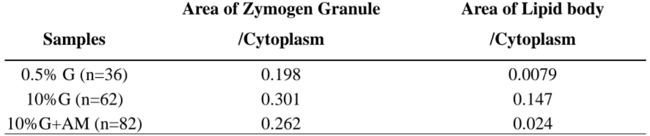
Two cross-fractured replicas of the fungal cytoplasm are shown in Figs. III-5B and C. Two kinds of organelles increased in volume: cytoplasmic vesicles (V) and lipid bodies (L). The median proportions of cytoplasmic area covered with each organelle were 0.198 (V) and 0.0079 (L) for 0.5% glucose (N=36), and 0.301 (V) and 0.147 (L) for 10% glucose (N=62), with significant differences (V, P<0.01; L, P<0.001; median test). The data are shown in Table III. Looking at Fig. III-5B, the corpulency observed in Fig. III-5A appears to be due to the accumulation of large amounts of lipid bodies in the cytoplasm. Also, in Fig. III-5C, the increased cytoplasmic vesicles have coalesced to form a shape resembling a bunch of grapes.
After extensive EM observations, we reached the following conclusions: (1) No external needle-shaped crystals could be found. (2) Either the cells secreted lipids in amounts too small to form crystals or lipid secretion itself was inhibited.
Glucose-fortified fungal cells treated with algal suspension medium
Fungi with large amounts of lipid bodies were cultured on an agar medium containing 10% glucose. The fungi shown in Fig. III-5 were obtained from a 15-mm colony after 3 months of continuous culturing. The large amount of lipid accumulation was conjectured to be due to an inhibition of lipid secretion. To stimulate these cells to secrete sufficient lipid to form crystals, we soaked the 15-mm colony with various materials reported to promote lipid secretion, such as -cyclodextrin and albumin (Bird et al., 1996; Kabakibi et al., 1998; Kamp and Hamilton, 1992 and Kamp and Hamilton, 1993; Kamp et al., 2003; Kilsdonk et al., 1995).However, we failed. We were unable to observe lipid crystals outside. Finally, after removing the fungal colony onto a normal agar medium containing 0.5% glucose, we soaked the colony with a few drops of algal suspension medium. The drops of algal suspension medium were prepared as follows: normal suspension medium in which algae alone had been growing for 5 months was filtrated through pores of 0.22mm in order to remove algal cells (conditioned medium). The colonies were soaked with a few drops (300mL/15-mm colony) of this filtrate for 1–14 days before the fungal cells in the colony were used for chemical analysis and for EM observations.
Although the replicas taken from the 3-day-soaked samples had no crystals outside, those of 9-day-soaked cells, which are shown in Fig. III-7, contained crystals
First, the fungi shown in Fig. III-7A had no deep invaginations on the P-faces. IMP occupied most of the P-face without the presence of worm-like places from where the particles had cleared off. The fungi cultured alone in the normal medium always displayed these configurations, and the medium, when tested by TLC, contained a negligible amount of secreted lipids (data not shown).
Second, in contrast to the observation just made, the fungus shown in Fig. III- 7B had very extensive invaginations of a deep and very serpentine shape. And in the cytoplasm (Fig. III-7C), the vesicles that had many IMP coalesced into one gigantic aggregate, suggesting that cellular exocytosis had been inhibited.
Third, in Fig. III-7D, the fungal cytoplasm was occupied by medium-sized vesicles (0.3–0.4mm), which appeared to be ready for exocytosis. The medium-sized vesicles shown here were apparently pinched off from the gigantic aggregate, as indicated by the arrow in Fig. III-7C. The cytoplasm contained lipid droplets, but the amount, when compared to that of the bodies shown in Fig. III-5B, was reduced (median proportion of cytoplasmic area covered with lipid bodies 0.024, N=82, P<0.001, median test; see Table III). The micrograph reminded us of exocrine pancreatic cells (Ichikawa, 1965), especially when looking at the vesicles in continuity (arrow).
In Fig. III-7E, the configuration of exocytosis of one of these medium-sized vesicles is clearly shown. Observing the round holes on the P-face, one is convinced that the cell is now actively secreting the contents of the vesicle, the secretory proteins. The large arrow indicates a typical snake-like invagination on the P-face. The small arrow indicates a reverse configuration of a "frog swallowing snake." The secretory granule fused with the membrane at the worm-like plasma membrane site from where the IMP had cleared off.
In this algal-medium-soaked specimen (Fig. III-7), we observed the following: (1) crystals of atranorin present outside the fungal cell, suggesting the secretion of a large amount of lipids; (2) the typical exocytotic features of the secretory vesicles, indicating the start of extensive protein secretion; and (3) no indication of how the lipids exited the cell. We could detect only the reciprocal relationship between the number of the lipid bodies inside and that of the lipid crystals outside.
Quantitative analysis of atranorin by means of LC
It was usual to find multiple lipid bodies in cultured fungal cells around where no crystals were found (Fig. III-5). It was also usual that fungal cells growing in the wild and being accompanied by crystals had very few lipid bodies (Fig. III-2). The morphological characteristics of lipid bodies in the fungal cells on replicas were similar to those of the bodies made of neutral lipid. Although it was easy to observe the shape of atranorin extracellulary, it was impossible to find any guarantee of atranorin in the cytoplasm by EM, even though its location should surely be in the cytoplasm. We considered the following: (1) Atranorin does not easily leak out of the cell. (2) Lipid bodies increase in size after receiving feedback information from the production line of atranorin. To clarify the above points, we employed microanalysis of LC. The colonies of fungal cells that had been fortified with 10% glucose were extracted with chloroform–methanol and the quantity of atranorin was measured. The samples clearly indicated the existence of atranorin (Fig. III-8, 0 day).
We also measured the amount of atranorin from the algal-medium-soaked colonies of fungal cells that had been fortified with 10% glucose. As shown in Fig. III-8, the concentration of atranorin was increased and reached a plateau in 3 days. After 6
days of treatment the concentration decreased gradually until Day 9 of incubation. We also picked several samples for EM from this experimental line. Although the samples soaked for 9 days contained atranorin crystal (Fig. III-7), those soaked for 3 days had no crystals, even though the concentration of atranorin had already reached a plateau.
Discussion
According to observations of the morphology of wild and cultured samples with EM, the following results were obtained in this study: (1) The dynamic morphological change of the plasma membrane of the fungi occurred in parallel with the activities of lipid secretion. (2) The zymogen granules (or protein vesicles) contained in the fungal cytoplasm exocytose during lipid secretion. (3) The number of lipid bodies contained in the fungi inversely changed with those of the crystal deposition outside the cell.
We considered that if we could promote the cultured fungi into actively lipid- secreting cells, and if we could find crystals around the cells, we would have an opportunity to understand the entire scheme of how the cell secretes lipid and the significance of symbiosis of lichens. Along with this consideration, we cultured fungi in medium with a higher concentration of sugar than usual. Hamada (1993) reported that under these conditions fungi synthesize large amounts of lipid. However, we did not detect crystal by EM or the existence of atranorin by TLC. In the fungal cytoplasm cultured in this sugar-fortified medium, multiple lipid bodies together with numbers of zymogen granules were observed. The appearance of these accumulations of multiple organelles suggested the cellular inhibition of secretions of both lipids and proteins. To release the inhibition, we soaked the fungi growing in this sugar-fortified culture in a few drops of the medium in which the algae had been cultured alone. The soaking caused the fungi to release the inhibition and made them actively exocytose zymogen granules. The plasma membrane changed shape, with many invaginations of omega appearances. The crystals were found outside the cells.
The modes of lipid secretion so far observed in the long course of histology are as follows: (1) simple diffusion, (2) exocytic secretion, and (3) endoplasmocrine secretion. Simple diffusion is the mechanism by which lipid in the cytoplasm diffuses freely through the lipidic plasma membrane. Secretion by exocytosis is the mechanism by which lipids bound by the biological membrane secrete themselves, similar to the mechanism of protein secretion. Rhodin (1971) has interpreted the endoplasmocrine secretion of lipids. The lipid body encircled by the tubules of smooth ER makes contact with the plasma membrane and the intact lipid body is exocytosed outside.
"Exocytosis" of lipid bodies
Rhodin (1971) has suggested direct "exocytosis" (endoplasmocrine secretion) of lipid bodies encircled by tubules of sER in the adrenal cortex cells of rats. However, it is difficult to suppose that all the steps of steroid hormone synthesis take place inside the lipid body. It is difficult also to suppose that the direct conversion of the body to an active hormone could occur in the extracellular space.
Having added 10% glucose to the fungal culturing medium, we were able to make the number and size of lipid bodies increase in the cytoplasm. But in this culturing medium, no crystals were found by EM observations. Also, when cells contained large amounts of lipid bodies, we could not detect atranorin in the culturing medium by means of TLC or LC.
When observing replicas of fungal cells containing large lipid bodies, we often encountered damaged cells, whose lipid bodies had already been cast out of the cytoplasm. Comparing the appearance of these bodies, no difference was detected between those inside the cell and those outside. On the replicas, both appear with the usual neutral lipid features (data not shown). This strongly indicates that lipid bodies are
not the direct source of the atranorin crystals occupying the extracellular space of lichens. We do not endorse the theory of "endoplasmocrine secretion" in which lipid bodies secrete atranorin. In addition, the nature of the contents of lipid bodies of Saccharomyces cerevisiae grown under the presence of glucose has been reported (Schaffner and Matile, 1981). The contents consisted of sterol derivatives (62.9%) and triacylglycerols (32.8%). These results support the idea that the lipid bodies in our fungal cells might also be reservoirs of excess carbon and a source to use in the future production of atranorin via acetyl-CoA.
Polyketide synthase (PKS) synthesizes atranorin and both are located
in the cytoplasm
It is well known that PKS interacts to produce many fungal polyketides such as brefeldin (BFA), lovastatin, aflatoxin, and other antibiotics (Child et al., 1996; Fujii et al., 2000; Hopwood, 1997; Hopwood and Sherman, 1990; Kennedy et al., 1999; Spencer and Jordan, 1992). It was presumed that atranorin may also be produced via the interaction of PKS.
PKS was first isolated from Penicillium patulin by Dimroth et al. (1970), employing exactly the same method used to isolate fatty acid synthase (FAS), a well- known cytosolic enzyme. The cytosolic location of PKS has been discussed in the previous article. The final polyketide products, however, were not synthesized by PKS alone. According to Kennedy et al. (1999), polyketides produced by PKS interaction must be further processed by accessory proteins during the final synthesis of the substrate. Accessory proteins bind with both PKS and the substrate. Because the location of the PKS appeared almost certainly to be in the cytoplasm, it was presumed that the final polyketide product, atranorin, should also be located in the cytoplasm, as we already mentioned in the previous article (see Arakawa-Kobayashi et al., 2004).
If atranorin translocates into the lumen of rER, its next step in moving to the outside should be along the usual exocytosis pathway. In fungal cells of all functional stages, elements of Golgi membranes were difficult to observe. However, the fact that a considerable amount of exocytosis of zymogen vesicles did occur indicated the existence of trans-Golgi membranes making zymogen vesicle. The quantity of zymogen vesicles in the fungi cultured alone in normal medium was the same as that in wild fungi. The median proportion of cytoplasmic area covered with zymogen vesicles of fungi cultured alone (see Table III) was 0.198 (N=36), and that of the fungi in the wild was 0.199 (N=183). TLC indicated that the fungi in the wild contained plenty of atranorin, whereas the fungi cultured alone contained no detectable amount. We are certain that the atranorin secretion could never have a chance to use exocytotic pathways.
Exocytotic secretion
After the addition of 10% glucose, the increased materials other than lipid bodies were zymogen vesicles. These vesicles had numerous IMP on their E-faces, and tiny,
atranorin and the lipid bodies were completely extracted by alcohol dehydration, while electron-opaque material contained in the vesicle was never extracted (Fig. III-2 and Fig. III-9). This indicates that the nature of the content of the vesicle could never be lipidic rather than proteinous.
The fungal cells that had been treated with 10% glucose were supplemented with a conditioned medium. The following changes were observed. (1) Actual crystals of atranorin were observed in the extracellular space. (2) Invaginations of plasma membrane showed deeper and wider trenches. (3) Exocytosis of vesicles resembling those in exocrine pancreatic cells had appeared. (4) Huge proteinous vesicles had fused with many vesicles to form a shape like a bunch of grapes.
After the addition of 10% glucose, cells synthesized proteins other than lipids at a higher rate than usual. It was obvious that the resultant proteinous vesicles were very similar in appearance to those of the rER of castrated or thyroidectomized anterior pituitary cells (Farquhar and Rinehart, 1954a and Farquhar and Rinehart, 1954b). As fungal cells had limited dimensions of Golgi apparatus, the transport of secretory proteins would have stopped before they entered the cis-Golgi membranes. The zymogen vesicles, however, had numerous IMP on the E-face, while those on the E-face of rER were scarce (see Fig. III-2D). Moreover, the vesicles had no ribosomes studded on the membranes. It is safe to assume that these vesicles were the usual secretory granules derived from trans-Golgi membranes, and that the "bunch of grapes" shape resulted from the exocytosis activity being inhibited in its final stage. Looking at Figs. III-7D and E, it appears that the size of the vesicles is related to their ability to undergo exocytosis. The size of the vesicles captured under exocytosis (Fig. III-7E) appears small and constant (0.3–0.4 mm) when compared to that of the vesicles in the "bunch of grapes" shape (over 1.6 mm). We can conclude that the bunched vesicles are a feature resulting from the cellular inhibition of exocytosis.
Secretion by simple diffusion
While the IMP represent membrane protein, the particle-cleared area appearing on freeze replicas of biological membranes consists only of membrane lipids (Elgsaeter et al., 1976). Lipids, at least in uncharged form, are generally considered to penetrate easily through this particle-cleared area of biological membranes (Hamilton, 2003).
We observed that the serpentine ditch on the P-face of fungal cells was often in continuity with the serpentine structure having no IMP. We presumed that the serpentine structures without particles on the P-face were hallmarks of fungal cells cultured alone in normal medium (Fig. III-4B), and this medium showed no atranorin by TLC and by LC (data not shown). The crystals of atranorin appeared only after extensive exocytosis had occurred. The exocytotic vesicles started from deep inside the cytoplasm and moved toward the particle-free region, fusing the two membranes (Fig. III-7E). The dense concentration of protein seemed necessary to pull atranorin out of/or through the membrane. This may explain the site of exocytosis, which is occurring always at places where the IMP have cleared.
Atranorin release from atranorin-containing liposome
The interesting report by Kabakibi et al. (1998) helps in understanding the enigmatic fungal lipid secretion that we observed for the first time using the quick-frozen electron microscopy of living cells. Kabakibi et al. demonstrated the release of fatty acid ethyl