Roles of IP
3signaling in Wnt-regulated
asymmetric divisions of C. elegans
epithelial stem cells
Naoki YOSHIDA
Doctor of Philosophy
Multicellular Organization Laboratory, Structural Biology Center, National Institute of Genetics
Department of Genetics, School of Life Science
The Graduate University for Advanced Studies
2014 (School Year)
List of Contents
Abbreviations ... 3
Abstract ... 5
1 Introduction ... 9
1-1 Asymmetric cell division ... 9
1-2 Asymmetric cell division in C. elegans ... 9
1-3 The Wnt/β-catenin asymmetric (WBA) pathway ... 10
1-4 IP3 signaling pathway ... 10
1-5 New findings of this study ... 11
2 Materials and Methods ... 14
2-1 Strains ... 14
2-2 Genotyping of C. elegans using PCR ... 14
2-3 Lineage analysis of seam cells ... 15
2-4 Confocal microscopy and data analysis ... 15
2-5 Time-lapse microscopy ... 16
2-6 Statistical Analysis ... 16
2-7 cDNA library: RNA isolation from C. elegans and RT-PCR ... 16
2-8 Construction of Plasmids ... 17
2-8-1 Translational genes ... 17
2-8-2 A transcriptional fusion gene ... 18
3 RESULTS ... 20
3-1 ppk-1/PIP5K is required for asymmetric cell division ... 20
3-2 ppk-1 regulates localizations of the WBA pathway components 21 3-3 ppk-1 localizes to the cell cortex ... 23
3-4 ppk-1 functions in the IP3 signaling pathway ... 23
3-5 plc-4 is highly expressed in seam cells ... 24
3-6 Ca2+ imaging in seam cells ... 25
3-7 cdc-42 may be a downstream effector of itr-1 ... 25
3-8 ppk-1 genetically interacts with cwn-2/Wnt ... 26
3-9 ppk-1 genetically interacts with lin-18/Ryk and mom-5/Frizzled
... 27
3-10 Overexpression of CWN-2 promotes the reversed polarity in ppk-1 mutants ... 28
3-11 Overexpression of LIN-18 promotes the reversed polarity in ppk-1 mutants ... 28
4 Discussion ... 31
4-1 Summary of results ... 31
4-2 ppk-1 is linked to IP3 signaling to regulate the ACD ... 31
4-3 A model for PPK-1 function ... 32
4-4 Downstream components of IP3 signaling to regulate the ACD . 33 4-5 Why do ppk-1 mutants show the reversal phenotype only in specific cells? ... 33
4-6 IP3 signaling and cell polarity in other organisms ... 34
4-7 The significance of this study ... 35
5 Figures ... 37
References ... 58
My publications ... 63
Acknowledgements ... 64
Abbreviations
APC Adenomatous polyposis coli APR-1 APC related protein
ajm Apical junction molecule
AP Anterior-posterior
C. elegans Caenorhabditis elelgans Ca2+ Calcium ion 2+
cam-1 CAN cell migration defective cdc-42 Cell division cycle 42
cDNA complementary DNA
csnk-1 Casein kinase 1 gamma cwn-1/-2 C. elegans Wnt1/2
DV Dorsal-ventral
DG Diacylglycerol
DNA Deoxyribonucleic acid
ER Endoplasmic reticulum
egl-20 Egg laying variant 20
Fz Frizzled
GCaMP GFP-Calmodulin-M13 Peptide
gf Gain-of-Function
GFP Green fluorescent protein Hyp7 Hypodermal 7 syncytium
itr-1 Inositol 1,4,5-triphosphate receptor 1 IP3 Inositol 1,4,5-triphosphate
IP3R Inositol 1,4,5-triphosphate receptor L1/L2/L3/L4 Larval 1/2/3/4
lin-17/-18 abnormal cell lineage -17/-18 LSM confocal laser scanning microscopy mCherry Monomeric Cherry
mg Microgram
min Minute
ml Microliter
mm Micrometer
mom-5 More of Ms
NGM Nematode growth medium
PCP Planer cell polarity PCR Polymerase chain reaction PH domain Pleckstrin homology domain
PI Phosphatidylinositol
PI(4,5)P2/PIP2 Phosphatidylionositol-4,5-bisphosphate plc-1/-2/-3/-4 Phospholipase C 1/2/3/4
PIP5K Phosphatidylinositol-4-phosphate 5-kinase
PKC Protein kinase C
POP-1 Posterior pharynx defect
ppk-1 Phosphatidylinositol phosphate kinase 1
RNA Ribonucleic Acid
RNAi RNA interference
rpm Rotation per minute
Ror Receptor-related
RT-PCR Reverse transcription-polymerase chain reaction Ryk Receptor-like tyrosine kinase
sec second
SSD Salmon sperm DNA
TCF T cell factor
unc Uncoordinated
UV Ultra Violet
Wnt Wingless-Int
WntR Wnt Receptor
WRM-1 Worm armadillo 1
Abstract
During development, asymmetric cell division is an important mechanism to generate cellular diversity and to maintain self-renewing stem cells. In C. elegans development, most somatic cells are polarized and divide asymmetrically to produce daughter cells with distinct fates. These asymmetric cell divisions are regulated by the Wnt/β-catenin asymmetry pathway that utilizes components similar to those of the canonical Wnt pathway. These components localize asymmetrically in the cell. For example, APR-1/APC localizes to the anterior cell cortex and regulates asymmetric nuclear localization of POP-1/TCF that localizes to the anterior daughter nucleus. Most somatic cells show the same POP-1 asymmetry and, hence, have the same polarity orientation. However, the mechanism to orient cell polarity is a mystery.
To reveal the mechanism that orients cell polarity, I focused on inositol 1,4,5-trisphoshate (IP3) signaling that plays roles in regulating cell polarity. In this signaling, phosphatidylinositol-4-phosphate 5-kinase (PIP5K) produces phosphatidylinositol 4,5-bisphosphate (PIP2), PIP2 is hydrolyzed by phospholipase C (PLC) to produce IP3. IP3 binds to IP3 receptor (IP3R) causing to release of Ca2+ from endoplasmic reticulum (ER). This increased cytoplasmic Ca2+ in turn activate several cellular processes, resulting in regulation of cell polarity that is required for the direction of cell migration, the formation of cellular protrusions, and cell-cell adhesion. However, the relationship between asymmetric cell division and IP3 signaling has not been studied directly.
I examined the ppk-1/PIP5K functions in seam cells which are epithelial stem
cells that repeatedly undergo self-renewing asymmetric cell divisions. Each seam cell is polarized and divides asymmetrically giving rise to an anterior daughter that is
terminally differentiated to the hypodermal cell and a posterior one that remains in the seam cell fate. I found that ppk-1(ok1411) mutants had a defect in asymmetric cell divisions of seam cells by observing fates of daughter cells using elt-3::GFP (a hypodermal cell marker). Their daughter cell fates were swapped with each other compare to those in wild type (reversal phenotype). Furthermore, I found that the orientation of POP-1 asymmetry is also reversed in ppk-1 mutants. These results indicate that that ppk-1 regulates the polarity orientation of the seam cells. The reversal phenotype in ppk-1 mutants was fully rescued by expression of PPK-1 under a seam cell specific promoter, suggesting that PPK-1 functions cell autonomously to regulate the cell polarity. I found that mCherry-tagged PPK-1 localizes symmetrically to the cell cortex and regulates the asymmetric localization of cortical APR-1/APC, judged by GFP-tagged APR-1. I revealed that IP3 signaling components (PLC-4/PLC and ITR-1/IP3R) act downstream of PPK-1 by analyzing genetic interactions among these components. Based on these results, I conclude PPK-1 regulates the APR-1 polarity as a component of the IP3 signaling pathway.
To further understand ppk-1 function, I analyzed the genetic interactions between ppk-1 and three Wnt genes. I found that the reversal phenotype in the ppk-1 mutants is suppressed by loss-of-function mutations of cwn-2/Wnt. Furthermore, the reversal phenotype in ppk-1 mutants was enhanced by the overexpression of CWN-2. These results suggest that the reversed polarity in ppk-1 mutants is caused by cwn-2. In
contrast, in wild type, cwn-2 promotes the normal orientation of cell polarity,
suggesting that cwn-2 has two functions promoting both the normal polarity orientation and the reversed polarity orientation. How does cwn-2 have the two opposite functions? To answer this question, I analyzed the genetic interactions between ppk-1 and six Wnt receptor genes. The reversal phenotype in ppk-1 mutants was suppressed by each of lin-18/Ryk and mom-5/Frizzled mutations, suggesting that each of lin-18 and mom-5 is necessary to promote the reversal phenotype in ppk-1 mutants. From these results, I conclude that cwn-2 has two functions promoting both the reversed polarity orientation through the Wnt receptors (LIN-18 and MOM-5) and the normal polarity orientation depending on PPK-1 activities. Based on these results, I propose ppk-1 is a switch that determines the CWN-2 function. Since IP3 signaling and Wnt signaling are highly conserved among metazoans, similar mechanisms may also regulate proper cell polarity in other organisms.
Chapter
1
Introduction
1 Introduction
1-1 Asymmetric cell division
Asymmetric cell division (ACD) has an important role to generate cellular diversity during development and tissue regeneration. This division generates two daughter cells with distinct fates (Horvitz and Herskowitz, 1992). Because there are many examples of ACDs in developmental biology and stem cell biology, The ACD is not unique to animal development, to stem cells. In fact, bacterial and yeast cells divide asymmetrically, as do many cells in developing embryos (Amon, 1996; Goley et al., 2009; Laub et al., 2007). Therefore, the ACD is a ubiquitous feature of all organisms. However, many unanswered questions remain to be solved to understand mechanisms of ACDs.
1-2 Asymmetric cell division in C. elegans
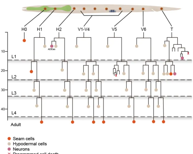
C. elegans is one of the best-understood model systems for ACDs. During larval development, epithelial stem cells, called seam cells, undergo repeated self-renewing ACDs. Each of these ACDs produces a posterior daughter that remains as the seam cell and an anterior daughter that adapts terminally differentiate fate that fuses with the hyp7 syncytium (Sulston and Horvitz, 1977). One of the most important concepts in these ACD is that these cells have the same polarity orientation (Fig.1). However, mechanisms to orient the cell polarity are mystery.
1-3 The Wnt/β-catenin asymmetric (WBA) pathway
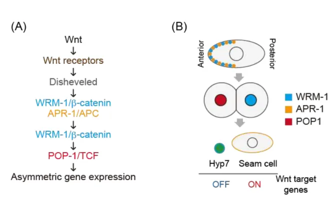
In C. elegans, most of ACDs are regulated by the Wnt/β-catenin asymmetry pathway (Mizumoto and Sawa, 2007; Siegfried et al., 2004; Walston et al., 2004). Each seam cell is polarized and divide asymmetrically, giving rise to an anterior daughter that terminally differentiates and a posterior one that remains in a seam cell fate. Our laboratory previously reported that the Wnt/β-catenin asymmetry pathway is essential for establishing ACDs of seam cells (Mizumoto and Sawa, 2007; Takeshita and Sawa, 2005; Yamamoto et al., 2011). This pathway utilizes components similar to those of the canonical Wnt pathway. In the WBA pathway, WRM-1/β-catenin and APR-1/APC localized to the anterior cell cortex to regulate the nuclear localization of WRM-1, resulting in its preferential localization to the posterior nucleus. This nuclear WRM-1 removes POP-1/TCF from the posterior nucleus, causing that POP-1 accumulates in the anterior nucleus. These asymmetric localizations are regulated by Wnt and Frizzled proteins (Fig. 2). Based on these asymmetries, all seam cells show the same POP-1 asymmetry and, hence, have the same polarity orientation. However, the mechanism to orient the cell polarity is a mystery.
1-4 IP3 signaling pathway
Inositol 1,4,5-trisphoshate (IP3) signaling is involved in regulation of cell polarity (Wei et al., 2009). In this signaling, phosphatidylinositol-4-phosphate 5-kinase (PIP5K) produces PIP2, PIP2 is hydrolyzed by phospholipase C (PLC) to produce IP3. IP3 binds to IP3 receptors (IP3Rs) causing release of Ca2+ from endoplasmic reticulum
(ER) (Fig. 3). This increased cytoplasmic Ca2+ in turn regulates many cellular processes, including cell polarization that are required for directed cell migration (Wei et al., 2009), the formation of cellular protrusions (Evans and Falke, 2007), cell-cell adhesion (Clapham, 2007; Pilipiuk et al., 2009), and convergent extension (Wallingford et al., 2001). In C. elegans, IP3 signaling regulates a wide variety of physiological functions, including developmental morphogenesis (Pilipiuk et al., 2009; Thomas-Virnig et al., 2004), neuronal signaling (Walker et al., 2009), as well as rhythmic behaviors such as pharyngeal pumping (Walker et al., 2004), oocyte fertilization (Corrigan et al., 2005), defecation (Dal Santo et al., 1999), and male mating (Gower et al., 2005). However, relationships between IP3 signaling and the ACD has not been reported.
1-5 New findings of this study
Here, I show that IP3 signaling regulates the CWN-2/Wnt functions to correctly orient the seam cell polarity. First, Our laboratory performed RNAi screening to identify novel regulators of the WBA pathway. ppk-1 was isolated as a result of its knockdown phenotype causing distal tip cell (DTC) hyperplasia which is also regulated by the WBA pathway. I found that a ppk-1/PIP5K mutation causes a reversed polarity orientation of seam cells, suggesting that ppk-1 is a new regulator that orients the polarity. PPK-1 localizes symmetrically to the cell cortex and regulates the asymmetric localization of APR-1/APC at the cortex. I found that IP3 signaling components (PLC-4/PLC and ITR-1/IP3 receptor) act downstream of PPK-1. Meanwhile, the ppk-1 phenotype was suppressed by a mutation in cwn-2/Wnt, suggesting that cwn-2 promotes
the reversed polarity in ppk-1 mutants. In contrast, in wild type, cwn-2 promotes the normal polarity, suggesting that cwn-2 has two functions promoting both the normal and reversed polarity orientation. Based on my results, I conclude that ppk-1 regulates the CWN-2 functions via IP3 signaling to correctly orient the cell polarity.
Chapter
2
Materials and methods
2 Materials and Methods 2-1 Strains
Strains were derived from wild-type N2 Bristol strain and were cultured at 22.5°C on NGM plates and were fed with OP50 bacteria as described (Brenner, 1974). Double-mutant strains were constructed using standard genetic methods without additional marker mutations, except for itr-1(sy290) which marked with unc-24(e138) mutation. Mutations in the double mutants were confirmed by PCR and DNA
sequencing.
In this study, the following alleles were mainly used: ppk-1(ok1411), itr-1(sy290), plc-1(rx1), plc-2(ok1761), plc-3(tm1340), plc-4(ok1215), cwn-1(ok546) cwn-2(ok895), egl-20(n585), lin-17(n3091), cam-1(ks52), cfz-2(ok1201), mig-1(e1787), mom-5(ne12), lin-18(e620), cdc-42(gk388), unc-24(e138) and unc-76(e911).
Molecular information of tm allele are described in http://www.shigen.nig.ac.jp/c.elegans/
Molecular information of all alleles except for tm allele is described in http://www.cbs.umn.edu/CGC/index.html
2-2 Genotyping of C. elegans using PCR
To detect DNA mutations, worms were transferred into individual PCR tube with 20 µl worm lysis buffer containing Proteinase K (Wako) and incubate at 50 °C for one hour, which resulted in lysis of worms and release of genomic DNA. Proteinase K
was inactivated at 95 °C for 15 min. The worm lysate was used as templates for PCR with primers designed to distinguish between wild type and mutant alleles by producing two DNA fragments different in sizes or sequences.
2-3 Lineage analysis of seam cells
Lineage analyses were performed as described previously (Sulston and Horvitz, 1977). Seam cell nuclei were distinguished from hypodermal nuclei based on their morphology in addition to the expression of markers: elt-3::GFP(vpIs1), scm::GFP(wIs51), or ajm-1::GFP+scm::GFP(wIs79). Since the V5.pa cell produces only neural cells that do not express these markers, I did not analyze polarity of the V5.pa division. Samples were prepared by immobilizing L2 stage hermaphrodites with 50 mM sodium aside and by placing them on 2% agarose pads. A Zeiss LSM700 microscope equipped with a 60x objective was used to examine cell fates of Vn.p cell daughters. Each strain was scored twice independently.
2-4 Confocal microscopy and data analysis
Confocal-microscopic images were obtained with Zeiss LSM700 equipped with a 488 nm and 561 nm lasers. Images of whole worms were complied using Adobe Photoshop CS5 and ImageJ (http://imagej.nih.gov/ij/). Intensities of GFP in nuclei were measured by imageJ (http://imagej.nih.gov/ij/).
2-5 Time-lapse microscopy
For live observations of GCaMP animals, a focal plane through the center of the seam cells was chosen. Samples were prepared by immobilizing L2 stage hermaphrodites with 2 mM levamisole in M9 buffer by placing them on 5% agarose pads. For imaging, Zeiss AxioImager Z1 equipped with a confocal scanner unit (CSU-W1, Yokogawa Electric Corporation) and an sCMOS camera (Neo, Andor Technology) was used. Image processing was performed with ImageJ (http://imagej.nih.gov/ij/).
2-6 Statistical Analysis
Statistical analysis was performed with the Fisher exact test.
2-7 cDNA library: RNA isolation from C. elegans and RT-PCR
For C. elegans total RNA isolation, wild-type (N2) worms were collected from non-starved plates by washing them with M9 buffer several times and were frozen at -80 °C. To extract RNAs, I used PureLink TM RNA Mini Kit (Life technologies). All buffers and regents were form this kit. An equal amount of glass beads (212-300 mm, SIGMA, G9143) was added to the frozen worm pellets, and the tubes were put in a Micro Smash (SM-100, Tomy Seiko) three times for 15 sec at 3000 rpm, while keeping them always on ice. RNA purification was carried out using the PureLinkTM RNA Mini Kit (Life technologies).
The second step, conversion of RNA to DNA, was carried out using the superscript III Reverse Transcriptase (Life technologies) according to the manufacture’s protocol. After the reverse transcriptase reaction was completed, the generated cDNA from the original single stranded mRNA was used as template in a standard PCR.
2-8 Construction of Plasmids 2-8-1 Translational genes
A full-length ppk-1 cDNA was cloned from a C. elegans cDNA library (see 2-7) by PCR amplification using PrimeSTAR Max (Takara). Primers were designed based on the predicted WormBase sequence of F55A12.3. Full-length translational mCherry reporters for PPK-1 and PHPLCδ (from Addgene) were generated using Infusion
technique (Invitrogen) and inserted into Fire vector modified pPD95.75 vector. mCherry was fused to the N-terminal of the proteins. Expression of mCherry::PPK-1 and mChery::PHPLCδ were driven by a 9 kb fragment of seam cell specific promoter (Koh and Rothman, 2001). Transgenic worms were generated by injecting DNAs into unc-76(e911) worms with DNA (Mello et al., 1991). A unc-76-rescuing plasmid (Bloom and Horvitz, 1997) was used as a transformation marker, and co-injected with the PPK-1 and PHPLCδ plasmids. The mCherry::PPK-1 and mChery::PHPLCδ plasmids were injected at 10 ng/ml together with 50 ng/ml of the unc-76 plasmid and 40 ng/ml of salmon sperm DNA (SSD) (life technologies). Transgenic animals produced by
injection typically carry large extrachromosomal arrays that contain many copies of the co-injected DNAs.
Extrachromosomal arrays can be integrated into a chromosome. Integrated lines of worms expressing mCherry::PPK-1 and mCHerry::PHPLCδ were generated by exposing 50 transgenic animals to a dose of 30,000 µJ/cm2 of UV lights in CL-1000 Ultraviolet Crosslinker (Uutra-Violet products). Irradiated animals were grown until
their stervation on 9 cm plates five times. After this, one hundred nonUnc offspring were isolated. Each of integrated lines, osIs91 (Pscm::mCHerry::PPK-1; unc-76) and osIs90 (Pscm::mCherry::PHPLCδ; unc-76) were then identified by segregation of 100% nonUnc and mCherry-positive animals.
2-8-2 A transcriptional fusion gene
plc-4::Venus construct. 1.855 kb of upstream plc-4 promoter and 2.981 kb of plc-4 gene were amplified from wild-type genomic DNA using the following primers: plc-4-F: 5’-GTGCCCAAAGCACTCTAATCGG-3’. And
plc-4-R: 5’-GTACTCTTCCTCCATTGCAATTCGTAC-3’.
By using Infusion technique (life technologies), the PCR fragment was cloned into modified-pPD95.75 vector in which GFP was replaced by Venus. The
plc-4::Venus plasmid was injected at 10 ng/ml together with 50 ng/ml of unc-76 and 40 ng/ml of SSD. The resulting array is osEx523.
Chapter
3
Results
3 RESULTS
3-1 ppk-1/PIP5K is required for asymmetric cell division
Lateral seam cells are specialized epithelial stem cells. During each larval stage, each of these seam cells is polarized and divides asymmetrically to give rise to an anterior daughter that fuses with hyp7 epidermal syncytium and a posterior daughter that remains as a seam cell (Fig. 1).
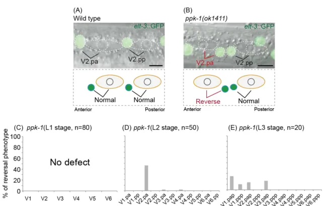
To understand roles of IP3 signaling in asymmetric division of seam cells, I examined ppk-1/PIP5K mutants. To analyze fates of the seam cell daughters, I used elt-3::GFP(vpIs1), which is expressed in the hyp7 epidermal syncytium but not in seam cells (Gilleard et al., 1999; Koh and Rothman, 2001) (Fig. 4A). I then analyzed the lineage of seam cells during each larval stage. Although ppk-1(ok1411) mutants showed normal daughter fates of seam cells during the L1 stage (Fig. 4C), At the L2 and L3 stages, I found that ppk-1(ok1411) mutants had a defect in ACDs of some seam cells (Fig. 4 DE). This defect is that the fates of the daughter cells were swapped with each other compare to those in wild type (Fig. 4B). Hereafter I call this phenotype as
‘reversal phenotype’. The reversal phenotype was observed in specific seam cells at each stage. At the L2 stage, reversal phenotype was observed in V2.pa cell, but not in V2.pp cell, even though these cells are V2.p cell daughters and have same cell lineages (Fig. 4D). At the L3 stage, the reversal phenotype was observed in seam cells that are located in anterior side of animal body (Fig. 4E). I could not analyze the phenotype at the L4 stage, because ppk-1(ok1411) mutants arrest their development at the late L3 stage. In contrast to the reversal phenotype, ppk-1(ok1411) mutants did not show
symmetric cell divisions, suggesting that ppk-1 is important to orient the cell polarity but not to generate it. Because the V2.pa cell division showed the strongest reversal phenotype, hereafter I focused on the ACD of the V2.pa cell (Fig. 4 DE).
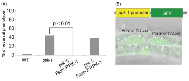
I next conducted a cell-specific rescue experiment to examine the cell autonomy of the ppk-1 function. The reversal phenotype was fully rescued by the expression of PPK-1 under the seam cell-specific promoter (scm promoter) (Fig. 5A). No rescue was observed when PPK-1 was expressed with the pharynx-specific promoter (myo-2 promoter) (Fig. 5A). It was reported previously that GFP was expressed under the control of ppk-1 promoter, Pppk-1::GFP(gqEx33), is expressed in seam cells in adult hermaphrodites (Weinkove et al., 2008). I found that Pppk-1::GFP(gqEx33) is also expressed in seam cells at the L2 stage (Fig. 5B). These results indicate that ppk-1 functions cell autonomously in the seam cells to regulate ACDs.
3-2 ppk-1 regulates localizations of the WBA pathway components
In C. elegans, ACDs of seam cells are regulated by the WBA pathway. Components of this pathway include WRM-1/β-catenin, APR-1/APC and a POP-1/TCF transcription factor (Mizumoto and Sawa, 2007) (Fig. 2A). During cell divisions, WRM-1 and APR-1 are localized asymmetrically to the anterior cortex in the mother cell (Mizumoto and Sawa, 2007; Nakamura et al., 2005). After a cell division, nuclei of the anterior and posterior daughters have more POP-1 and WRM-1 than the other nuclei, respectively (Fig. 2B).
As shown above, ppk-1 is important in asymmetric divisions of some seam
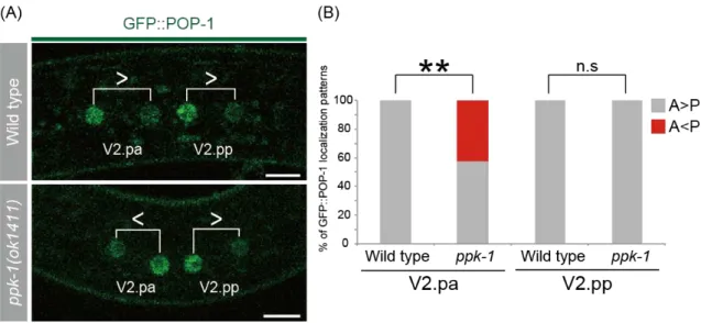
cells. However, ppk-1 has not been implicated in the WBA pathway previously. To examine whether ppk-1 regulates the localizations of the WBA pathway components, I first analyzed the POP-1/TCF localization. I used a transgene GFP::POP-1(qIs74) (Siegfried et al., 2004). POP-1 normally accumulates in the anterior daughter nucleus in wild type. In ppk-1 mutants, I found that GFP::POP-1 asymmetry was reversed about 42% in V2.pa daughters in ppk-1(ok1411) mutants (Fig. 6AB). These results indicate that ppk-1 controls the cell polarity via the WBA pathway.
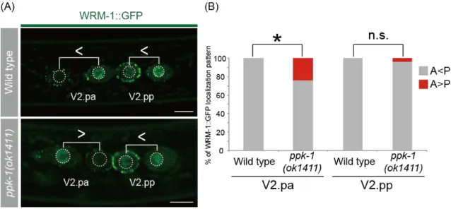
Because the asymmetric localization of GFP::POP-1 requires nuclear WRM-1/β-catenin (Lo et al., 2004), I observed the asymmetric localization of WRM-1 in daughter nuclei by using WRM-1::GFP (osIs5) (Mizumoto and Sawa, 2007). WRM-1 normally accumulates in the posterior daughter nucleus in wild-type animals (Nakamura et al., 2005; Takeshita and Sawa, 2005) (Fig. 7A). This asymmetry was reversed about 20% in ppk-1(ok1411) mutants (Fig. 7AB), suggesting that ppk-1 functions upstream of nuclear WRM-1.
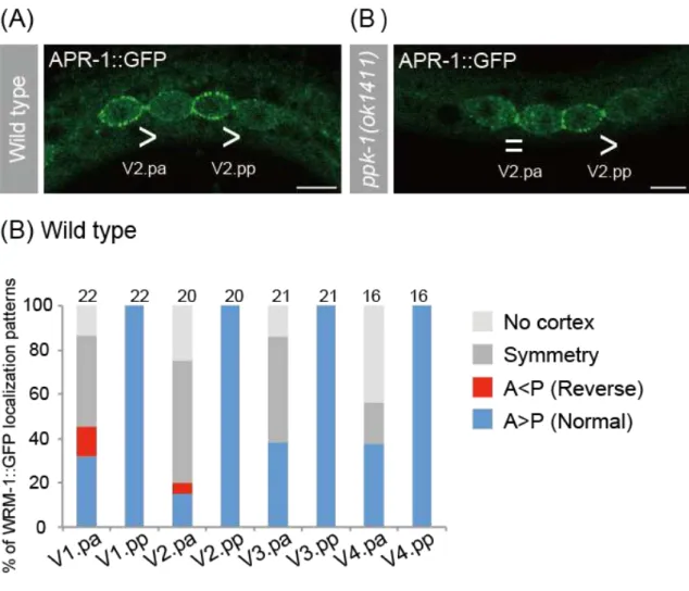
Because the asymmetric localization of WRM in daughter nuclei requires the anterior cortical localization of WRM-1 and APR-1 in their mother cell (Mizumoto and Sawa, 2007). I observed the localization of cortical APR-1 by using
APR-1::GFP(osIs13) (Mizumoto and Sawa, 2007). While APR-1::GFP normally accumulated on the anterior-daughter cortex of seam cells in wild-type animals (Fig. 8A), APR-1::GFP was not asymmetrically localized in V2.pa cell in ppk-1(ok1411) mutants (Fig. 8B). Next I observed the localization of WRM-1 by using
WRM-1::GFP(osIs5). I could not distinguish the discrepancy of the cortical WRM-1
localization between wild type and ppk-1(ok1411) mutants, because WRM-1::GFP localization was randomized in Vn.pa cells including V2.pa cell in wild type (Fig. 8C). I conclude that ppk-1 regulate at least cortical polarity of APR-1 in V2.pa cell.
3-3 ppk-1 localizes to the cell cortex
To further characterize the role of ppk-1 in Wnt-regulated ACDs, I analyzed PPK-1 subcellular localization in seam cells. I expressed a mCherry-tagged PPK-1 in seam cells by using the scm promoter. mCherry-tagged PPK-1 is functional, because its expression can rescue the reversal phenotype of ppk-1 mutants (Fig. 5A). In all
cell-cycle including the interphase, prophase, metaphase and telophase,
PPK-1::mCherry is localized uniformly to the cell cortex (Fig. 9ABC). Thus, the subcellular localization of PPK-1 is consistent with its role in regulation of cortical polarity.
3-4 ppk-1 functions in the IP3 signaling pathway
To understand the molecular mechanism underlying the regulation of the cortical APR-1 asymmetry mediated by ppk-1, I analyzed genetic interactions between ppk-1 and IP3 signaling components. Since a gain of function (gf) mutation of itr-1/IP3R is able to suppress the sterility of ppk-1(RNAi), itr-1 is downstream effector of ppk-1 (Fig. 3) (Xu et al., 2007). This itr-1(sy290gf) allele carries a missense mutation in IP3 binding domain of ITR-1/IP3R (Clandinin et al., 1998).
To examine whether the asymmetric division of the seam cell is linked to the
IP3 signaling pathway, I observed the ppk-1(ok1411) phenotype in the itr-1(sy290gf) background. ppk-1(ok1411); itr-1(sy290gf) mutants displayed weaker reversal phenotype (15%) than ppk-1 single mutants (46%) (Fig. 10), suggesting that the reversal phenotype in ppk-1(ok1411) mutants is suppressed by the itr-1(sy290gf) mutation and that ppk-1 functions through IP3 signaling.
Phospholipase C (PLC) is necessary for producing IP3 from PIP2. I hypothesized that the disruption of the PLC function should result in the reversal phenotype of the seam cell as in ppk-1 due to the decrease of IP3 signaling. I tested functions of four genes encoding PLCs (plc-1, plc-2, plc-3, and plc-4) (Fig. 11). Among the four PLCs, a mutation of only the plc-4 gene showed the reversal phenotype in the seam cell. Thus, plc-4 activity is required for the proper ACD in the seam cell. Depletion of plc-1, plc-2, and plc-3 had no effects on the ACD in seam cells. To elucidate the relationship between ppk-1 and plc-4, I analyzed cell fates of ppk-1; plc-4 double mutants. ppk-1; plc-4 double mutants did not suppress, nor enhanced the reversal phenotype compare to those in ppk-1 mutants, suggesting that both ppk-1 and plc-4 function in the IP3 signaling pathway (Fig. 11). Based on these results, it is suggested that ppk-1 functions in the ACD in through IP3 signaling.
3-5 plc-4 is highly expressed in seam cells
To examine the expression pattern of plc-4, I generated transgenic worms expressing full-length PLC-4 fused to Venus. This expression was driven by a 1.9-kb fragment of the plc-4 promoter located immediately upstream of the start codon (Fig 12A).
Prominent expression of PLC-4::Venus was detected in hypodermal seam cells, intestine, body wall muscles, and excretory cells in L2 larvae (Fig. 12B). Weaker expression was seen in the pharynx and some neurons. Similar expression pattern was partially described in previous study (Vazquez-Manrique et al., 2010). PLC-4::Venus localized to the cortex of seam cells (Fig. 12C). Thus, the subcellular localization of PLC-4 is consistent with its role in the regulation of polarity orientation of the seam cell.
3-6 Ca2+ imaging in seam cells
Based on the genetic interaction between ppk-1 and the calcium channel ITR-1, I expected involvements of Ca2+ in the ACD. To analyze Ca2+ dynamics in seam cells, I used a Ca2+ sensor GCaMP3 which has been used in C. elegans neurons and hypodermal cells to image calcium transients (Tian et al., 2009; Xu and Chisholm, 2011).
I established transgenic worms expressing GCaMP3 under the control of the scm promoter. Although I succeeded to observe Ca2+ dynamics only in three animals, in all cases, I found that GCaMP3 fluorescence repeatedly spreads from V1.pa cell to V6.pp cell like waves (n=3) (Fig. 13). This result raises a possibility that Ca2+ dynamics orients the polarity of seam cells.
3-7 cdc-42 may be a downstream effector of itr-1
In the Xenopus embryo, Cdc42 functions downstream of IP3 signaling which
activates IP3R to express Ca2+ to cytoplasm. An increase of intracellular Ca2+ activates PKC (Sheldahl et al., 2003) that regulates small GTPase Cdc42 (Schlessinger et al., 2007). In addition, Cdc42 is a major regulator of cell polarity from yeast to mammalian cells (Etienne-Manneville, 2004). From these data, I hypothesized that cdc-42 regulates cell polarity as a down stream effector of IP3 signaling.
I found that cdc-42(gk388) animals showed the reversal phenotype similar to ppk-1(ok1411) animals (Fig. 14AB). To examine whether cdc-42 is a downstream effector of itr-1, I analyzed cdc-42(gk388); itr-1(sy290gf) double mutants. itr-1(sy290gf) fails to suppress the reversal phoenotype defect of cdc-42(gk388) (Fig. 14C). This result may suggest that cdc-42 is a downstream effector of itr-1 to orient the polarity orientation of the seam cell.
3-8 ppk-1 genetically interacts with cwn-2/Wnt
The polarity orientation of seam cells at L1 stages is redundantly regulated by Wnts (cwn-1, cwn-1, and egl-20) (Yamamoto et al., 2011). My results suggest that ppk-1 also regulates the polarity orientation of the V2.pa cell. I next examined genetic interactions between ppk-1 and three Wnt genes (cwn-1, cwn-2, and egl-20). I found that the reversal phenotype of ppk-1(ok1411) mutants was suppressed by cwn-2(ok895) or cwn-2(RNAi), but not by mutations in the other Wnt genes (cwn-1(ok546) and egl-20(n585)) (Fig. 15). These epistatic relationships suggest that ppk-1 functions upstream of or in parallel to cwn-2. It is unlikely, however, that ppk-1 functions upstream of cwn-2. As described above, ppk-1 functions cell-autonomously, whereas
Wnt proteins including CWN-2 are believed to act in a non-cell autonomous manner (Clevers, 2006). In addition, CWN-2 expressed specifically in the pharynx can regulate the polarity of seam cells (Yamamoto et al., 2011). Therefore, these observations
suggest that ppk-1 functions in parallel to cwn-2 signaling rather than downstream of it. The reversal phenotype in ppk-1(ok1411) mutants was enhanced by the
overexpression of CWN-2 (Fig. 15). These results suggest that the reversed polarity in ppk-1 mutants depends on CWN-2. Therefore, CWN-2 functions to promote reversed orientation of the polarity in ppk-1 mutants. In contrast, in the presence of normal ppk-1 function, cwn-2 promotes the normal orientation of the polarity at least in the L1 stage (Yamamoto et al., 2011). These results suggest that cwn-2 has two functions promoting normal and reversed polarity orientation, depending on the PPK-1 activities.
3-9 ppk-1 genetically interacts with lin-18/Ryk and mom-5/Frizzled
How does cwn-2 have the opposite functions? I hypothesized that these cwn-2 functions are mediated by different Wnt receptors. To examine this hypothesis, I
analyzed genetic interactions between ppk-1(ok1411) and six Wnt receptor genes (mom-5(ne12), cfz-2(ok1201), lin-17(n3091), mig-1(e1787), cam-1(ks52), and
lin-18(e620)). I found that the reversal phenotype in ppk-1 mutants was suppressed in the lin-18 or mom-5 backgrounds. Mutations of other Wnt receptor genes did not modify the reversal phenotype of ppk-1 (Fig. 16). These results suggest that lin-18 and mom-5 are necessary to promote the reversal phenotype in ppk-1 mutants.
3-10 Overexpression of CWN-2 promotes the reversed polarity in ppk-1 mutants In L2 stage, only V2.pa cell showed reversal phenotype in ppk-1 mutants (Fig. 17D). As shown above, this reversal phenotype is suppressed by cwn-2 mutation in ppk-1 mutants. To examine whether overexpression of CWN-2 causes the reversal phenotype in other seam cells. I expressed CWN-2 in the anterior side of the animals by using Pcwn-2::CWN-2::Venus(osIs23) in ppk-1 mutants. I found that overexpression of CWN-2 in ppk-1 mutants caused the reversal phenotype in the anterior seam cells (V1.pa and V3.pa cells) that are not affected in ppk-1 single mutants. Next, I expressed CWN-2 in the posterior side of animals by using Pegl-20::CWN-2::Venus(osEx398) in ppk-1 mutants to find that V6.pa and V4.pa cells located in the posterior side of the animals showed the reversal phenotype (Fig. 17AC). These results suggest that the CWN-2 has a potential at least weakly to reverse the polarity of some seam cells in ppk-1 mutants. Pegl-20::CWN-2::Venus rescued the polarity defect of all seam cells in triple Wnt mutants at the L2 stage (Fig. 18AB), as reported for L1 seam cells (Yamamoto et al., 2011). However, Pegl-20::CWN-2::Venus(osEx398) did not strongly promotes the reversal phenotype of seam cells in ppk-1 mutants (Fig. 17C). These results suggest that CWN-2 is not sufficient to reverse the polarity orientation in ppk-1 mutants.
3-11 Overexpression of LIN-18 promotes the reversed polarity in ppk-1 mutants CWN-2 is not sufficient to reverse the polarity orientation in ppk-1 mutant, suggesting that other components are also required for the reversal phenotype in ppk-1
mutants. As shown above, I found that lin-18 and mom-5 are required for the reversal phenotype of the V2.pa cell in ppk-1 mutants (Fig. 16). At the L2 stage,
mom-5::Venus(osEx524) is expressed in all seam cells (Fig. 19A). In contrast,
lin-18::GFP(syIs75) is expressed in V1.pn, V2.pn and V3.pn cells that are located in the anterior-side of the animals at the L2 stage (Fig. 19B). In other words, lin-18 is not expressed in the posterior side of the animals. I hypothesized that animals that express LIN-18 in all seam cells should cause the reversal phenotype in all seam cells of ppk-1 mutants. To examine this hypothesis, I expressed LIN-18 in all seam cells by expressing it under the scm promoter in ppk-1 mutants. These animals showed the reversal
phenotype (in addition to rare symmetric divisions) in V1.pa, V3.pa and V5.pa cells that did not show the phenotype in ppk-1 single mutants (Fig. 19C). This result suggests that expression of LIN-18 is important for the reversal phenotype in ppk-1 mutants. Because CWN-2 is expressed only in the anterior side of the animals, Next I expressed
Pegl-20::CWN-2::Venus and Pscm::LIN-18::Venus in ppk-1 mutants. These animals showed that the reversal phenotype and symmetric cell division, but these phenotypes are at low frequencies (Fig. 19D). These results suggest that CWN-2 and LIN-18 are not sufficient for the reversal phenotype in ppk-1 mutants, and that some downstream components might be required for CWN-2 to cause this phenotype.
Chapter
4
Discussion
4
Discussion4-1 Summary of results
My results establish ppk-1 as a new regulator of the ACD regulated by the WBA pathway. ppk-1(ok1411) mutants showed the reversal phenotype, suggesting that ppk-1 regulates the polarity orientation of the ACD. The experiments of molecular imaging revealed that ppk-1 localizes to the plasma membrane and regulates the cortical polarity of APR-1. From genetic interaction analyses, I found that IP3 signaling
components (plc-4/PLC and itr-1/IP3R) act downstream of ppk-1, suggesting that ppk-1 regulates the APR-1 polarity by regulating the function of the IP3 signaling pathway. Further genetic interaction analyses revealed that ppk-1 interacts genetically with cwn-2/CWN-2, lin-18/Ryk and mom-5/Frizzled, suggesting that these components promote the reversed polarity. All my results suggest that ppk-1 has a novel function to regulate the polarity orientation in seam cells.
4-2 ppk-1 is linked to IP3 signaling to regulate the ACD
ppk-1(ok1411) induced the reversal phenotype was suppressed by the itr-1(sy290gf) background (fig. 10). The sy290 mutation is a gain-of-function causing ITR-1/IP3R to be constitutively active or more sensitive to IP3 than wild type (Clandinin et al., 1998). This result suggests that ITR-1 activity is important for regulating the polarity orientation in the ppk-1 function to regulate the cell polarity orientation. Activated ITR-1 releases Ca2+ from the smooth ER, causing an elevated cytoplasmic Ca2+ concentration. Therefore the increased Ca2+ most likely contribute to orient the
proper polarity of the seam cell in PPK-1 function.
4-3 A model for PPK-1 function
My results revealed, depending on the ppk-1 activities, CWN-2 has two functions promoting normal and reversed polarity of the seam cell. I call these functions as ‘normal CWN-2 function’ and ‘reversed CWN-2 function’ (Fig. 20A). The reversed CWN-2 function was observed in ppk-1(ok1411) mutants, but not in wild type. These results suggest that the CWN-2 functions are regulated by PPK-1 to correctly orient the polarity. How does cwn-2 have the two opposite functions? My results suggest that each of lin-18 and mom-5 is necessary to promote the reversal phenotype in ppk-1 mutants. In contrast, in wild type, lin-17, cam-1, and mom-5 are redundantly required for the normal polarity at least L1 stage (Yamamoto et al., 2011). How does PPK-1 control the polarity orientation? My results suggest that ppk-1 functions via IP3 signaling to regulate seam cell polarity. IP3 signaling regulates Ca2+ expression from ER. Then Ca2+ expression may be decreased in ppk-1 mutants. Based on these data, I propose a model to explain the ppk-1 function. In this model, there are two pathways. One pathway is that increased Ca2+ concentration activates the normal CWN-2 function through the Wnt receptors (LIN-17, CAM-1, and MOM-5) (Fig. 20A: Pathway 1). In this case, the activity of the normal CWN-2 function is higher than the reversed CWN-2 function. Then the cell shows the normal polarity in its ACD. In ppk-1 mutants, the activity of the normal CWN-2 function is downregulated. Then these animals showed the reversal phenotype. The second pathway is that increased Ca2+ concentration inhibits the
reversed CWN-2 function through Wnt receptors (LIN-18 and MOM-5) (Fig. 20A: Pathway 2). In this model, only the normal CWN-2 function is activated by CWN-2. In ppk-1 mutants, both CWN-2 functions are activated. Then these animals showed the reversal phenotype.
4-4 Downstream components of IP3 signaling to regulate the ACD
This study demonstrated that ppk-1 is linked to itr-1 and the WBA pathway, suggesting that ppk-1 mediates IP3 signaling to regulate the ACD. ITR-1 is required for Ca2+ expression from ER in response to IP3. It is unclear that how ITR-1 modulates the polarity orientation of the cell. In Xenopus embryo, small GTPase Cdc42 functions downstream of IP3 signaling. An increase of intracellular Ca2+ activates cPKC (Sheldahl et al., 2003). Then, ITR-1 positively regulates Cdc42 (Schlessinger et al., 2007). Cdc42 is the key effector of the planer cell polarity (PCP) pathway to remodel actin cytoskeleton (Schlessinger et al., 2007). My results showed that cdc-42 caused small percentage of the reversal phenotype in seam cells and that itr-1(sy290gf) fails to suppress the reversal phenotype of cdc-42, implicating that cdc-42 is a downstream effector of itr-1 to regulate polarity of seam cells.
4-5 Why do ppk-1 mutants show the reversal phenotype only in specific cells? The reversal phenotype of ppk-1 mutants was observed in specific cells at the L2 and L3 stages, but not at the L1 stage. At the L3 stage, seam cells that located anterior side of animals showed this phenotype (V1.pap, V1.ppp, V2.pap, and V3.pap)
(Fig. 4E). At the L2 stage, only V2.pa cell showed this phenotype (Fig. 4D). All the cells that showed the reversal phenotype expressed LIN-18. Furthermore, all the cells that showed the normal polarity did not express LIN-18, suggesting that lin-18 is necessary to promote the reversed polarity. LIN-18 was previously reported to re-orient P7.p and to reverse the AP pattern of nuclear TCF/POP-1 levels in P7.p daughters (Deshpande et al., 2005; Inoue et al., 2004), suggesting that LIN-18 have a potential to reverse the polarity. This is consistent with my finding that LIN-18 is necessary to promote the reversal phenotype in ppk-1 mutants. Whether is LIN-18 sufficient to reverse the polarity orientation? Ectopic expression of LIN-18 in seam cells promotes the reversed polarity in some Vn.pa cells in ppk-1 mutants, but not in all seam cells (Fig. 19C). This result suggests that LIN-18 is not sufficient for the reversal phenotype in ppk-1 mutants. To clearly explain why ppk-1 mutants show the reversal phenotype only in specific cells, further analysis is necessary to identify the components to promote the cell polarity.
4-6 IP3 signaling and cell polarity in other organisms
IP3 signaling regulates the Ca2+ release from ER. This increased cytoplasmic Ca2+ in turn activate several cellular processes, resulting to regulate cell polarity. This polarity regulates the direction of cell migration (Wei et al., 2009), the formation of cellular protrusions (Evans and Falke, 2007), cell-cell adhesion (Clapham, 2007), and convert extension (Wallingford et al., 2001). Then IP3 signaling may be a ubiquitous regulator of the cell polarity, Since IP3 signaling and Wnt signaling are highly
conserved among metazoans, similar mechanisms may also regulate proper cell polarity in other organisms.
4-7 The significance of this study
The ACD is a ubiquitous feature of all organisms. In C. elegans, the WBA pathway orients the cell polarity of seam cells to promote proper ACDs. This work provides new insight into the mechanism that orients the cell polarity. I showed that IP3
signaling components is required for the proper localization of cortical APR-1. As shown above, IP3 signaling is involved in regulation of the cell polarity in many developmental events. IP3 signaling may be important regulator of cell polarity orientation in animal development.
Chapter
5
Figures
5
FiguresFigure 1. Epithelial stem cells in C. elegans
Division patterns of epithelial stem cells during larval stages. Hours post hatching and larval stages are indicated on the left.
Figure 2. Introduction of the Wnt/β-catenin asymmetry (WBA) pathway (A) A scheme of the WBA pathway.
(B) A schematic drawing of the localization of the WBA components during the seam cell division.
Figure 3. IP3 signaling
A scheme of IP3 signaling. Components of C. elegans homologs are noted in brackets.
Figure 4. Analyses of the seam cell polarity in ppk-1 mutants
(A, B) seam cells of the late L2 stage hermaphrodite visualized by elt-3::GFP.
(A) Wild type. The two seam cells divide asymmetrically. Daughter cells have the same polarity orientation.
(B) ppk-1(ok1411). The division of the V2.pa cell is reversed (the fates of their daughter cells are exchanged with each other). Scale bars are 5 µm.
(C, D, E) Frequencies of the reversal phenotype in ppk-1 mutants. (A) L1 stage. (B) L2 stage. (C) L3 stage.
Figure 5. PPK-1 functions cell-autonomously and expressed in seam cells
(A) Frequencies of the reversal phenotype of V2.pa cell in wild type (n=81), ppk-1(ok1411) (n=53), ppk-1; Pscm::mCherry::PPK-1 (n=20), and ppk-1; Pmyo-2::mCherry::PPK-1 (n=21).
(B) Pppk-1::GFP was expressed in seam cells. Scale bar is 5 µm.
Figure 6. POP-1 localization patterns in daughter nuclei in wild type and ppk-1(ok1411) mutants
(A) Confocal images showing the localization of GFP::POP-1 in wild type (top), and ppk-1(ok1411) (bottom). Pairs of daughter nuclei are shown with brackets in each panel. The inequality signs indicate relative intensities of GFP::POP-1 levels between two daughters. Scale bars are 5 µm.
(B) Frequencies of the localization patterns of GFP::POP-1in wild type (n=20) and ppk-1 (n=26). A, intensity of GFP::POP-1 at the anterior nucleus; P, intensity of GFP::POP-1 at the posterior nucleus. **, P<0.01. n.s., not significant.
Figure 7. WRM-1 localization patterns in daughter nuclei in wild type and ppk-1(ok1411) mutants
(A) Confocal images showing the localization of WRM-1::GFP in wild type (top), and ppk-1(ok1411) (bottom). Pairs of daughter nuclei are shown with brackets for each panel. The inequality signs indicate relative intensities of WRM-1::GFP level between two daughters. Scale bars are 5 µm.
(B) Frequencies of the localization patterns of WRM-1::GFP in wild type (n=26) and ppk-1 (n=25). A, intensity of WRM-1::GFP at the anterior nucleus; P, intensity of WRM-1::GFP at the posterior nucleus. *, P<0.05. n.s., not significant. Scale bar is 5 µm.
Figure 8. Cortical localization patterns of APR-1 and WRM-1
(A, B) Confocal images showing the localization of APR-1::GFP in the V2.pa and V2.pp cells of wild type(A, n=22) and ppk-1(ok1411)(B, n=11). The inequality signs indicate relative intensities of APR-1::GFP level between two daughters just after the division. Scale bars are 5 µm.
(B) Frequencies of the localization patterns of cortical WRM-1::GFP. The number of samples is shown above each column. The inequality signs indicate relative intensities of cortical WRM-1::GFP level between two daughters. No cortex means that WRM-1::GFP signal is absent from the cortex of the cells. Symmetry means that WRM-1::GFP localizes uniformly to the cortex.
Figure 9. Intracellular localization patterns of PPK-1
(A-C) Confocal images showing the localization of mCherry::PPK-1 in the V2.pa and V2.pp cells of wild type at (A) interphase, (B) prophase, and (C) telophase. Scale bars are 5 µm.
Figure 10. The itr-1(gf) mutation rescues reversal phenotype of V2.pa cell in ppk-1 mutants
Frequencies of the reversal phenotype in V2.pa cell of wild type (n=81), ppk-1(ok1411) (n=50), and ppk-1(ok1411); itr-1(sy290,gf) (n=20). *, P<0.05.
Figure 11. PLC-4 affects the ACD of the V2.pa cell.
Frequencies of the reversal phenotype in V2.pa cell of ppk-1(ok1411) (n=50), plc-1(rx1) (n=20), plc-2(ok1761) (n=21), plc-3(tm1340) (n=20), plc-4(ok1215) (n=35), and ppk-1(ok1411); plc-4(ok1215) (n=30). n.s., not significant.
Figure 12. plc-4::Venus reporter experiment (A) A schematic drawing of Pplc-4::plc-4::Venus.
(B) plc-4 is expressed at high levels in seam cells, excretory cells, intestine, body wall muscles, and P cells. Scale bars are 5 µm.
(C) A magnified image of the seam cells that shows localization of PLC::Venus to the cell cortex.
Figure 13. Calcium dynamics in seam cells
(A) Ten individual frames from time-lapse imaging of GCaMP3 wave in seam cells. 0s means start of Ca2+ wave.
Figure 14. cdc-42 may be a downstream effector of itr-1
(A) Frequencies of the reversal phenotype in ppk-1(ok1411) mutants at L2 stage (n=50). (B) Frequencies of the reversal phenotype in cdc-42(gk388) mutants at L2 stage. (n=31) (C) Frequencies of the reversal phenotype in V2.pa cell of cdc-42(gk388) (n=31) and cdc-42(gk388); itr-1(sy290gf) (n=56) . n.s., not significant
Figure 15. Genetic interaction analyses between ppk-1 and Wnt genes
(A) A frequencies of the reversal phenotype in wild type, ppk-1, ppk-1; cwn-1, ppk-1; egl-20, ppk-1; cwn-2, ppk-1; cwn-2(RNAi), ppk-1;Pcwn-2::CWN-2::Venus, cwn-1, egl-20, cwn-2, cwn-2(RNAi), and Pcwn-2::CWN-2::Venus. Numbers of samples are shown above each column.
Figure 16. ppk-1 phenotype partially depends on the amount of LIN-18.
Top: frequency of the reversal phenotype in ppk-1(ok1411) animals carrying mom-5(ne12), cfz-2(ok1201), lin-17(n3091), mig-1(e1787), cam-1(ks52), and lin-18(e620) mutations. Bottom: frequency of the reversal phenotype in mom-5(ne12), cfz-2(ok1201), lin-17(n3091), mig-1(e1787), cam-1(ks52), and lin-18(e620). **, P<0.01. Numbers of samples are shown above each column.
Figure 17. Overexpressed CWN-2 promotes the reversed polarity in ppk-1 mutants (A) A schematic drawing of positions in which CWN-2 is overexpressed
(B) Frequencies of the reversal phenotype in ppk-1(ok1411) animals carrying Pcwn-2::CWN-2::Venus (n=24)
(C) Frequencies of the reversal phenotype in ppk-1(ok1411) animals carrying Pegl-20::CWN-2::Venus (n=22)
(D) Frequencies of the reversal phenotype in ppk-1 mutants (n=50)
Figure 18. Pegl-20::CWN-2::Venus rescued the cell fate defects of triple Wnt mutants
(A) Frequencies of cell fate defects in triple Wnt mutants (cwn-1(ok546); egl-20(n585) cwn-2(ok895)). (n=21)
(B) Frequencies of cell fate defects in the triple Wnt mutants carrying Pegl-20::CWN-2::Venus. (n=23)
Figure 19
Figure 19. Overexpressed LIN-18 promotes the reversed polarity in ppk-1 mutants (A) Confocal images showing the expression pattern of Pmom-5::mom-5::Venus(osEx 524). MOM-5 is expressed in all seam cells.
(B) Confocal images showing the expression pattern of Plin-18::lin-18::GFP(syIs75) (Inoue et al., 2004). LIN-18::GFP is expressed in seam cells that are located in the anterior side of the animals (from V1.pa to V3.pp cells) at the L2 stage.
(C) Frequencies of the cell fate defects in ppk-1(ok1411) animals carrying Pscm::LIN-18::Venus (n=24).
(D) Frequencis of the reversal phenotype in ppk-1(ok1411) animals carrying cwn-2(ok895) and Pegl-20::CWN-2::Venus (n=22).
(E) Frequencies of the reversal phenotype in ppk-1 mutants (n=50).
Figure 20. A model for the role of PPK-1 in the ACD
(A) CWN-2 promotes both normal and reversed polarities. PPK-1 is thought to have two functions. Pathway 1 is that PPK-1 promotes the normal polarity. Pathway 2 is that PPK-1 inhibits the reversed polarity.
(B and C) The details of Pathway 1 and Pathway 2.
References
Amon, A. (1996). Mother and daughter are doing fine: asymmetric cell division in yeast. Cell 84, 651-654.
Bloom, L., and Horvitz, H.R. (1997). The Caenorhabditis elegans gene unc-76 and its human homologs define a new gene family involved in axonal outgrowth and
fasciculation. Proceedings of the National Academy of Sciences of the United States of America 94, 3414-3419.
Brenner, S. (1974). The genetics of Caenorhabditis elegans. Genetics 77, 71-94.
Clandinin, T.R., DeModena, J.A., and Sternberg, P.W. (1998). Inositol trisphosphate mediates a RAS-independent response to LET-23 receptor tyrosine kinase activation in C. elegans. Cell 92, 523-533.
Clapham, D.E. (2007). Calcium signaling. Cell 131, 1047-1058.
Clevers, H. (2006). Wnt/beta-catenin signaling in development and disease. Cell 127, 469-480.
Corrigan, C., Subramanian, R., and Miller, M.A. (2005). Eph and NMDA receptors control Ca2+/calmodulin-dependent protein kinase II activation during C. elegans oocyte meiotic maturation. Development 132, 5225-5237.
Dal Santo, P., Logan, M.A., Chisholm, A.D., and Jorgensen, E.M. (1999). The inositol trisphosphate receptor regulates a 50-second behavioral rhythm in C. elegans. Cell 98, 757-767.
Deshpande, R., Inoue, T., Priess, J.R., and Hill, R.J. (2005). lin-17/Frizzled and lin-18 regulate POP-1/TCF-1 localization and cell type specification during C. elegans vulval development. Developmental Biology 278, 118-129.
Etienne-Manneville, S. (2004). Cdc42--the centre of polarity. Journal of Cell Science
117, 1291-1300.
Evans, J.H., and Falke, J.J. (2007). Ca2+ influx is an essential component of the positive-feedback loop that maintains leading-edge structure and activity in
macrophages. Proceedings of the National Academy of Sciences of the United States of America 104, 16176-16181.
Gilleard, J.S., Shafi, Y., Barry, J.D., and McGhee, J.D. (1999). ELT-3: A
Caenorhabditis elegans GATA factor expressed in the embryonic epidermis during morphogenesis. Developmental Biology 208, 265-280.
Goley, E.D., Toro, E., McAdams, H.H., and Shapiro, L. (2009). Dynamic chromosome organization and protein localization coordinate the regulatory circuitry that drives the bacterial cell cycle. Cold Spring Harbor Symposia on Quantitative Biology 74, 55-64.
Gower, N.J., Walker, D.S., and Baylis, H.A. (2005). Inositol 1,4,5-trisphosphate signaling regulates mating behavior in Caenorhabditis elegans males. Molecular Biology of the Cell 16, 3978-3986.
Horvitz, H.R., and Herskowitz, I. (1992). Mechanisms of asymmetric cell division: two Bs or not two Bs, that is the question. Cell 68, 237-255.
Inoue, T., Oz, H.S., Wiland, D., Gharib, S., Deshpande, R., Hill, R.J., Katz, W.S., and Sternberg, P.W. (2004). C. elegans LIN-18 is a Ryk ortholog and functions in parallel to LIN-17/Frizzled in Wnt signaling. Cell 118, 795-806.
Koh, K., and Rothman, J.H. (2001). ELT-5 and ELT-6 are required continuously to regulate epidermal seam cell differentiation and cell fusion in C. elegans. Development 128, 2867-2880.
Laub, M.T., Shapiro, L., and McAdams, H.H. (2007). Systems biology of Caulobacter. Annual review of Genetics 41, 429-441.
Lo, M.C., Gay, F., Odom, R., Shi, Y., and Lin, R. (2004). Phosphorylation by the beta-catenin/MAPK complex promotes 14-3-3-mediated nuclear export of TCF/POP-1 in signal-responsive cells in C. elegans. Cell 117, 95-106.
Mello, C.C., Kramer, J.M., Stinchcomb, D., and Ambros, V. (1991). Efficient gene transfer in C.elegans: extrachromosomal maintenance and integration of transforming sequences. EMBO J 10, 3959-3970.
Mizumoto, K., and Sawa, H. (2007). Cortical beta-catenin and APC regulate asymmetric nuclear beta-catenin localization during asymmetric cell division in C. elegans. Developmental Cell 12, 287-299.
Nakamura, K., Kim, S., Ishidate, T., Bei, Y., Pang, K., Shirayama, M., Trzepacz, C., Brownell, D.R., and Mello, C.C. (2005). Wnt signaling drives WRM-1/beta-catenin asymmetries in early C. elegans embryos. Genes & Development 19, 1749-1754.
Pilipiuk, J., Lefebvre, C., Wiesenfahrt, T., Legouis, R., and Bossinger, O. (2009). Increased IP3/Ca2+ signaling compensates depletion of LET-413/DLG-1 in C. elegans epithelial junction assembly. Developmental Biology 327, 34-47.
Schlessinger, K., McManus, E.J., and Hall, A. (2007). Cdc42 and noncanonical Wnt signal transduction pathways cooperate to promote cell polarity. The Journal of Cell Biology 178, 355-361.
Sheldahl, L.C., Slusarski, D.C., Pandur, P., Miller, J.R., Kuhl, M., and Moon, R.T. (2003). Dishevelled activates Ca2+ flux, PKC, and CamKII in vertebrate embryos. The Journal of Cell Biology 161, 769-777.
Siegfried, K.R., Kidd, A.R., 3rd, Chesney, M.A., and Kimble, J. (2004). The sys-1 and sys-3 genes cooperate with Wnt signaling to establish the proximal-distal axis of the Caenorhabditis elegans gonad. Genetics 166, 171-186.
Sulston, J.E., and Horvitz, H.R. (1977). Post-embryonic cell lineages of the nematode,
Caenorhabditis elegans. Developmental Biology 56, 110-156.
Takeshita, H., and Sawa, H. (2005). Asymmetric cortical and nuclear localizations of WRM-1/beta-catenin during asymmetric cell division in C. elegans. Genes &
Development 19, 1743-1748.
Thomas-Virnig, C.L., Sims, P.A., Simske, J.S., and Hardin, J. (2004). The inositol 1,4,5-trisphosphate receptor regulates epidermal cell migration in Caenorhabditis elegans. Current Biology : CB 14, 1882-1887.
Tian, L., Hires, S.A., Mao, T., Huber, D., Chiappe, M.E., Chalasani, S.H., Petreanu, L., Akerboom, J., McKinney, S.A., Schreiter, E.R., et al. (2009). Imaging neural activity in worms, flies and mice with improved GCaMP calcium indicators. Nature Methods 6, 875-881.
Vazquez-Manrique, R.P., Legg, J.C., Olofsson, B., Ly, S., and Baylis, H.A. (2010). Improved gene targeting in C. elegans using counter-selection and Flp-mediated marker excision. Genomics 95, 37-46.
Walker, D.S., Ly, S., Gower, N.J., and Baylis, H.A. (2004). IRI-1, a LIN-15B homologue, interacts with inositol-1,4,5-triphosphate receptors and regulates gonadogenesis, defecation, and pharyngeal pumping in Caenorhabditis elegans. Molecular Biology of the Cell 15, 3073-3082.
Walker, D.S., Vazquez-Manrique, R.P., Gower, N.J., Gregory, E., Schafer, W.R., and Baylis, H.A. (2009). Inositol 1,4,5-trisphosphate signalling regulates the avoidance response to nose touch in Caenorhabditis elegans. PLoS Genetics 5, e1000636.
Wallingford, J.B., Ewald, A.J., Harland, R.M., and Fraser, S.E. (2001). Calcium signaling during convergent extension in Xenopus. Current Biology : CB 11, 652-661.
Walston, T., Tuskey, C., Edgar, L., Hawkins, N., Ellis, G., Bowerman, B., Wood, W., and Hardin, J. (2004). Multiple Wnt signaling pathways converge to orient the mitotic
spindle in early C. elegans embryos. Developmental Cell 7, 831-841.
Wei, C., Wang, X., Chen, M., Ouyang, K., Song, L.S., and Cheng, H. (2009). Calcium flickers steer cell migration. Nature 457, 901-905.
Weinkove, D., Bastiani, M., Chessa, T.A., Joshi, D., Hauth, L., Cooke, F.T., Divecha, N., and Schuske, K. (2008). Overexpression of PPK-1, the Caenorhabditis elegans Type I PIP kinase, inhibits growth cone collapse in the developing nervous system and causes axonal degeneration in adults. Developmental Biology 313, 384-397.
Xu, S., and Chisholm, A.D. (2011). A Galphaq-Ca2+ signaling pathway promotes actin-mediated epidermal wound closure in C. elegans. Current Biology : CB 21, 1960-1967.
Xu, X., Guo, H., Wycuff, D.L., and Lee, M. (2007). Role of
phosphatidylinositol-4-phosphate 5' kinase (ppk-1) in ovulation of Caenorhabditis elegans. Exp Cell Res 313, 2465-2475.
Yamamoto, Y., Takeshita, H., and Sawa, H. (2011). Multiple Wnts redundantly control polarity orientation in Caenorhabditis elegans epithelial stem cells. PLoS Genetics 7, e1002308.
My publications
NMR analyses of the interaction between the FYVE domain of early endosome antigen 1
(EEA1) and Phosphoinositides embedded in a lipid bilayer.
Yokogawa M, Kobashigawa Y, Yoshida N, Ogura K, Harada K, Inagaki F.
J Biol Chem. 2012 Oct 12;287(42):34936-45.
Phosphoinositide-incorporated lipid-protein nanodiscs: A tool for studying protein-lipid
interactions.
Kobashigawa Y, Harada K, Yoshida N, Ogura K, Inagaki F.
Anal Biochem. 2011 Mar 1;410(1):77-83.
Solution structure of a novel Cdc42 binding module of Bem1 and its interaction with Ste20 and
Cdc42.
Takaku T, Ogura K, Kumeta H, Yoshida N, Inagaki F.
J Biol Chem. 2010 Jun 18;285(25):19346-53.
Acknowledgements
The work described in this thesis has been carried out under the direction of Professor Hitoshi Sawa. I would like to extend my deep gratitude to Professor Hitoshi Sawa for his mentorship, suggestion, and encouragement for whole work.
I am truly thankful to Professor Takehiko Kobayashi and Associate Professor Daiju Kitagawa for their understanding and support for the completion my study. I would like to express my appreciation to Professor Hiroyuki Arai, Professor Yasushi Hiromi, Associate Professor Shinya Miyagishima, and Professor Yumiko Saga for valuable suggestions about my work. I wish to thank Associate Professor Shinji Ihara for valuable suggestions and for teaching me many experimental techniques about C. elegans. I also thank to Dr. Sohei Nakayama for enthusiastic discussions and for teaching me the many types of fluorescent proteins.
This thesis could not have been realized without the considerable help from the following people: Dr. Shuhei So, Mr. Kyohei Uemura, Mr. Kiyotaka Matsuzawa, Dr. Yinhua Jin, Ms. Masako Yokoo, Ms. Kayo Nakajima, and Ms. Yuriko Konagaya. The author is grateful to all the past and present members of the Department of Multicellular Organization Lab. , National Institute of Genetics.
Finally, I express special thanks to my parents and friends who have always supported me.
March 2015