PHYSIOLOGICAL SIGNIFICANCE OF
AUTOPHAGY IN PROTEIN TURNOVER
JUN ONODERA
DOCTOR OF PHILOSOPHY
Department of Molecular Biomechanics
School of Life Science
The Graduate University for Advanced Studies
2004 (School Year)
ACKNOWLEDGEMENTS
I wish to express deepest appreciation to Professor Yoshinori Ohsumi for giving a chance of this study in his laboratory, his constant supervision, extensive advice, insightful discussion and encouragement throughout this study. I am grateful to Dr. Misuzu Baba for her excellent electron microscopic analyses. I would like to thank Misses Shigemi Takami (Center for Analytical Instruments, National Institute for Basic Biology) and Hideko Yomo (Suntory Limited) for their assistance in amino acids analysis. I also express my thanks to present and past members in Professor Yoshinori Ohsumi's laboratory for their continuous supports and good company along my stay in National Institute for Basic Biology, Okazaki.
June 30, 2004
Jun Onodera
The Graduate Uni versity for Advance d Studies
National Institute for B asic Biology
TABLE OF CONTENTS
SUMMARY 1
INTRODUCTION 4
Protein Degradation 4
Autophagy in the Yeast 5
The Cytoplasm to Vacuole Targeting Pathway 8 One of Problem to Be Solved in Autophagy 11
MATERIALS AND METHODS 15
Yeast Strain and Media 15
Plasmid Construction 15
Two-dimensional PAGE 17
Peptide Mass Finger-printing 18
Antibodies 18
Immuno-blot of Whole-cell Lysates 19 Pulse-chase Experiments and Immuno-precipitation 19
Light Microscopy 20
Immuno-electron Microscopy 20
Subcellular Fractionation 20
Proteinase K Protection Assay 21 Determination of Cell Viability 21 Assay of Ald6p Enzymatic Activity 21
Northern Blot 22
Amino Acid Analysis of Whole Cell 22 Assay of in vivo Protein Synthesis 22
RESULTS 26
I. Studies on Degradation of Ald6p, a Preferential Substrate of Autophagy 26 Screen for Proteins Reduced under Nitrogen Starvation 26 Ald6p, Cytosolic Acetaldehyde Dehydrogenase 27
Proteins Required for the Reduction of Ald6p 28 Reduced Ald6p Levels Implied a Rapid Degradation 29
Ald6p Was Degraded in the Vacuole with Autophagic Body 30 Ald6p Was Preferentially Transported to the Vacuole
via the Autophagosome 31
Phenotype of ALD6 Disruptant Cells 32 Ald6p Enzymatic Activity May Be Disadvantageous
during Nitrogen Starvation 33
II. Amino Acids Supply from Autophagy Is Essential for Protein Synthesis 59 Several Proteins Increased under Nitrogen Starvation 59 Autophagy Contributes to the Maintenance of Amino Acids Pool 60 Transient Accumulation of Cysteine during Nitrogen Starvation 61 Protein Synthesis Require Amino Acids Pool 62
DISCUSSION 75
Rate of Ald6p Degradation 75
Molecular Mechanism of Preferential Ald6p Segregation 75
Physiological Significance of Autophagic Ald6p Degradation 76 Metabolic Dynamics in Early Phase of Starvation 78 Autophagy Is Essential for the Formation of Amino Acids Pool 79
REFERENCES 83
LIST OF FIGURES AND TABLES
Table I. New nomenclature of autophagy-related genes. 10 Figure 1. Model of autophagy and the Cvt transport to the vacuole. 13 Figure 2. Immuno-staining image of alcohol dehydrogenase in the yeast cell. 14
Table II. Yeast strains used in this study. 24
Table III. Ald6p activity of overexpression and C306S mutant. 35 Figure 3. Two-dimensional PAGE of cell lysate in before and
after nitrogen starvation. 36
Figure 4. Illustration of procedures of peptide mass finger-printing. 37 Figure 5. Three expression patterns of protein spots
before/after nitrogen starvation. 38
Figure 6. Cytosolic acetaldehyde dehydrogenase involves
in a production of cytosolic acetyl-CoA and NADPH. 39 Figure 7. Preparation of anti-Ald6p antibodies. 40 Figure 8. Time course of Ald6p reduction under nitrogen starvation. 41 Figure 9. Proteins required for the reduction of Ald6p. 42 Figure 10. Proteasome degradation does not require
for the Reduction of Ald6p. 43
Figure 11. Pulse-chase analysis of Ald6p and ADH during nitrogen starvation. 44 Figure 12. Detection of Ald6p in the autophagic bodies. 45 Figure 13. Visualization of vacuolar transport of Ald6p-GFP. 46
Figure 14. Immunological detection of Ald6p. 47 Figure 15. Illustrated of collection of the autophagosome-enriched fraction. 48
Figure 16. Ald6p was preferentially sequestered in the autophagosome. 49 Figure 17. Proteinase K protection assay of API and Ald6p in each
subcellular fraction. 51
Figure 18. Ald6p is not degraded under carbon starvation,
or nitrogen and carbon starvation. 52
Figure 19. Cell viability and growth curve of ∆ald6 cells. 53 Figure 20. Cell viability and growth curve of ∆ald4 cells. 54
Figure 21. Ald6p does not decrease in their overexpression cells. 55 Figure 22. Cell viability phenotype of Ald6p overexpressing cells. 56 Figure 23. Site-direct mutagenesis of Ald6p to yield enzymatic
activity deficient mutant. 57
Figure 24. Cell viability phenotype of ald6C306Scells. 58 Table IV. Cycloheximide inhibited TCA-insoluble [14C]valine uptake. 64
Figure 25. Autophagy is required for the synthesis of starvation
induced proteins. 65
Figure 26. Expression of nitrogen starvation induced gene, ARG1 and HSP26. 67 Figure 27. Change in the total content of free amino acids
during nitrogen starvation. 68
Figure 28. Change in the individual content of free amino acid
during nitrogen starvation. 69
Figure 29. Change in cysteine content during nitrogen or sulfur starvation. 72 Figure 30. Protein synthesis during nitrogen starvation. 73 Figure 31. Protein synthesis in supplement of amino acids ahead of the assay. 74 Figure 32. Model of physiological roles of the preferential Ald6p degradation. 81 Figure 33. Model of contribution of autophagic degradation for the amino
acids pool formation in yeast cells under nitrogen starvation. 82
ABBREVIATIONS
A600 absorbance at 600 nm ADH alcohol dehydrogenase API aminopeptidase I ATP adenosine triphosphate CPY carboxypeptidase I DNA deoxyribonucleic acid DTT dithiothreitol
EDTA ethylenediamine tetraacetic acid G6PDH glucose-6-phosphate dehydrogenase GFP green fluorescent protein
GST glutathione S-transferase
HA hemagglutinin
MALDI-TOF matrix associated laser deionization - time of flight
NAD+ oxidized form of reduced form of nicotinamide adenine dinucleotide NADH reduced form of nicotinamide adenine dinucleotide
NADP+ oxidized form of nicotinamide adenine dinucleotide phosphate NADPH reduced form of nicotinamide adenine dinucleotide phosphate ORF open reading frame
PAGE polyacrylamide gel electrophoresis PBS phosphate-buffered saline
PCR polymerase chain reaction PGK phosphoglycerate kinase PMSF phenylmethanesulfonylfluoride RNA ribonucleic acid
SD synthetic dextrose SDS sodium dodecyl sulfate TBS Tris-buffered saline
SUMMARY
Cellular activities require the maintenance of a balance between the synthesis and degradation of proteins. Macroautophagy (hereafter referred to as autophagy) is an intracellular bulk degradation system, which is well conserved in eukaryotes; autophagy transports cytoplasmic components to the lysosome/vacuole for degradation. This degradation is a cellular response to starvation and also plays a role in the recycling of cytoplasmic components, which may be important for cellular remodeling, development and differentiation. A total of 16 genes, which are essential for autophagy, and which are named APG and AUT (current nomenclature is ATG), have been identified by genetic screens in the yeast, Saccharomyces cerevisiae. Much progress has been made in the functional analysis of these genes.
Autophagy is initiated by the sequestration of cytoplasmic components in a double-membrane structure termed the autophagosome. Immuno-electron microscopy has shown that ribosomes and typical cytosolic marker enzymes are present in the autophagosome and autophagic bodies at the same densities as in the cytosol. It is indicated that autophagy is a nonselective degradation. If degradation of long-lived proteins is exclusively mediated by autophagy, all proteins might be expected to have similar lifetimes. However, long-lived proteins have a variety of lifetimes; therefore, the autophagic pathway might have some selectivity.
To investigate the possibility of selective autophagic degradation, I attempted to compare the amounts of each intracellular protein under growth and starvation conditions in the yeast, S. cerevisiae. I performed a systematic analysis using two-dimensional PAGE and MALDI-TOF mass spectrometry to detect the autophagy dependent degradation of intracellular proteins. During this analysis, I detected that the Mg2+- and NADPH-dependent cytosolic acetaldehyde dehydrogenase (Ald6p) decreased under nitrogen starvation. This enzyme catalyzes the conversion of acetaldehyde to acetate in the cytosol (acetaldehyde + NADP+ → acetate + NADPH). As assessed by immuno-blot, Ald6p was reduced by greater than 82% after 24 h of nitrogen starvation. This reduction was dependent on Atg/Apg proteins and vacuolar
proteases, but was not dependent on the proteasome degradation, the Cvt pathway or the Vid protein.
I hypothesized that the decrease in Ald6p levels was the result of degradation during nitrogen starvation. To examine this possibility, the kinetics of Ald6p degradation was measured by pulse-chase experiments, which suggest that Ald6p is degraded much more rapidly than typical cytosolic proteins. Ald6p was visualized by Ald6p-GFP fusion protein and immuno-electron microscopy analyses. In ∆pep4 vacuolar proteinase deficient cells, Ald6p or Ald6p-GFP was localized in autophagic bodies in the vacuole under nitrogen starvation. These results indicate that Ald6p is degraded in the vacuole under nitrogen starvation. Furthermore, using subcellular fractionation and pulse-chase experiments, I also demonstrated that Ald6p was preferentially transported to the vacuole via autophagosome.
To address the physiological significance of this preferential degradation, I analyzed cells of Ald6p over-producer and its disruptant. ∆atg7 ∆ald6 double mutant cells were able to maintain higher rates of viability than ∆atg7 cells under nitrogen starvation, and ALD6 overexpressing cells were not able to maintain high rates of viability. Furthermore, the Ald6pC306S mutant, which lacks enzymatic activity, had viability rates similar to ∆ald6 cells. Ald6p enzymatic activity may be disadvantageous for survival under nitrogen starvation; therefore, yeast cells may preferentially eliminate Ald6p via autophagy.
These results show that Ald6p is one example of a preferential substrate for autophagic degradation. Ald6p was the only major protein on the two-dimensional PAGE gel to decrease during starvation; however, it is still possible that other minor proteins behave like Ald6p. If further studies were able to find such proteins, it would help clarify the molecular mechanisms of selective autophagy and the physiological significance of the preferential degradation.
I also found several specific proteins are induced under nitrogen starvation on the above-mentioned screening using two-dimensional PAGE. These proteins included typical proteins of environment stress responses (Eno1p/Hsp48p and Hsp26p), enzyme of amino acid biosynthesis (Arg1p), quenching enzyme of reactive oxygen species
(Sod2p) and so on. These proteins did not increase in ∆atg7 mutant cells; however, their mRNA levels were high as wild-type cells under nitrogen starvation. Thus, it is possible that these proteins synthesis are inhibited in the translational step.
It is generally thought that autophagic protein degradation supplies significant amounts of free amino acids under nitrogen starvation. From this perspective, I quantified the free amino acids in yeast cells. Wild-type cells could maintain the constant level of amino acids pool during nitrogen starvation, while ∆atg7 cells depleted free amino acids after a few hours starvation. This result may indicate that ∆atg7 cells cannot keep free amino acids enough to synthesize starvation-induced proteins. To ensure this consequence, I assessed in vivo protein synthesis using [14C]valine; protein synthesis of ∆atg7 cells was even lower level than that of wild-type cells after 6 h starvation. However, when nitrogen starved cells fed free amino acids beforehand, protein synthesis of ∆atg7 cells was high level as well as that of wild-type cells. These results suggest that the pool size of free amino acids should limit the protein synthesis.
It is often presumed that the ubiquitin-proteasome degradation is the most important for protein turnover in all phase. In this study, I showed the direct evidence that autophagy is essential for protein turnover and formation of amino acids pool under nitrogen starvation condition.
INTRODUCTION
Protein Degradation
It is now well known that every cellular activity requires the maintenance of a balance between the synthesis and degradation of proteins. Every protein has its own lifetime of wide range, from a few minutes to more than ten days (Schimke and Doyle, 1970; Goldberg and Dice, 1974; Goldberg and John, 1976). We do not know yet the determinants of the lifetime of each protein and the exact meanings of protein turnover, but dynamic state of equilibrium itself must be crucial for maintenance of life. Recently it was realized that proteins are not degraded spontaneously but are degraded rather by active processes.
There are two major pathways of intracellular protein degradation. First, the ubiquitin-proteasome system in the cytosol is involved in degradation of short-lived, damaged or misfolded proteins (Hochstrasser, 1996; Hershko and Ciechanover, 1998). Target proteins to be degraded is first tagged with a small protein, ubiquitin and then digested by a huge proteinase complex, proteasome. Both ubiquitination and cleavage processes require ATP hydrolysis, and undergoes with strict recognition of target proteins by the sophisticated ubiquitin ligase system and proteasome. Short-lived proteins play crucial roles in important cellular events such as transcriptional regulation and cell cycle control.
Long-lived proteins are believed to be degraded within a specific compartment, lysosome/vacuole. So far, several delivery routes to this lytic compartment are proposed. Process of degradation of own intracellular components in lysosomes is generally called autophagy in contrast to heterophagy of extracellular materials (Mortimore and Poso, 1987). Macroautophagy is a major pathway in autophagy, and initiates by enwrapping a portion of cytoplasm by membrane sac called isolation membrane, to form a double membrane structure, autophagosome (Seglen and Bohley, 1992). Autophagosome then fuses with lysosome and turns to be autolysosome and its inner membrane and contents are digested for reuse. Microautophagy is a process in which the lysosome or vacuole directly engulfs a portion of cytoplasm. Hereafter in this thesis, macroautophagy will be
referred as simply autophagy.
Autophagy is characterized as nonselective and bulk degradation of cellular proteins. More than 99% of cellular proteins are long lived proteins, thus turnover of long-lived proteins is important to understand the control of cell growth, because their degradation mainly contributes to the pools of amino acids and other building blocks for biosynthesis. Bulk protein degradation is also play roles in the process of starvation response, cellular remodeling, development, differentiation and some aspects of organelle homeostasis (Tsukada and Ohsumi, 1993; Doelling et al., 2002; Hanaoka et al., 2002; Otto et al., 2003; Otto et al., 2004; Melendez et al., 2003; Levine and Klionsky, 2004).
Already more than a half-century has passed since the lytic organelle; lysosome was discovered by de Duve using cell fractionation procedures (de Duve, 1959). Since then many electron micrographs showing autophagy have been reported in a variety of cells from different organs and cultured cells, and it is now generally accepted that autophagy is ubiquitous cellular activity of eukaryotic cells. However, mammalian lysosomes are so dynamic and complicated that a general picture of the autophagic process was difficult to draw. Moreover, there was no specific marker for monitoring autophagy or no good quantitative assay system for autophagy in mammalian cells. Therefore, any genes specifically involved in autophagy have not been identified so long time, and the molecular basis of autophagy has been remained to be uncovered. These years, genetical and molecular biological approaches using the yeast, Saccharomyces cerevisiae have begun to unravel a molecular dynamics of autophagy.
Autophagy in the Yeast
In early 1990s, Takeshige et al. found that the yeast, S. cerevisiae, cell induces autophagy under various nutrient starvation conditions by light microscopy. When vacuolar proteinase-deficient mutants grown in a rich medium were transferred to nitrogen-depleted medium, spherical structures appeared in the vacuole after 30–40 min, accumulated and almost filled the vacuole up to 10 h (Takeshige et al., 1992). These structures, named autophagic bodies, were mostly single membrane-bound structures
containing a portion of cytoplasm (Takeshige et al., 1992; Figure 1). Then double membrane structures, autophagosomes, were found in the cytosol of the starved cells. Autophagosome in yeast was about 300–900 nm in diameter and contained ribosomes and occasionally other various cellular structures including mitochondrion and rough endoplasmic reticulum (Takeshige et al., 1992; Baba et al., 1994; Figure 1).
Thus, autophagy in yeast is easily monitored by the accumulation of autophagic bodies under light microscopy. Based on loss of the accumulation of autophagic bodies, Tsukada and Ohsumi isolated a group of autophagy defective mutants, called apg (autophagy) mutants, in the yeast, S. cerevisiae (Tsukada and Ohsumi, 1994). The original apg mutant fell into 14 complementation groups, and all APG genes (APG 1–10 and APG 12–15) were already cloned. In addition, the APG16 and APG17 genes were added to the original APG genes by two-hybrid screening with APG12 and APG1, respectively (Mizushima et al., 1999; Kamada et al., 2000). The APG16 gene was revealed to be identical to APG15 (Okazaki et al., 2004). Because all the apg mutants are defective in formation of autophagosomes, these gene products function before or at the step of membrane biogenesis of autophagosomes (M. Baba and Y. Ohsumi, unpublished results). Thumm et al. also isolated a series of autophagy-defective mutants, named aut (autophagocytosis) mutants. Fatty acid synthase is degraded in the vacuole under starvation depending on autophagy. The isolation of aut mutants was performed with based on retention of the synthase in the cytoplasm (Thumm et al., 1994). Barth et al. screened about 5,000 deletion mutants of non-essential gene to clone further autophagy gene, and identified an autophagy gene AUT10 (Barth et al., 2001). The APG/AUT genes are novel except APG6, which turned out to be allelic to VPS30 required for vacuolar protein sorting (Kametaka et al., 1998).
The APG/AUT gene products have been characterized. Apg/Aut proteins has identified two ubiquitin-like conjugation systems utilizing approximately half of the Apg/Aut proteins (Ohsumi, 2001). These conjugation processes is essential for autophagy. Apg12p, an ubiquitin-like protein, is covalently attached to Apg5p to form an Apg12p-Apg5p conjugate controlled by the serial action of Apg7p and Apg10p (Mizushima et al., 1998). Apg7p, a member of the ubiquitin E1-like activating enzymes,
from a thioester linkage with Apg12p (Tanida et al., 1999; Kim et al., 1999; Yuan et al., 1999). Apg12p subsequently form a thioester intermediate with Apg10p, ubiquitin E2-like conjugating enzyme (Shintani et al., 1999). Apg12p is then linked to lysine-149 of Apg5p through an isopeptide bond with C-terminal glycine. Apg16p then interact through its C-terminal coiled-coil region to form homo-oligomers following the direct interaction with the Apg12p-Apg5p conjugate (Mizushima et al., 1999). Apg12p-Apg5p conjugate and Apg16p form an approximately 350-kDa complex in the cytosol (Kuma et al., 2002).
Aut7p/Apg8p, a second ubiquitin-like protein involved in autophagy, is conjugated to phosphatidylethanolamine by the serial action of three Apg/Aut proteins, Aut2p/Apg4p, Apg7p and Aut1p/Apg3p (Kirisako et al., 2000; Ichimura et al., 2000). The C-terminal arginine-117 of Aut7 is removed through the action of Aut2p, a cysteine protease (Kirisako et al., 2000), to expose glycine-116. Following activation by Apg7p (E1), the processed Aut7p is transferred to Aut1p (E2). Aut7p is then covalently linked to phosphatidylethanolamine. Aut2p further cleaves Aut7p-phosphatidylethanolamine, releasing Aut7p, an essential step in the normal progression of autophagy (Kirisako et al., 2000). Aut7p is the first protein to localize to autophagosome and intermediate structures (Kirisako et al., 1999; Huang et al., 2000); this characteristic of Aut7p allows us to use Aut7p to monitor autophagosome formation.
Additional Apg/Aut proteins, not known to participate in ubiquitin-like systems, are also required for autophagy. Vps30p/Apg6p, in addition to a role in autophagy, function in vacuolar protein sorting (Kametaka et al., 1998). Vps30p form a specific phosphoinositide-3-kinase complex required for autophagy, consisting of Vps30p, Apg14p, Vps34p and Vps15p. This data suggests that the dynamic membrane events mediated by the phosphoinositide-3-kinase complex are necessary for autophagy (Kihara et al., 2001).
Tor-kinase negatively regulates the induction of autophagy; this kinase activity is inhibited by rapamycin (Noda and Ohsumi, 1998). As the inactivation of Tor activity causes a rapid dephosphorylation of Apg13p (Kamada et al., 2000; Abeliovich et al., 2000). Apg13p and Apg17p associate with Apg1p protein kinase form the Apg1p
protein kinase complex, an essential component of autophagy (Kamada et al, 2000). Binding of dephosphorylated Apg13p to this complex enhances the kinase activity of Apg1p (Kamada et al., 2000).
The Cytoplasm to Vacuole Targeting Pathway
The cytoplasm to vacuole targeting (Cvt) pathway is a selective transport for aminopeptidase I (API) and α-mannosidase, and occur constitutively in S. cerevisiae. The classical model for delivery of hydrolases to the vacuole is via a portion of the secretory pathway. Proteins transit from the endoplasmic reticulum through the Golgi complex, are sorted away from other proteins in the secretory pathway, diver to the endosome, and then to the vacuole. Analysis of API and α-mannosidase, which are vacuolar soluble hydrolases, indicate that they are not transported via the secretory pathway (Yoshihisa and Anraku, 1990; Klionsky et al., 1992). It is constitutively synthesized as an inactive precursor form in the cytosol. It is targeted to the vacuole and then processed by vacuolar proteinase B at its N-terminal region to become mature (Segui-Real et al., 1995).
After synthesis, precursor API rapidly oligomerizes in the cytosol into a dodecamer (Kim et al., 1997). Then the complex (the Cvt complex) are specifically enwrapped by the Cvt vesicle, and delivered to the vacuole (Figure 1). The morphological studies of the Cvt pathway using electron microscopy showed that the Cvt complex is enclosed by autophagosome-like membrane, and the Cvt bodies inside the vacuole was detected in vacuolar proteinase deficient cells (Figure 1). The membrane topology between autophagosome and the Cvt vesicle is quite similar, although they are made in different sizes. The size of autophagosome is 300–900 nm, whereas that of the Cvt vesicle is 140–160 nm (Takeshige et al., 1992; Baba et al., 1997). Under starvation condition, since the Cvt complex is enriched in the autophagosome, the autophagy carries out the transport of precursor API.
Harding et al. initially isolated the complementation genes of cvt mutants (Harding et al., 1995). Most of APG/AUT genes are also required for the Cvt pathway (Scott et al., 1996; Table I). This substantial genetic overlap between autophagy and the
Cvt pathway suggests a mechanistic similarity between them.
Nomenclature of autophagy and its related genes has added confusion for that APG, AUT and CVT gene was overlapped intricately each other. Accordingly, following discussion at the first Gordon Research Conference on "Autophagy in Stress, Development, and Diseases", the different researcher working on these genes have recently decided to adopt a unified gene and protein nomenclature (Klionsky et al., 2003). The new gene and protein names will be ATG and Atg, respectively, which stand for "autophagy-related" (Table I). Hereafter in this thesis autophagy-related genes (APG, AUT and CVT) and proteins (Apg, Aut and Cvt) will be referred as ATG and Atg, respectively.
Table I. New nomenclature of autophagy-related genes.
Current name APG AUT CVT
ATG1 1 3 10
ATG2 2 8 –
ATG3 3 1 –
ATG4 4 2 –
ATG5 5 – –
VPS30/ATG6 6 – –
ATG7 7 – 2
ATG8 8 7 5
ATG9 9 9 7
ATG10 10 – –
ATG11 – – 9
ATG12 12 – –
ATG13 13 – –
ATG14 14 – 12
ATG15 – 5 17
ATG16 16 – 11
ATG17 17 – –
ATG18 – 10 18
ATG19 – – 19
ATG20 – – 20
ATG21 – – 21 MAI1
ATG22 – 4 –
ATG23 – – 23 MAI2
SNX4/ATG24 – – 13
ATG25 – – – PDD1
ATG26 – – – UGT51
ATG27 – – 24 ETF1
One of Problems to Be Solved in Autophagy
Much progress has been made in the functional analysis of Atg/Apg proteins. One of the interesting research subjects remained is the substrate selectivity in autophagy. It was suggested that Atg/Apg proteins function in the steps of autophagosome formation, so that we may continue paying attention to an autophagosome membrane and its associated proteins. Inside autophagosome, the cargo of autophagy is not analyzed in detail. Our laboratory previously indicated that autophagy transports cytoplasmic components to the vacuole non-selectively in morphological and biochemical experiments. Immuno-electron microscopy has shown that ribosomes and typical cytosolic marker enzymes, such as alcohol dehydrogenase (ADH) and phosphoglycerate kinase (PGK), are present in the autophagosome and autophagic bodies at the same densities as in the cytosol (Baba et al., 1994; Figure 2). The measurement of the enzymatic activities of ADH, PGK, glucose-6-phosphate dehydrogenase (G6PDH) and glutamate dehydrogenase also supports this conclusion (Takeshige et al., 1992; T. Noda and Y. Ohsumi., unpublished results).
If degradation of long-lived proteins is exclusively mediated by autophagy, all proteins might be expected to have similar lifetimes. However, long-lived proteins have a variety of lifetimes; therefore, the autophagic pathway might have some selectivity. It is known that precursor API and α-mannosidase (via the Cvt pathway), fructose-1, 6-bisphosphatase (via the vacuolar import and degradation, the Vid pathway; Chiang and Schekman, 1991; Hoffman and Chiang, 1996; Klionsky and Ohsumi, 1999) and the peroxisome (via macropexophagy; Tuttle and Dunn, 1995; Klionsky and Ohsumi, 1999) are selectively transported from the cytoplasm to the vacuole using autophagy-like vesicular machinery. Recently, Chiba et al. observed that autophagosome-like structure selectively sequestered to the part of chloroplast to the exclusion of thylakoid membranes termed Rubisco containing bodies in wheat leaf cells during natural senescence (Chiba et al., 2003). Microautophagy (Muller et al, 2000) may have the substrate selectivity potentially, in fact, peroxisome and cell nucleus were imported to the vacuole by microautophagy process (via microautophagy; Tuttel and Dunn, 1995; Sakai et al., 1998; Klionsky and Ohsumi, 1999, via piecemeal microautophagy; Roberts
et al., 2003).
To investigate the possibility of selective autophagic degradation, I attempted to compare the amounts of each intracellular protein under growth and starvation conditions in the yeast, S. cerevisiae. I performed a systematic analysis using two-dimensional PAGE and MALDI-TOF mass spectrometry to detect the autophagy dependent degradation of intracellular proteins. If this study were able to clear the selective autophagy, it would help clarify another physiological roles of autophagy in the yeast cell.
Figure 1. Model of autophagy and the Cvt transport to the vacuole. There are many shared mechanistic features for the two pathways. prAPI, precursor of aminopeptidase I; mAPI, mature form of aminopeptidase I.
Autophagosome
Vacuole
Autophagic body
Cvt vesicle prAPI
mAPI Cytosol Autophagy
(starvation condition)
Cvt pathway (nutrient condition)
Cvt body Autophagosome
Vacuole
Autophagic body
Cvt vesicle prAPI
mAPI Cytosol Autophagy
(starvation condition)
Cvt pathway (nutrient condition)
Cvt body
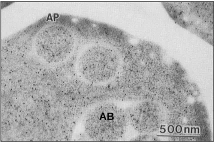
Figure 2. Immuno-staining image of alcohol dehydrogenase in the yeast cell.
Vacuolar proteinase deficient cell contains autophagic bodies in the vacuole and autophagosomes in cytosol (Baba et al., 1994). AP, autophagosome; AB, autophagic body; Bar = 500 nm.
AB
MATERIALS AND METHODS
Yeast Strain and Media
The yeast strains used in this study are listed in Table II. Standard techniques were used for yeast manipulation (Burke et al., 2000). Yeast cells are grown in YPD medium (1% yeast extract, 2% polypeptone and 2% glucose), SD medium (0.17% yeast nitrogen base withoutamino acids and ammonium sulfate, 0.5% ammonium sulfate and 2% glucose), SD + CA medium (0.17% yeast nitrogen base withoutamino acids and ammonium sulfate, 0.5% casamino acid, 0.5% ammonium sulfate and 2% glucose) supplementedwith 0.002% adenine sulfate, 0.002% uracil, and 0.002% tryptophanif necessary. For nitrogen starvation, SD(-N) medium (0.17% yeastnitrogen base without ammonium sulfate and amino acids, and 2%glucose), for carbon starvation, S(-C) medium (0.17% yeastnitrogen base without ammonium sulfate and amino acids, and 0.5%ammonium sulfate), for nitrogen and carbon starvation, S(-N, C) medium (0.17% yeast nitrogen base without ammonium sulfate and amino acids), and for sulfur starvation, SD(-S) medium (0.17% yeast nitrogen base without sulfur source, and 2% glucose) were used.
Plasmid Construction
DNA manipulations were performed using standard methods (Sambrook et al., 1989). To create the glutathione S-transferase (GST)-Ald6p fusion construct (pJO1), the ORF of ALD6/YPL061w lacking the initiation codon (1.5 kb) was amplified by genomic PCR using the following primers:
5'-CGCGGATCCACTAAGCTACACTTTGACACTGC-3', and 5'-CCGCTCGAGCAACTTAATTCTGACAGCTTTTACTTC-3'.
This strategy incorporated novel BamHI and XhoI sites into the resulting DNA fragment, which was then cloned into the BamHI and XhoI sites of pGEX-4T-1 (Amersham Bioscience) to yield pJO1.
To create the Ald6p-green fluorescent protein (GFP) genome integration vector (pJO402), novel XbaI sites were added to the terminator sequence (0.5 kb) of ALD6 by
genomic PCR amplification using the following primers: 5'-GCTCTAGATGTACCAACCTGCATTTCTTTC-3', and 5'-GCTCTAGACGAAGAAGGATGTTATTATATG-3'.
Novel XhoI and BamHI sites were added to the ALD6 promoter region (0.3 kb) and the ALD6 ORF lacking the stop codon (1.5 kb) by genomic PCR amplification using the following primers:
5'-CGCTCGAGCACCGACCATGTGGGCAAATTC-3', and 5'-CGCGGATCCCAACTTAATTCTGACAGCTTTTAC-3'.
BamHI sites were added to the ORF of modified GFP (S65T) lacking the initiation codon by PCR amplification using the following primers:
5'-CGCGGATCCGGTAAAGGAGAAGAACTTTTCACTGG-3', and 5'-CGGGATCCTTACTTGTATAGTTCATCCATG-3'.
The resulting DNA fragments were cloned into the pRS306 integration vector (Sikorski and Hieter, 1989) to yield pJO402.
To create the Ald6p overexpression construct (pJO203), XhoI and BamHI sites were added to a sequence containing the ALD6 ORF (1.5 kb), the promoter region (0.3 kb), and the terminator sequence (0.5 kb) by genomic PCR amplification using the following primers:
5'-CCGCTCGAGCACCGACCATGTGGGCAAATTC-3', and 5'-CGCGGATCCCGAAGAAGGATGTTATTATATGATCTC-3'.
The resulting DNA fragment was cloned into the BamHI and XhoI sites of the pRS426 multicopy plasmid (Sikorski and Hieter, 1989) to yield pJO203.
A QuikChangeTM Site-Directed Mutagenesis Kit (Stratagene) was used to create the Ald6pC306S mutant overexpression construct (pJO213). To generate pJO213, the pJO203 plasmid was amplified by PCR with the following primers:
5'-AGAACGCTGGTCAAATTTCTTCCTCTGGTT-3', and 5'-AACCAGAGGAAGAAATTTGACCAGCGTTCT-3'.
The site of mutagenesis in pJO213 was confirmed by automated DNA sequencing. To create the expression vectors of Arg1p- or Hsp26p- 3 × hemagglutinin (HA) tagging protein, novel XbaI sites were added to the terminator sequence of ALD6 (0.3
kb) by genomic PCR amplification using the following primers: 5'-GCTCTAGATGTACCAACCTGCATTTCTTTC-3', and 5'-GCTCTAGACGAAGAAGGATGTTATTATATG-3'.
And novel SpeI sites were added to the coding sequence of HA lacking the initiation codon (0.1 kb) by PCR amplification using the following primers:
5'-GACTAGTTACCCATACGATGTTCCTGAC-3', and 5'- GACTAGTTTAAGCGTAATCTGGAACGTC-3'.
Novel XhoI and BamHI sites were added to the each promoter region and the ORF lacking the stop codon (ARG1/YOL058w 2.2 kb, and HSP26/YBR072w 1.6 kb) by genomic PCR amplification using each following primers:
ARG1: 5'-CCGCTCGAGAGGTTGCCACATACATGGCCAAG-3', and 5'-CGGGATCCCAAAGTCAACTCTTCACCTTTG-3';
HSP26: 5'-CCGCTCGAGCGTTGGACTTTTTTTAATATAAC-3', and 5'-CGGGATCCGTTACCCCACGATTCTTGAGAAG-3'.
The resulting DNA fragments were cloned into the pRS316 centrometric plasmid Sikorski and Hieter, 1989) to yield pJO128 (Arg1pHA) and pJO129 (Hsp26pHA).
Two-dimensional PAGE
Yeast cell lysates were prepared by breaking cells with glass beads in lysis buffer (50 mM Tris-HCl, pH 8.0, 1 mM EDTA, 1 mM PMSF and protease inhibitor cocktail, Roche Applied Science). The lysates were centrifuged at 100,000 × g for 1 h, and the supernatant was desalted with NAP-10TM (Amersham Bioscience) gel filtration column. Protein concentration was determined by BCA assay kit (Pierce), and 300 µg of each protein was applied to the gel. Isoelectrofocusing was performed with IPGphor and ImmobilineTM DryStrip pH 4–7, 13 cm (Amersham Bioscience) as described (O'Farrell, 1975; Görg et al., 2000). Strips were re-hydrated together with the isoelectrofocusing buffer (8 M urea, 0.5% v/v IPG Buffer, Amersham Bioscience, and 65 mM DTT) contained sample proteins and isoelectrofocusing was performed for 10 h at 30 V, for 1 h at 200 V, for 1 h at 500 V, for 1 h at 1000 V and for 40000 Vh at 8000 V, at 50 µA/strip and 20˚C. Strips were equilibrated by SDS equilibration buffer (50 mM
Tris-HCl, pH 8.8, 6 M urea, 30% v/v glycerol and 2% SDS) with 70 mM DTT for 15 min and then with 140 mM iodoacetamide and 0.004% bromphenol blue for 15 min. Equilibrated gel was subjected to SDS-PAGE (12.5% acrylamide concentration) and stained with Coomassie brilliant blue R-250.
Peptide Mass Finger-printing
Protein spots on two-dimensional PAGE gel were picked, washed with 100 mM ammonium bicarbonate, dehydrated with acetonitrile, and dried in an evaporator. Spots were digested in the gel with 0.5 mg/ml of trypsin (Promega) with 100 mM ammonium bicarbonate for 12 h at 30˚C. Digested peptides were extracted from the gel with 10% formic acid and 50% acetonitrile, and desalted with the ZipTipTM C-18 (Millipore). The samples andα-cyano-4-hydroxy-cinnamic acid (Fluka) were mixedat a 1:1 ratio and analyzed by matrix associated laser deionization - time of flight (MALDI-TOF) mass spectrometry, REFLEX III (Bruker). Proteins were identified by searching the ProFounddatabase (http://129.85.19.192/).
Antibodies
Ald6p specific antibodies were prepared as follows. The pJO1plasmid was transformed into Escherichia coli (DH5α), and transformants were grown in LB medium containing 50 µg/mlampicillin to A600 = 0.6. Recombinant protein expression was inducedwith 1 mM isopropyl-β-D-thiogalactopyranoside for an additional6 h at 37°C. The recombinant protein was separated by SDS-polyacrylamide gel electrophoresis and simultaneously stained withGel code (Pierce). The protein band was excised from the gel and eluted with an electric current. The eluted protein-dye complex was used to immunize rabbits. Anti-Ald4p/6p antibodies were purchased from Rockland. Anti-PGK antibody was purchased from Molecular Probes. Anti-ADH antibodies have been described previously (Baba et al., 1994). Anti-API antibodies have been described previously (Hamasaki et al., 2003). Anti-HA epitope antibody (16B12) has purchased from BabCo.
Immuno-blot of Whole-cell Lysates
Immuno-blot was performed as previously described (Kirisako et al., 1999). Whole-cell lysates were prepared by disrupting cells with glass beads in lysis buffer (50 mM Tris-HCl, pH 8.0, 1 mM EDTA, 1 mM PMSF and the protease inhibitor cocktail, Roche Applied Science). Cell lysates were boiled with SDS-sample buffer for 5 min, and these proteins content was determined by BCA assay kit (Pierce). Total protein (10 µg) was subjected to SDS-PAGE (Laemmli, 1970), and transferred to polyvinylidene fluoride membrane (ImmobilonTM-P, Millipore) and detected with a combination of each antibody and peroxidase-conjugatedgoat anti-rabbit IgG antibody (Jackson) or peroxidase-conjugated goat anti-mouse IgG antibody (Jackson) by the ECL system (Amersham Bioscience).
Pulse-chase Experiments and Immuno-precipitation
Pulse-chase experiments were performed as previously described (Ishihara et al., 2001). Yeast cells were cultured in YPD medium to A600 = 1.0 at 30°C and were then washed twice and suspended in SD(-N) medium. Cells were pulse labeled for 30 min by adding 1 MBq of [35S]methionine (Perkin-Elmer) / A600 unit and chased by adding 0.004% methionine and 0.003% cysteine at 30°C. Each sample was collected at the incubate time
Immuno-precipitation was performed as follows. Cell lysates were prepared by breaking cells with the glass beads in TBS (50 mM Tris-HCl, pH 7.4 and 150 mM NaCl) and 1% SDS. Moreover, they were boiled 5 min, diluted by adding two fold volume TBS and 2% Triton X-100, and centrifuged at 15,000 × g for 1 min to remove insoluble materials. Cell lysate, 0.1% v/v anti-Ald6p or anti-ADH antibodies, 1.25% v/v protein A-SepharoseTM beads (Amersham Bioscience) and the protease inhibitor cocktail (Roche Applied Science) were incubated at 4˚C for 2 h. Sample were centrifuged for 1 min precipitated beads were washed once with IP buffer (TBS, 5 mM EDTA, 0.2% SDS and 1% and Triton X-100), once with urea buffer (IP buffer and 2 M Urea), high salt buffer (50 mM Tris-HCl, pH 7.4, 500 mM NaCl, 5 mM EDTA, 0.2% SDS and 1% Triton X-100), once with detergent free buffer (TBS and 5 mM EDTA).
Proteins were eluted in SDS-sample buffer with 5 min boil, and were analyzed by SDS-PAGE (Laemmli, 1970) and autoradiographed with BAS-2000 analyzer (Fuji Film).
Light Microscopy
Fluorescence microscopy was performed using a Delta Vision microscope (Applied Precision) as described (Suzuki et al., 2001). To observe GFP-tagged proteins under a fluorescence microscope, I employed a FITC filter.
Immuno-electron Microscopy
Yeast cells were subjected to rapid freezing and freeze-substitution fixation, and observed as previously reported (Baba et al., 1997). For immuno-electron microscopy, ultrathin sections were collected onto formvar-coated nickel grids and blocked in PBS containing 2% BSA at room temperature for 15 min. Incubations were carried out by floating grids on a 20 µl drop of a 1:1,000 dilution of anti-Ald6p antiserum, at room temperature for 1.5 h. After washing, the grids were incubated for 1 h with 5 or 10 nm gold-conjugated goat anti-mouse IgG (Bio Cell Lab.). The grids were washed several times in PBS followed by several drops of distilled water and fixed with 1% glutaraldehyde for 3 min. The sections were stained with 4% uranyl acetate for 7 min and examined.
Subcellular Fractionation
Subcellular fractionation was performed as previously described (Ishihara et al., 2001). Growing or nitrogen starved cells (50 A600 unit) were harvested, washed with 100 mM Tris-HCl, pH 9.0, 40 mM 2-mercaptethanol, and were converted to supheroplasts in 1 ml of SD + CA or SD(-N) supplemented with 1.4 M sorbitol and 20 mM Tris-HCl, pH 7.5. After the addition of 0.5 mg of Zymolyase-100T (Seikagaku Corporation), the cell suspensions were incubated at 30ºC for 30 min. The supheroplasts were harvested, washed with 1 M sorbitol, and re-suspended at 50 A600 unit/ml in a lysis buffer (1 M sorbitol, 0.5% FicollTM 400 and 1 mM MgCl2). The lysates were passed
through a polycarbonate filter with 3 µm pores (Nucleopore, Whatman). Cleared lysate (Total) was generated by two consecutive centrifugations at 500 × g for 5 min. The lysates were spun at 13,000 × g for 15 min to separate the pellet (P13), and the supernatant was centrifuged again at 100,000 × g for 1 h to generate a pellet (P100) and supernatant (S100). P13 and P100 were re-suspended in lysis buffer equal to the original volume.
Proteinase K Protection Assay
Proteinase K protection assay was performed as previously described (Ishihara et al., 2001). To examine proteinase K-sensitivity, each Subcellular fraction without protease inhibitors was treated with 2 mg/ml proteinase K (Roche) on ice for 30 min with or without 1% Triton X-100. The samples were precipitated with 10% trichloroacetic acid, washed once with cold acetone, re-suspendedin SDS-sample buffer, and analyzed by SDS-PAGE (Laemmli, 1970) and immuno-blot.
Determination of Cell Viability
Determination of yeast cell viability was performed as previously described (Tsukada and Ohsumi, 1994). Cell viability was measured by phloxine B (final concentration 2 µg/ml; Sigma) stain, and fluorescence microscopy with a blue filter. Brightly fluorescent cells were counted as dead cells.
Assay of Ald6p Enzymatic Activity
The activity of NADP+- and Mg2+-dependent acetaldehyde dehydrogenase (Ald6p) was determined by monitoring the NADPH production of NADP+ using fluorophotometer (Hitachi F-3010), as described (Dickinson, 1996) with some modifications. The assay mixture containing 50 mM Tris-HCl, pH 8.0, 1 mM acetaldehyde, 0.5 mM NADP+, 15 mM MgCl2, 1 mM pyrazole and a preparation of enzyme.
Northern Blot
Total RNA isolation was performed RNeasy Mini Kit (QIAGEN) according to the appended protocol. Total RNA (5 µg) separated on 1% agarose containing 2.2 M formaldehyde by electrophoresis, and transferred to BIODYNE A membrane (Pall). DIG-labeled probes (0.5 kb) for ARG1, HSP26 and ACT1/YFL039c mRNA were prepared from each ORF fragment using PCR DIG Probe Synthesis Kit (Roche Applied Science) according to the appended protocol, and PCR amplification using each following primers:
ARG1: 5'-TCTAAGGGAAAAGTTTGTTT-3', and 5'-TGGTTTGGGCGACGGGAATA-3';
HSP26: 5'-TCATTTAACAGTCCATTTTT-3', and 5'-TGTCTGCATCCACACCTGGG-3';
ACT1: 5'-CGGTAGAGATTTGACTGACT-3', and 5'-TTGTTGGAAGGTAGTCAAAG-3'.
The mRNA-transferred membrane was incubated with each probe at 50°C overnight, and signals were detected by Anti-Digoxigenin-AP (Roche Applied Science), CDP-Star (Roche Applied Science) and LAS-1000 system (Fuji Film).
Amino Acid Analysis of Whole Cell
Determinate quantity of free amino acids in whole cell was performed as previously described (Ohsumi et al., 1988; Kitamoto et al., 1988). Yeast cells of 10 A600
unit were harvested and washed twice with distilled water. The cells were suspended in 500 µl of distilled water and boiled for 15 min. The suspension was centrifuged for 3 min at 5,000 rpm, and the supernatant was collected as extraction of whole-cell amino acid. This extract was analyzed with an amino acid analyzer (Hitachi L-8500A).
Assay of in vivo Protein Synthesis
Yeast cells of 1 A600 unit were washed in SD(-N) medium twice, and were suspended in SD(-N) medium containing [14C]valine (Moravec MC277) at a final concentration of 74 kBq/ml and 10 µM. The cultures were labeled for 0, 2 and 4 min at
30°C and were then added 10 volumes cold distilled water or 11% w/v TCA solution. In TCA suspension, it kept at 90°C, 10 min for peptidyl-tRNA degradation, following it kept at 4°C, 30 min for protein precipitation. Both distilled water and 10% TCA suspensions were then filtered with 0.45 µm pore membranes (MFTM-membrane, Millipore). These membranes were dried up and were determined radioactivity in a liquid scintillation counter (Packard TRI-CARB 2700TR).
Table II. Yeast strains used in this study.
Strain Genotype Source
SEY6210 MATα his3∆200 leu2-3,112 lys2-801
trp1∆901 ura3-52 suc2∆9 GAL Robinson et al., 1988 KVY118 SEY6210; ∆atg7::HIS3 Kirisako et al., 2000 JOY67 SEY6210; ∆atg7::kanMX4 This study
JOY676 SEY6210; ∆atg7::HIS3 ∆ald6::kanMX4 This study JOY674 SEY6210; ∆atg7::HIS3 ∆ald4::kanMX4 This study JOY617 SEY6210; ∆atg17::HIS3 This study
TVY1 SEY6210; ∆pep4::LEU2 Gerhardt et al., 1998 JOY6p4 SEY6210; ∆pep4::kanMX4 This study JOY6005 SEY6210; ∆pep4::LEU2
∆ald6::ALD6-GFP This study
JOY6006 SEY6210; ∆pep4::LEU2 ∆atg7::HIS3
∆ald6::ALD6-GFP This study JOY66 SEY6210; ∆ald6::kanMX4 This study JOY64 SEY6210; ∆ald4::kanMX4 This study JOY69 SEY6210; ∆atg11::URA3 This study JOY622 SEY6210; ∆vid22::kanMX4 This study
KVY4 SEY6210; ∆ypt7::LEU2 Kihara et al., 2001 YAK1 SEY6210; ∆ypt7::HIS3 ∆atg1::LEU2 This study
X2180-1B MATα SUC2 mal mel gal2 CUP1 Y. G. S. C. JOY27 X2180-1B; ∆atg7::kanMX4 This study
MT13-3A MATα SUC2 mal mel gal2 CUP1 atg1-1 Tsukada and Ohsumi, 1994 MT2-4-1 MATa SUC2 mal mel gal2 CUP1 atg2-4-1 Tsukada and Ohsumi, 1994 WCG4a MATa leu2-3,112 ura3 his3-11,15 Heinemeyer et al, 1993 WCG4-11a MATa leu2-3,112 ura3 his3-11,15 pre1-1 Heinemeyer et al, 1993 BY4741 MATa his3∆1 leu2∆0 met15∆0 ura3∆0 Brachmann et al., 1998 Y00753 BY4741; ∆ald2::kanMX4 Brachmann et al., 1998
Y00752 BY4741; ∆ald3::kanMX4 Brachmann et al., 1998 Y01671 BY4741; ∆ald4::kanMX4 Brachmann et al., 1998 Y00213 BY4741; ∆ald5::kanMX4 Brachmann et al., 1998 Y02767 BY4741; ∆ald6::kanMX4 Brachmann et al., 1998 Y. G. S. C., Yeast Genetic Stock Center.
RESULTS
I. Studies on Degradation of Ald6p, a Preferential Substrate of Autophagy
Screen for Proteins Reduced under Nitrogen Starvation
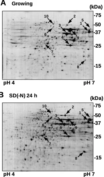
To investigate the possibility of selective autophagic degradation, I attempted to compare the amounts of each intracellular protein under nutrient growth and nitrogen starvation conditions in the yeast, S. cerevisiae. I investigated the expression profiles of soluble proteins using two-dimensional PAGE. Using this method, I expected to be able to identify cellular proteins whose levels decreased during nitrogen starvation. Yeast cells were grown at 30˚C in YPD medium, were harvested at middle logarithmic phase (A600 = 1.0), and were washed twice with starvation medium. The cells were then transferred to SD(-N) medium and incubated for 24 h. I chose a long-term stress period of 24 h in order to observe obvious differences in protein expression; importantly, most of the cells were still viable at this time point (Tsukada and Ohsumi, 1994).
In my two-dimensional PAGE experiments, the soluble fraction of cell lysate separated approximately 800–1,000 spots on a Coomassie brilliant blue R-250 stained the gel. Nitrogen starved wild-type cells (SEY6210) showed the protein spots more than nutrient growing cells (Figure 3). However, nitrogen starved autophagy defective
∆atg7/apg7 cells (KVY118) showed the same number of protein spots as growing cells (data not shown). Trabalzini et al. recently detected many protein fragments on the two-dimensional PAGE gel in the yeast cells of late stationary phase. These fragments did not appear in the present of 1 mM PMSF, inhibitor of vacuolar proteinase B (Trabalzini et al., 2003). Thus, the part of increased spots may come from fragmentation of abundant proteins.
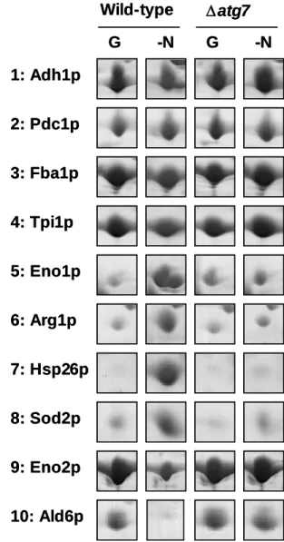
The intense protein spots by tryptic digestion and MALDI-TOF mass spectrometry analyses (see Materials and Methods; Figure 4) allowed the identification of several proteins. In both wild-type (SEY6210) and autophagy defective ∆atg7 (KVY118) yeast cells, most proteins showed little change after starvation (Figure 5
lanes 1–4).
In figure 5 lanes 5–8, several proteins showed increased levels after nitrogen starvation, including typical proteins of environment stress responses (Eno1p/Hsp48p, enolase I; Hsp26p, heat shock protein of 26-kDa), enzyme of amino acid biosynthesis (Arg1p, argininosuccinate synthetase) and quenching enzyme of reactive oxygen species (Sod2p, mitochondrial manganese superoxide dismutase). However, these specific proteins did not increase by nitrogen starvation in autophagy-defective cells (Figure 5 lanes 5–8), suggesting that starvation-induced up-regulation of these proteins requires the supply of amino acids produced by autophagy. I will describe particulars about this phenomenon in "Result – II. Amino Acids Supply from Autophagy Is Essential for Protein Synthesis".
In contrast, only few as proteins exhibited the apparent decrease during starvation in wild-type cells (Figure 5 lanes 9 and 10). Among them, cytosolic acetaldehyde dehydrogenase (Ald6p) showed the most distinctive difference between the wild-type and ∆atg7 mutants. Therefore, I focused on this protein for further analysis.
Ald6p, Cytosolic Acetaldehyde Dehydrogenase
Ald6p is Mg2+- and NADPH-dependent cytosolic acetaldehyde dehydrogenase, which catalyzes the conversion of acetaldehyde to acetate in the cytosol (EC 1.2.1.3; acetaldehyde + NADP+ → acetate + NADPH; Meaden et al., 1997). The S. cerevisiae genome encodes five or more different members ofthe aldehyde dehydrogenase family. Ald4p is the major K+- and NAD+-dependent mitochondrial acetaldehyde dehydrogenase (Tessier et al., 1998) and Ald5p is a minor K+-dependentmitochondrial acetaldehyde dehydrogenase, which is induced when cells are grown in ethanol containing medium (Kurita and Nishida, 1999). Ald2p and Ald3p are closely related cytosolic enzymes that are required for in vivo pantothenic acid biosynthesis via conversion of 3-aminopropanol to β-alanine (White et al., 2003). Ald4p, Ald5p and Ald6p functionin the conversion of acetaldehyde to acetate, which is a key intermediate during fermentation of sugars and growth on ethanol,and are consequently important
for acetyl-CoA production (Saint-Prix et al., 2004). In contrast, Ald2p and Ald3p may not contribute to the oxidation of acetaldehyde in vivo (Saint-Prix et al., 2004). Therefore, Ald6p is the only cytosolic acetaldehyde dehydrogenase in the yeast cell.
During fermentative growth in yeast, pyruvate is decarboxylated into acetaldehyde by pyruvate decarboxylase, which is, in its turn, reduced into ethanol in the cytosol by ADH (Murray et al., 2003; Figure 6). During respiratory metabolism in yeast, pyruvate can enter the mitochondria by a specific carrier and is decarboxylated and oxidized into acetyl-CoA by pyruvate dehydrogenase, a multi-enzyme complex located in the matrix (Murray et al., 2003). In addition, a pyruvate dehydrogenase bypass located in the cytosol converts pyruvate into acetyl-CoA by the action of the following enzymes: pyruvate decarboxylase, Ald6p, Ald4p and acetyl-CoA synthetases (Gounaris et al., 1971; van den Berg and Steensma, 1995; Dicinson, 1996; van den Berg et al., 1996; Meaden et al., 1997; Boubekeur et al., 1999; Figure 6). Acetyl-CoA synthesized in the cytosol is either directly used for the fatty acid biosynthetic pathway or enters the mitochondria via the carnitine acetyltransferase system (Kispal et al., 1991; Kispal et al., 1993; Murray et al., 2003; Figure 6). Acetyl-CoA can only move from the cytosol into mitochondria (Kispal et al., 1991). Ald6p contributes to the productions both cytosolic acetyl-CoA and NADPH in the yeast cell.
Proteins Required for the Reduction of Ald6p
I could purchase anti-yeast aldehyde dehydrogenase polyclonal antibodies as a commercial product (Rockland). However, these commercial antibodies cross-reacted both Ald4p and Ald6p (Figure 7A). I also prepared Ald6p specific antibodies (see Material and Methods). Prepared antiserum detected Ald6p nicely much more than Ald4p at immuno-blot (Figure 7B). In this study, I used two types antibodies both anti-Ald4p/6p and Anti-Ald6p as appropriate.
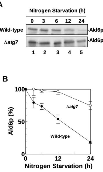
Using immuno-blot analyses, I attempted to determine which proteins are required for the reduction of Ald6p. In wild-type cells (SEY6210), the amount of Ald6p decreased in a near-linear manner, and was ultimately reduced to 18% of the original level after 24 h starvation (Figure 8). In contrast, Ald6p levels decreased only slightly in
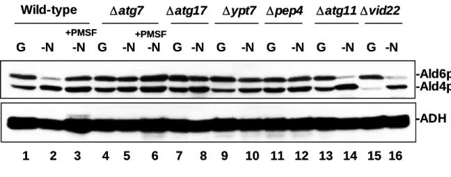
∆atg7 mutant cells (KVY118). I next investigated whether the amount of Ald6p was reduced in various yeast strains that are defective in various steps of autophagy. ∆atg7 (KVY118), ∆atg17/apg17 (JOY617) and all ∆atg/apg mutant cells tested showed a similar defect in the loss of Ald6p (parts shown in Figure 9 lanes 1, 2, 4, 5, 7 and 8). The decrease of Ald6p also required Ypt7p, a protein that is essential for the fusion of autophagosomes to vacuoles (Kirisako et al., 1999), and Pep4p, vacuolar proteinase A (Figure 9 lanes 1, 2 and 9–12). In the present of 1 mM PMSF, the decrease of Ald6p was inhibited under nitrogen starvation (Figure 9 lanes 1–6).
The selective transport of vacuolar enzymes (via the Cvt pathway), such as API and α-mannosidase, is known to utilize all of the Apg/Atg proteins except Atg17p (Kamada et al., 2000). Atg11p/Cvt9p and Atg19p/Cvt19p function only in the Cvt vesicle formation, and do not play a role in autophagosome formation (Kim et al., 2001; Scott et al., 2001). In ∆atg11 (JOY69) and ∆atg19 mutant cells, Ald6p was reduced in a similar manner to wild-type cells under nitrogen starvation (Figure 9 lanes 1, 2, 13 and 14; data for ∆atg19 not shown). As expected, another system of vacuolar transport, the Vid pathway (Hoffman and Chiang, 1996; Klionsky and Ohsumi, 1999; Brown et al., 2002) was not involved in this phenomenon (Figure 9 lanes 1, 2, 15, 16).
One mutant allele of the proteasome subunit PRE1 is pre1-1, which is frequently used for the following reasons: the pre1-1 mutation causes a defect in the degradation of short-lived proteins, ubiquitinated proteins (Heinemeyer et al., 1991; Heinemeyer et al., 1993) and N-end rule substrates (Richer-Ruoff et al., 1992; Seufert and Jentsch, 1992) at 30°C. In pre1-1 mutant cells (WCG4-11a), Ald6p was decreased similarly to wild-type cells (WCG4a) under nitrogen starvation, indicating that Ald6p is not a substrate for proteasome-mediated degradation (Figure 10). Taken together, these mutant studies indicate that the reduction of Ald6p requires all of the Atg/Apg proteins and the processes of vacuolar proteolysis. However, Atg/Cvt proteins, Vid proteins, and proteasomal degradation are not involved in this phenomenon.
Reduced Ald6p Levels Implied a Rapid Degradation
I hypothesized that the decrease in Ald6p levels was the result of rapid
degradation during nitrogen starvation. To examine this possibility, the kinetics of Ald6p degradation was measured by pulse-chase experiments. I sought an optimal ratio of yeast cells lysate per antibodies using non-radioactive immuno-precipitation and immuno-blot to accomplish quantitative pulse-chase experiments. Since Ald6p and ADH exist abundantly in yeast cells, "the ratio of 0.1% antibodies in 1 A600 unit cell lysate" did not saturated the binding capacity of antibodies (data not shown). The binding capacity of both anti-Ald6p and anti-ADH antibodies was saturated by "the ratio of 0.1% antibodies in 0.05 A600 unit cell lysate" (Figure 11A). These conditions were adopted for radioactive pulse-chase experiment.
Wild-type (SEY6210) and ∆atg7 (KVY118) cells were pulse-labeled for 30 min with [35S]methionine and chased with cold methionine and cysteine for 0, 3, 6 and 9 h. In wild-type cells, the Ald6p was rapidly degraded and was barely detectable after 6 h of chase (Figure 11B). In contrast, the degradation rate of Ald6p was clearly slower in
∆atg7 mutant cells. In addition, ADH, a known non-selective marker of autophagy (Baba et al., 1994), did not show rapid degradation like Ald6p (Figure 11B). The reduction of Ald6p levels implied a rapid degradation dependent on Atg7p during nitrogen starvation. These results suggest that Ald6p is transported to the vacuole and degraded much more rapidly than typical cytosolic proteins.
Ald6p Was Degraded in the Vacuole with Autophagic Body
The process of Ald6p vacuolar transport was detected by subcellular fractionation using ∆pep4 cells that accumulate autophagic bodies in the vacuole under nitrogen starvation. Cytosolic Ald6p was mostly recovered in the S100 fraction, and mitochondrial Ald4p was in the P13 fraction in the growing cell (Figure 12 lanes 1–4). In nitrogen-starved ∆pep4 cells (TVY1), API was fractionated as a precursor form in the P13 fraction, which is dependent on Atg7p (Figure 12 lanes 9–24). This suggests that a precursor form of API is in the autophagic bodies. In nitrogen-starved ∆pep4 cells, Ald6p behaved similarly (Figure 12 lanes 9–24), and was fractionated into the P13 fraction (Figure 12 lane 14). Ald6p was also expected to be in the autophagic bodies.
The process of Ald6p vacuolar transport was also visualized by expressing
physiological levels of an Ald6p-GFP fusion protein from the authentic ALD6 promoter. Upon starvation, the vacuoles gradually became fluorescent. In addition, in ∆pep4 cells (JOY6005), many bright dots, which were presumably autophagic bodies, were observed moving around in the vacuole (Figure 13). In ∆pep4 ∆atg7 double mutant cells (JOY6006), no fluorescence was observed in the vacuoles, but rather, the cytosol was evenly stained (Figure 13). Furthermore, I performed immuno-electron microscopy using anti-Ald6p and anti-ADH sera. In cells after 24 h starvation, gold particles for Ald6p were concentrated in autophagic bodies (Figure 14). Quantitative measurement revealed that the vacuole contained a 5.0 ± 0.6 (n = 8) fold greater signal than the cytosol. Because Ald6p was transported to the vacuole in autophagic bodies during nitrogen starvation, I hypothesized that transport of Ald6p from the cytosol to the vacuole occurred via the autophagosome.
Ald6p Was Preferentially Transported to the Vacuole via the Autophagosome
Our laboratory previously reported that ∆ypt7 cells accumulate autophagosomes in the cytosol under nitrogen starvation (Kirisako et al., 1999). Using precursor API as a selective cargo marker of autophagosomes, Ishihara et al. showed the low speed pellet (P13) fraction enriches the autophagosomes (Ishihara et al., 2001; Figure 15). So next, I studied the behavior of Ald6p in ∆ypt7 cells (KVY4). Under growing conditions precursor API was exclusively resided in the high speed supernatant (S100), but under nitrogen starvation conditions a significant portion was recovered in the P13 fraction as reported (Ishihara et al., 2001, Figure 16A). Similarly, Ald6p was recovered in the P13 fraction only under nitrogen starvation condition (Figure 16A). This fraction completely diminished in ∆ypt7 ∆atg1/apg1 mutant (YAK1, Figure 16B lanes 5–8), indicating that certain amount of Ald6p is in the autophagosomes. As shown in Figure 17, Ald6p and precursor API in P13 fraction were resistant to proteinase K treatment, but were digested in the presence of 1% Triton X-100. This also supported that Ald6p is sequestered into autophagosomes.
I also quantified the amount of Ald6p in the P13 fraction. Precursor API forms one or a few large complexes named the Cvt complex in the cytosol, and are taken up
by an autophagosome at once (Suzuki et al., 2002). PGK is shown to be distributed evenly in the autophagosome, autophagic bodies and cytosol (Baba et al., 1994). As shown in Figure 16C, Ald6p translocated to the P13 fraction much more efficiently than PGK (Recovery in P13 fraction; Ald6p = 38.2 ± 2.1% n = 5; PGK = 14.9 ± 1.5% n = 5, Figure 16C), but less than Precursor API (67.2 ± 5.9% n = 5). Taken together, I concluded that Ald6p is preferentially sequestered into autophagosome, possibly in a different manner with the substrates for the Cvt pathway.
Phenotype of ALD6 Disruptant Cells
All atg/apg mutants showed quite similar growth phenotypes; they grew normally just like wild-type cells. They failed to induce bulk protein degradation under various nutrient-depletion conditions. As expected homozygous diploid with any atg/apg cells could not perform sporulation (Tsukada and Ohsumi, 1993). This cell differentiation triggered by nitrogen depletion must require bulk protein degradation via autophagy for intracellular remodeling. Another characteristic feature of autophagy-defective mutants is loss of viability during nitrogen starvation. These mutants start to die after 2 days of starvation and almost completely lose viability after 5 days (Tsukada and Ohsumi, 1993). Under carbon starvation, they can maintain their viability even prolonged starvation. Unbalance of nitrogen and carbon sources may cause this phenotype. I interested whether Ald6p was also degraded under carbon starvation; however, the amount of Ald6p was not decreased under carbon or nitrogen/carbon starvation condition (Figure 18). From this result, I expected that the absence of Ald6p might be important for the survival under nitrogen starvation.
Using ∆ald6 mutant cells, I examined the physiological relevance of the preferential degradation of Ald6p during starvation. The growth of Atg+∆ald6 (JOY66) and ∆atg7 ∆ald6 (JOY676) cells were slower than wild-type (SEY6210) and ∆atg7 (KVY118) cells in YPD medium (Figure 19A; Meaden et al., 1996). ∆atg7 ∆ald6 mutant (JOY676) cells also started to die after 2 days of nitrogen starvation; but its viability decreased more slowly than that of ∆atg7 mutant cells (KVY118; Figure 19B). The viability of Atg+ ∆ald6 cells (JOY66) also improved slightly than that of wild-type
cells (Atg+ ALD6; SEY6210) under nitrogen starvation. However, disruption of a mitochondrial acetaldehyde dehydrogenase (Ald4p), Atg+ ∆ald4 (JOY64) and ∆atg7
∆ald4 (JOY674) cells had no effect on the viabilities of wild-type (SEY6210) and ∆atg7 (KVY118) cells, respectively (Figure 20B).
Furthermore, I also examined on the Ald6p overexpressing cells harboring multicopy plasmid. The overproducers expressed about three folds as much as wild-type in the enzymatic activity (Table III). Overexpressed Ald6p was not degraded fully by autophagy for 24 h nitrogen starvation (Figure 21). It is possible that the autophagic degradation for Ald6p may reach saturation, however, it is a suitable condition for the propose of the physiological relevance of Ald6p under nitrogen starvation. Wild-type cells (Atg+) expressing Ald6p via multicopy plasmid (JOY66 harboring on pJO203 multicopy plasmid) showed a defect in the maintenance of viability during nitrogen starvation (Figure 22). These results indicate that abundant Ald6p causes the decrease of viability, and absence of Ald6p improves viability under nitrogen starvation.
Ald6p Enzymatic Activity May Be Disadvantageous during Nitrogen Starvation
I next asked whether Ald6p enzymatic activity or the protein molecule itself is harmful to the cell under nitrogen starvation. To address it I constructed an inactive Ald6p mutant. Farres et al. isolated recombinant Aldh2C321S from Rattus norvegicus liver mitochondrial class-II aldehyde dehydrogenase (Aldh2). This highly conserved cysteine-321 is an active site residue whose thiol group binds to the aldehyde group of the substrate (Farres et al., 1995; Figure 25A). Ald6p cysteine-306, which corresponds to R. norvegicus Aldh2 cysteine-321, was changed to a serine residue by site-directed mutagenesis (Figure 25B). Ald6pC306S completely lost NADP+ and Mg2+-dependent acetaldehyde dehydrogenase activity (Table III), however, this mutant protein showed the stable expression (data not shown).
Overexpression of Ald6pC306S in Atg+ and ∆atg7 cells (JOY66 harboring pJO213 plasmid, and JOY676 harboring pJO213 plasmid) had no effect on viabilities of Atg+∆ald6 (JOY66) and ∆atg7 ∆ald6 (JOY676), respectively (Figure 24). These results indicate that the acetaldehyde dehydrogenase activity of cytosolic Ald6p may have a
disadvantageous effect on the survival of yeast cells during nitrogen starvation.
Table III. Ald6p activity of overexpression and C306S mutant.
Wild-type (SEY6210), ∆atg7 (KVY118), ∆ald6 (JOY66) and ∆atg7 ∆ald6 (JOY676) cells growing in YPD medium (A600 = 1.0) were used. Blank, harboring pRS426 multicopy plasmid; ALD6, harboring pRS426::ALD6 (pJO203) multicopy plasmid; ald6C306S, harboring pRS426::ald6C306S (pJO213) multicopy plasmid; Ald6p specific activity, NADP+ and Mg2+-dependent acetaldehyde dehydrogenase specific activity (µmol NADPH·min-1·mg protein-1).
Genotype Plasmid Ald6p specific activity Wild-type Blank 45.3 ± 1.9
∆atg7 Blank 43.1 ± 1.1
∆ald6 Blank Not detected
∆atg7 ∆ald6 Blank Not detected
∆ald6 ALD6 128.3 ± 2.9
∆atg7 ∆ald6 ALD6 128.1 ± 3.0
∆ald6 ald6C306S Not detected
∆ald6 ∆atg7 ald6C306S Not detected