This document is downloaded at: 2018-03-23T18:11:55Z
MS#AMN 07215
Impact of Gender on In-hospital Mortality in Patients with Acute Myocardial
Infarction in Nagasaki
Yuji Koide 1, 2, Seiji Koga 2, Takahiro Muroya 2, Hiroshi NaKashiMa 3, Masahiko ishizaKi 4, Naoto ashizawa 5,
Hideaki saKai 6, Takatoshi yoshitaKe 7, Shiro hata 8, Yoshihisa KizaKi 9, Toshihiko yaMasa10, Koji oKu11, Yoshito taNioKa12,
Kenji yaMaguchi13, Takuya izuMiKawa14, Jun FuKui15, Masami FuKahori16, Kota Negishi17, Kazuroh yoshida18,
Satoshi iKeda 1, 2, Hiroaki KawaNo 1, 2, Koji MaeMura 1, 2
1 Department of Cardiovascular Medicine, Nagasaki University Graduate School of Biomedical Sciences, Nagasaki, Japan 2 Cardiovascular Medicine, Nagasaki University Hospital, Nagasaki, Japan
3 Nagasaki Harbor Medical Center, Nagasaki, Japan 4 Kouseikai Hospital, Nagasaki, Japan
5 The Japanese Red Cross Nagasaki Genbaku Hospital, Nagasaki, Japan 6 Nagasaki Heart Clinic, Nagasaki, Japan
7 Nagasaki Memorial Hospital, Nagasaki, Japan 8 Sasebo City General Hospital, Nagasaki, Japan 9 Sasebo Chuo Hospital, Nagasaki, Japan 10 Nagasaki Rosai Hospital, Nagasaki, Japan
11 National Hospital Nagasaki Medical Center, Nagasaki, Japan 12 Omura City Municipal Hospital, Nagasaki, Japan
13 Isahaya General Hospital, Nagasaki, Japan 14 Izumikawa Hospital, Nagasaki, Japan 15 Hokusyo Central Hospital, Nagasaki, Japan 16 Nagasaki Goto Chuoh Hospital, Nagasaki, Japan
17 Nagasaki Prefecture Tsushima Izuhara Hospital, Nagasaki, Japan 18 National Hospital Nagasaki Kawatana Medical Center, Nagasaki, Japan
Acute myocardial infarction (AMI) is one of the leading causes of death in Japan. Immediate reperfusion therapy, including coronary intervention, improves patient prognosis. Despite this, females are said to be more prone to poor prognosis. A regional AMI registry in Nagasaki prefecture has been instituted recently that will evaluate whether female gender might predict short-term in-hospital death. Seventeen regional AMI centers enrolled all AMI patients from September 2014 through March 2016. A pro-pensity score (PS) was derived using logistic regression to model the probability of females as a total function of the potential confounding covariates. Two types of PS techniques were used: PS matching and PS stratiication. The consistency of in-hospi-tal death was determined between PS matched patients of both genders. Based on PS, patients were ranked and stratiied into ive groups for the PS stratiication. Out of 996 patients, 67 (6.7%) died during hospitalization: 31 (10.4%) out of 298 females and 36 (5.2%) out of 698 males (p < 0.0025). The proportion of cardiac and non-cardiac related death was almost same between genders (25 and 6 in female, 29 and 7 in male, respectively). Among 196 PS matched patients, there was a consistency between genders regarding in-hospital deaths (McNemar test, p = 0.6698). The 717 propensity scored patients had no signiicant
differ-ences between genders among propensity quintiles (Cochran-Mantel-Heanszel test, p = 0.7117). We found that gender alone is
not an indicator of short-term in-hospital death in acute myocardial infarction patients.
ACTA MEDICA NAGASAKIENSIA 61: 87−95, 2017
Key words: acute myocardial infarction, in-hospital death, gender difference
Address correspondence: Yuji Koide, 1-7-1 Sakamoto, Nagasaki 852-8501 Japan Tel: +81-(0)95-819-7288, Fax: +81-(0)95-819-7290, E-mail: [email protected]
88 Yuji Koide et al.: Gender gap of mortality in acute myocardial infarction
Introduction
Cardiovascular disease (CVD) is the primary global cause
of death in both genders, accounting for 17.3 million deaths
per year, a number that is expected to increase by 2030 1.
Therefore, the prevention of atherosclerotic CVD is a pressing issue both worldwide and nationwide. The prognosis of acute myocardial infarction (AMI) is markedly improved by early reperfusion therapy to the occluded culprit coronary artery with bare metal or drug-eluting coronary stents, though AMI is still a major life-threatening disease.
CVD or AMI is associated more closely to male gender
both in medical literature and in popular opinion 2. It may
cause some shock to hear that those ideas are not actually
supported by epidemiological data 2. Female gender is thought
to be at a higher risk of mortality following an AMI 3, 4, but
negative findings have been reported elsewhere 5, 6. Some
scholarly articles pointed out the age difference between the
two genders 7. As noted in the Framingham Heart Study,
females are approximately ten years older than males at the
time of a first coronary event 8. It remains uncertain if gender
makes a difference in mortality or if this observed difference is only attributable to different baseline characteristics or
other factors between females and males suffering from AMI 5.
The prevalence and incidence of CVD at all ages tends to
be higher in males than in females 9. But because the female
to male ratio of the elderly is higher, the absolute number of females discharged for CVD is greater than the number of
males beyond 75 years of age in the U.S.A 9. Therefore, those
large numbers of elderly female patients might mislead clinical impressions. We must be aware of the main differences between genders regarding CVD epidemiology
and clinical presentation 2. To further research into this topic,
the newly instituted regional AMI registry in Nagasaki
prefecture aims to find any trends in short-term in-hospital
death in AMI cases.
Materials and Methods
Study protocol approval and patient registry
This study protocol was reviewed by the Clinical Research
Center of Nagasaki University Hospital and approved by the Institutional Review Board of Nagasaki University Hospital. Seventeen regional AMI centers were selected to enroll all AMI patients from September 2014 through March 2016. All were regional cardiovascular centers that accepted all AMI patients.
Diagnosis of acute myocardial infarction
The standards of AMI diagnosis are based on the Third
Universal Definition of Myocardial Infarction10. The AMI
diagnosis is usually based on clinical features, including
electrocardiographic findings, elevated values of biochemical
markers (biomarkers) of myocardial necrosis, and imaging,
or may be confirmed by pathology10. There should be evidence
of myocardial necrosis, which is marked by a significant cardiac
biomarker rise and/or fall with at least one of the following:
symptoms of ischemia; new or presumed significant ST-segment-T
wave (ST-T) changes or new left bundle branch blocks; development of pathological Q waves in the electrocardiogram; imaging evidence of new loss of viable myocardium or new
regional wall motion abnormality; or identification of an
intracoronary thrombus by angiography or autopsy10.
Clinical data
Information relevant to the study collected by the AMI
centers included the following: basic clinical backgrounds and characteristics of AMI patients, medical history, cardiovascular risk factors, date and time of AMI, any in-hospital complications, and short-time clinical prognoses. All data was entered into a formatted spreadsheet at each
regional AMI center either by a physician or a physicianʼs
assistant. All collected data was sent to and received by Nagasaki University Hospital.
End points
The primary statistic required was whether or not the AMI
patient died in the hospital during index admission. Cardiac and non-cardiac deaths (determined by authorities at each AMI center) were also reported.
Statistical analysis
Continuous variables were presented as mean values with
standard deviations and compared with unpaired t-tests. Categorical data were presented in contingency tables, and
Pearsonʼs chi-square test or Fisherʼs exact test for small
categorical numbers was used for analysis. Multiple logistic regression analysis might be useful to reveal a predictor of in-hospital death, but multiple potential confounding factors and relatively small numbers of in-hospital deaths limited the number of confounding factors for multivariate analysis. Instead, propensity score (PS) was expected to reveal the
A PS was derived using logistic regression to model the probability of being a female as a total function of the potential confounding seven covariates, i.e., age, body mass index, cigarette smoking behavior, presence of dyslipidemia,
artificial hemodialysis, status of employment, and presence
of primary care physician. These covariates were selected
because of the significant differences between in-hospital death
and hospital discharge among baseline characteristics. The
area under the fitting curve for PS (i.e., c statistics) was 0.77,
indicating a relatively strong ability to differentiate between two genders. The value of PS ranged from 0.0119 to 0.6769 and represented the probability that a patient was female.
Two types of PS techniques were used to evaluate gender
differences: PS matching and PS stratification. PS matching
was performed with a 1:1 nearest-neighbor matching method of an add-in macro. If PS was within 0.212 of each other, then that was considered a match. Patients were excluded if they did not match the PS. Finally, we were able to match
196 females and 196 males. Between these PS matched patients, the consistency of in-hospital death was determined by the McNemar test. As the PS matching method would decrease the patient numbers because of its nature, PS
stratification was also performed to 717 propensity scored
patients. They were ranked and stratified into five groups
based on their PS. The Cochran-Mantel-Heanszel test was the chosen method for this type of analysis.
All analyses were performed using JMP Pro 13 software
(SAS Institute Japan Ltd., Tokyo, Japan). The significance
level was set at p < 0.05.
Results
Patient backgrounds and characteristics (Table 1)
A total of 996 AMI events were logged from September 1,
2014, through March 31, 2016, by 17 facilities in the Nagasaki
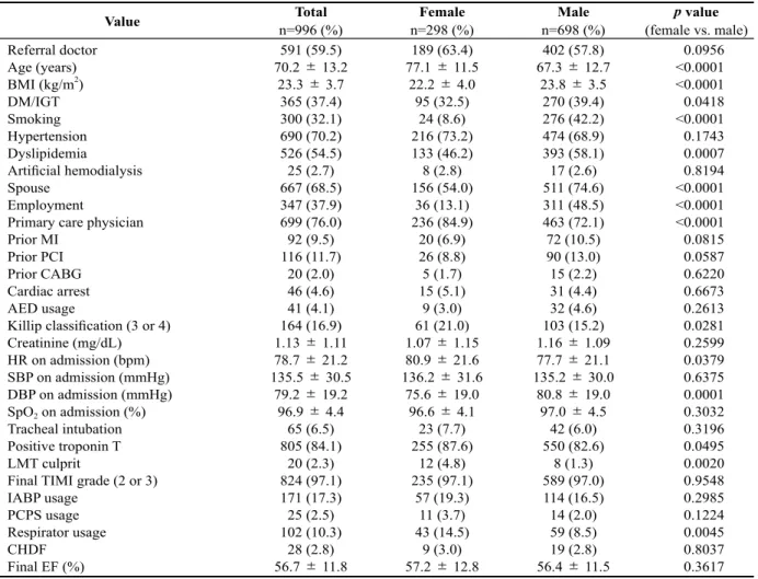
Table 1. Baseline backgrounds and characteristics of all registered patients with comparison between genders
Value Total Female Male p value
n=996 (%) n=298 (%) n=698 (%) (female vs. male) Referral doctor
Age (years) BMI (kg/m2
) DM/IGT Smoking Hypertension Dyslipidemia Artificial hemodialysis Spouse
Employment
Primary care physician Prior MI
Prior PCI Prior CABG Cardiac arrest AED usage
Killip classification (3 or 4) Creatinine (mg/dL) HR on admission (bpm) SBP on admission (mmHg) DBP on admission (mmHg) SpO2 on admission (%)
Tracheal intubation Positive troponin T LMT culprit
Final TIMI grade (2 or 3) IABP usage
PCPS usage Respirator usage CHDF
Final EF (%)
591 (59.5) 70.2 ± 13.2
23.3 ± 3.7 365 (37.4) 300 (32.1) 690 (70.2) 526 (54.5) 25 (2.7) 667 (68.5) 347 (37.9) 699 (76.0) 92 (9.5) 116 (11.7) 20 (2.0) 46 (4.6) 41 (4.1) 164 (16.9) 1.13 ± 1.11 78.7± 21.2 135.5 ± 30.5
79.2 ± 19.2 96.9 ± 4.4
65 (6.5) 805 (84.1) 20 (2.3) 824 (97.1) 171 (17.3) 25 (2.5) 102 (10.3) 28 (2.8) 56.7 ± 11.8
189 (63.4) 77.1 ± 11.5
22.2 ± 4.0 95 (32.5) 24 (8.6) 216 (73.2) 133 (46.2) 8 (2.8) 156 (54.0) 36 (13.1) 236 (84.9) 20 (6.9) 26 (8.8) 5 (1.7) 15 (5.1) 9 (3.0) 61 (21.0) 1.07 ± 1.15 80.9 ± 21.6 136.2 ± 31.6
75.6 ± 19.0 96.6 ± 4.1
23 (7.7) 255 (87.6) 12 (4.8) 235 (97.1) 57 (19.3) 11 (3.7) 43 (14.5) 9 (3.0) 57.2 ± 12.8
402 (57.8) 67.3 ± 12.7
23.8 ± 3.5 270 (39.4) 276 (42.2) 474 (68.9) 393 (58.1) 17 (2.6) 511 (74.6) 311 (48.5) 463 (72.1) 72 (10.5) 90 (13.0) 15 (2.2) 31 (4.4) 32 (4.6) 103 (15.2) 1.16 ± 1.09 77.7 ± 21.1 135.2 ± 30.0
80.8 ± 19.0 97.0 ± 4.5
42 (6.0) 550 (82.6) 8 (1.3) 589 (97.0) 114 (16.5) 14 (2.0) 59 (8.5) 19 (2.8) 56.4 ± 11.5
0.0956 <0.0001 <0.0001 0.0418 <0.0001 0.1743 0.0007 0.8194 <0.0001 <0.0001 <0.0001 0.0815 0.0587 0.6220 0.6673 0.2613 0.0281 0.2599 0.0379 0.6375 0.0001 0.3032 0.3196 0.0495 0.0020 0.9548 0.2985 0.1224 0.0045 0.8037 0.3617
90 Yuji Koide et al.: Gender gap of mortality in acute myocardial infarction
prefecture. Basic clinical characteristics and patient backgrounds were listed and then compared to gender (Table 1). Females were significantly older (p < 0.0001) and thinner (p < 0.0001) than males. Atherosclerotic risk factors, such as glucose
metabolism abnormality (p = 0.0418), dyslipidemia (p =
0.0007), and cigarette smoking behavior (p < 0.0001), were
significantly higher in males than females. Possession of
hypertension was the same (p = 0.1743) between genders.
Social status, such as marriage and employment, also had a
significant difference between genders. Killip classification
for acute heart failure from AMI was significantly higher
(p = 0.0281) in females. In females, heart rate at admission
was significantly higher (p = 0.0379) than that of males, and
diastolic blood pressure at admission was significantly lower
(p = 0.0001). Females showed significantly higher instances
of left main trunk culprit lesions (p = 0.0020) and need for
respirators (p = 0.0045).
In-hospital death (Table 2 and Table 3)
Out of 996 patients of both genders, 67 patients (6.7%)
died during index hospitalization: 31 (10.4%) of 298 females
Table 3. Comparison of parameters which might relate to in-hospital death in all registered patients
Value In-hospital death Hospital discharge p value
n = 67 (%) n = 929 (%) Referral doctor
Age (years) BMI (kg/m2
) DM/IGT Smoking Hypertension Dyslipidemia Artificial hemodialysis Spouse
Employment
Primary care physician Prior MI
Prior PCI Prior CABG Cardiac arrest AED usage
Killip classification (3 or 4) Creatinine (mg/dL) HR on admission (bpm) SBP on admission (mmHg) DBP on admission (mmHg) SpO2 on admission (%)
Tracheal intubation Positive troponin T LMT culprit
Final TIMI grade (2 or 3) IABP usage
PCPS usage Respirator usage CHDF
Final EF (%)
39 (59.1) 78.1 ± 12.5
22.1 ± 4.2 29 (44.6) 7 (13.0) 46 (76.8) 14 (26.4) 5 (8.8) 39 (62.9) 12 (19.4) 54 (90.0) 4 (6.7) 6 (9.5) 1 (1.6) 16 (24.2) 12 (17.9) 33 (54.1) 1.76 ± 1.92 88.6 ± 30.9 117.4 ± 31.8
67.5 ± 19.0 93.2 ± 8.1
24 (35.8) 60 (95.2) 4 (8.2) 37 (88.1) 25 (40.3) 13 (21.0) 30 (48.4) 11 (17.7) 44.9 ± 13.7
552 (59.4) 69.6 ± 13.0
23.4 ± 3.7 336 (36.8) 293 (33.3) 644 (69.8) 512 (56.1) 20 (2.3) 628 (68.9) 335 (39.2) 645 (75.0) 88 (9.7) 110 (11.9) 19 (2.1) 30 (3.2) 29 (3.1) 131 (14.4) 1.09 ± 1.00 78.0 ± 20.4 136.6 ± 30.1
79.9 ± 19.0 97.1 ± 4.0
41 (4.4) 745 (83.3) 16 (2.0) 784 (97.5) 146 (15.8) 12 (1.3) 72 (7.8) 17 (1.8) 57.1 ± 11.5
0.9500 <0.0001 0.0434 0.2107 0.0019 0.2579 <0.0001 0.0030 0.3286 0.0018 0.0086 0.4446 0.5778 0.7879 <0.0001 <0.0001 <0.0001 0.0057 0.0119 <0.0001 <0.0001 0.0009 <0.0001 0.0125 0.0049 0.0004 <0.0001 <0.0001 <0.0001 <0.0001 <0.0001
BMI, body mass index; DM, diabetes mellitus; IGT, impaired glucose tolerance; MI, myocardial infarction; PCI, percutaneous coronary intervention; CABG, coronary artery bypass graft; AED, automated external defibrillator;
HR, heart rate; SBP, systolic blood pressure; DBP, diastolic blood pressure; SpO2, saturation of peripheral oxyge n; LMT, left main trunk; TIMI, thrombolysis in myocardial infarction; IABP, intra-aortic balloon pumping; PCPS, percutaneous cardio pulmonary support; CHDF, continuous hemodiafiltration; EF, ejection fraction.
Table 2. In-hospital death and its detail of all registered patients with comparison between genders
Value Total Female Male p value
n=996 (%) n=298 (%) n=698 (%) (female vs. male) In-hospital death n (%) 67 (6.7) 31 (10.4) 36 (5.2) 0.0025 Cardiac death (%)
Non-cardiac death (%)
54 (5.4) 13 (1.3)
25 (8.4) 6 (2.0)
29 (4.2)
and 36 (5.2%) of 698 males died. Females had a significantly
higher (p = 0.0025) mortality rate than males before adjustment
(Table 2). Cardiac and non-cardiac deaths numbered 25 (8.4%) and 6 (2.0%) in females, respectively; and 29 (4.2%) and 7 (1.0%) in males, respectively. The proportion of cardiac to non-cardiac death was almost the same between genders
(p = 0.9926).
Univariate analyses of patient backgrounds and characteristics
regarding in-hospital death are listed in Table 3. Many factors were significant for in-hospital death, including age (p <
0.0001) and body mass index (p = 0.0434). Factors such as
cardiac arrest, automated external defibrillator (AED) usage,
and higher Killip classifications were more common in
in-hospital death patients than the patients who were discharged. Low blood pressure, high heart rate, high creatinine levels,
and low saturation of peripheral oxygen (SpO2) concentration
taken at admission were reported significant in in-hospital
death patients. Other parameters, such as mechanical circulation or respiratory support, culprit lesions in the left main trunk, low ejection fraction, and unsuccessful reperfusion were also dominant in in-hospital death.
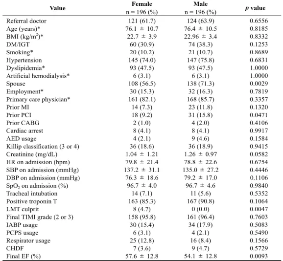
Backgrounds and characteristics of matched patients (Table 4)
Table 4 lists and compares the backgrounds and characteristics
of 196 matched patients of each gender. As mentioned above,
seven factors were used to derive PS (marked with*). PS
matching worked relatively well to uniform other factors. As a result, spouse, prior percutaneous coronary intervention
(PCI), and left main trunk (LMT) culprit remained significant,
and final ejection fraction (EF) became significant.
Table 4. Baseline backgrounds and characteristics of propensity score matched patients
Value Female Male p value
n = 196 (%) n = 196 (%) Referral doctor
Age (years)* BMI (kg/m2)* DM/IGT Smoking* Hypertension Dyslipidemia* Artificial hemodialysis* Spouse
Employment*
Primary care physician* Prior MI
Prior PCI Prior CABG Cardiac arrest AED usage
Killip classification (3 or 4) Creatinine (mg/dL) HR on admission (bpm) SBP on admission (mmHg) DBP on admission (mmHg) SpO2 on admission (%)
Tracheal intubation Positive troponin T LMT culprit
Final TIMI grade (2 or 3) IABP usage
PCPS usage Respirator usage CHDF
Final EF (%)
121 (61.7) 76.1 ± 10.7
22.7 ± 3.9 60 (30.9) 20 (10.2) 145 (74.0) 93 (47.5) 6 (3.1) 108 (56.5) 30 (15.3) 161 (82.1) 14 (7.3) 18 (9.2) 2 (1.0) 8 (4.1) 4 (2.1) 36 (18.6) 1.04 ± 1.21 79.8 ± 21.4 137.2 ± 31.1
76.3 ± 18.6 96.7 ± 4.0
14 (7.1) 163 (85.3) 8 (4.7) 158 (95.8) 30 (15.4) 6 (3.1) 25 (12.8) 7 (3.6) 57.6 ± 12.8
124 (63.9) 76.4± 10.5 22.96± 3.4 74 (38.3) 21 (10.7) 147 (75.8) 93 (47.5) 6 (3.1) 138 (71.3) 32 (16.3) 168 (85.7) 23 (11.8) 31 (15.8) 4 (2.0) 8 (4.1) 9 (4.6) 36 (18.9) 1.26 ± 0.97 78.8 ± 22.6 135.0 ± 27.2
79.2 ± 17.0 96.7 ± 4.6
11 (5.6) 167 (90.8) 0 (0.0) 161 (96.4) 34 (17.9) 4 (2.1) 16 (8.4) 9 (4.7) 54.1± 12.8
0.6556 0.8185 0.8332 0.1253 0.8689 0.6831 1.0000 1.0000 0.0029 0.7819 0.3357 0.1320 0.0471 0.4106 0.9917 0.1584 0.9415 0.0582 0.6754 0.4446 0.1106 0.9840 0.5352 0.1064 0.0047 0.7603 0.5083 0.5490 0.1566 0.5729 0.0093
BMI, body mass index; DM, diabetes mellitus; IGT, impaired glucose tolerance; MI, myocardial infarction; PCI, percutaneous coronary intervention; CABG, coronary artery bypass graft; AED, automated external defibrillator;
HR, heart rate; SBP, systolic blood pressure; DBP, diastolic blood pressure; SpO2, saturation of peripheral oxyge n; LMT, left main trunk; TIMI, thrombolysis in myocardial infarction; IABP, intra-aortic balloon pumping; PCPS, percutaneous cardio pulmonary support; CHDF, continuous hemodiafiltration; EF, ejection fraction.
92 Yuji Koide et al.: Gender gap of mortality in acute myocardial infarction
In-hospital death of matched patients (Table 5, Table 6, and Figure 1)
Age, body mass index (BMI), and presence of dyslipidemia
became significantly different between in-hospital deaths
and survivals in the matched 196 pairs (Table 5). Also, many parameters indicating respiratory, circulatory, or renal damage,
clearly and significantly demonstrated in bad health conditions
in in-hospital death. Many values were significantly worse
in in-hospital deaths compared to survivals.
In this study, PS matched pairs were assumed to have
similar backgrounds, excluding gender; consequently, if
gender had no influence, the short-term prognosis should
have consistency in each matched pair. Out of the 196 matched pairs, 26 patients (14 females and 12 males) died during hospitalization (Table 6), showing that there was
significant consistency between genders regarding
in-hospital deaths (McNemar test, p = 0.6698). There was no
statistical difference in in-hospital deaths between genders after propensity scores were matched (14 deaths out of 196
females versus 12 deaths out of 196 males, p = 0.6848).
The total number of scored patients was 717 (209 females
and 508 males). The numbers of scored patients in quintile (Q1, Q2, Q3, Q4, and Q5) numbered 7, 14, 46, 61, and 81 in females, respectively; and 136, 130, 97, 83, 62 in males, respectively; and, therefore, 143, 144, 143, 144, and 143 in subtotal, respectively. The Cochran-Mantel-Heanszel test was the best method for this type of analysis. The 717 propensity
scored patients had no significant differences between genders
among propensity quintiles (Figure 1,
Cochran-Mantel-Heanszel test, p = 0.7117).
Table 5. Comparison of parameters which might relate to in-hospital death in propensity score matched groups
Value In-hospital death Hospital discharge p value
n = 26 (%) n = 366 (%) Referral doctor
Age (years) BMI (kg/m2
) DM/IGT Smoking Hypertension Dyslipidemia Artificial hemodialysis Spouse
Employment
Primary care physician Prior MI
Prior PCI Prior CABG Cardiac arrest AED usage
Killip classification (3 or 4) Creatinine (mg/dL) HR on admission (bpm) SBP on admission (mmHg) DBP on admission (mmHg) SpO2 on admission (%)
Tracheal intubation Positive troponin T LMT culprit
Final TIMI grade (2 or 3) IABP usage
PCPS usage Respirator usage CHDF
Final EF (%)
16 (64.0) 81.5 ± 9.1 20.7 ± 3.4 7 (26.9) 3 (11.5) 20 (80.0) 5 8 (19.2) 1 (3.9) 16 (61.5) 2 (7.7) 24 (92.3) 2 (8.0) 5 (19.2) 0 (0.0) 3 (12.0) 1 (3.9) 16 (61.5) 1.90 ± 2.3 88.8 ± 27.8 114.9 ± 30.2
67.4 ± 17.1 93.1 ± 6.0
6 8 (23.1) 52 (95.7) 1 8 (5.6) 16 (88.9) 6 (27.3) 4 (18.2) 7 (31.8) 6 (27.3) 43.7 ± 15.0
229 (62.7) 75.9 ± 10.6
22.7 ± 3.7 127 (35.2) 38 (10.4) 272 (74.5) 181 (49.5) 11 (3.0) 230 (64.1) 60 (16.4) 305 (83.3) 35 (9.7) 44 (12.0) 6 (1.6) 13 (3.6) 12 (3.3) 56 (15.6) 1.10 ± 0.9 78.7 ± 21.5 137.5 ± 28.7
78.4 ± 17.7 96.9 ± 4.1
19 (5.2) 308 (87.5) 7 (2.2) 303 (96.5) 58 (16.0) 6 (1.7) 34 (9.3) 10 (2.8) 56.3 ± 12.6
0.8996 0.0050 0.0066 0.3927 0.8524 0.5412 0.0029 0.8100 0.7955 0.2400 0.2286 0.7838 0.2828 0.5106 0.0391 0.8801 <0.0001 0.0003 0.0284 0.0003 0.0048 <0.0001 0.0003 0.2438 0.3638 0.1056 0.1670 <0.0001 0.0009 <0.0001 0.0058
BMI, body mass index; DM, diabetes mellitus; IGT, impaired glucose tolerance; MI, myocardial infarction; PCI, percutaneous coronary intervention; CABG, coronary artery bypass graft; AED, automated external defibrillator;
Discussion
This study proved the hypothesis that gender is not a risk
factor of in-hospital death from AMI. Although females have a less positive CVD prognosis even after some covariates have been adjusted in some literatures 3, 4, we can also find
the opposite is true in other literatures 5, 6, and our study
supports the latter.
In the present study, AMI patients were enrolled from 17
regional AMI centers throughout the Nagasaki prefecture. All were regional cardiovascular centers that accepted all AMI patients. Biases related to omission of certain categories of patients were therefore unlikely. Thus, despite a relatively small number of enrollment, extrapolation of the results may
be more reliable than other previous reports.
Atherosclerotic cardiovascular disease is the dominant
cause of death in western and modern countries and its death
toll is expected to increase 1. Currently no difference is seen
in treatment guidelines for female patients for the vast
majority of atherosclerotic risk factors 2. In the United States
since the 1980s, the annual CVD mortality rate has remained higher in females than in males, as has the prevalence of
CVD11. In Brazil, from 1980 to 2012, there was a significant
reduction in cardiovascular mortality among males, particularly for CVD mortality, but no improvement was seen among
females12. Age of the patient could explain the difference 3, 7,
We need to look for the reason for the increase of atherosclerotic
disease and prepare for the fight against it. If female gender
Table 6. Distribution of in-hospital death and hospital discharge between propensity score matched females and males
Female
In-hospital death Hospital discharge Total
Male
In-hospital death Hospital discharge
2 12
10 172
12 184
Total 14 182 196
McNemar test for consistency demonstrates p = 0.6698; marginal frequencies are not different.
3 Propensity Quintiles
F M F M F M F M F M
Q1 Q2 Q3 Q4 Q5
P
ro
p
en
si
ty
sc
o
re
0.0 0.1 0.2 0.3 0.4 0.5 0.6 0.7
Figure 1. Each gender is divided into five sub classifications according to propensity scores. Cochran-Mantel-Heanszel analysis shows no significance (p = 0.6795).
94 Yuji Koide et al.: Gender gap of mortality in acute myocardial infarction
itself is a risk factor, we may not yet have useful tools to improve prognoses. Fortunately, our results proved that gender is not a risk factor of in-hospital death from AMI.
The patients who suffered AMI and whose information
was recorded in this registry had heterogynous backgrounds and characteristics. Simple univariate analysis makes gender look like a risk factor of in-hospital death. Two techniques,
PS matching and PS stratification, were used in this study. By
using the PS matching method, backgrounds of both genders became relatively homogenous as shown in Table 4. It is
interesting to note that when we adjust patientsʼ backgrounds
with some covariates, significant other differences disappeared
between genders. PS stratification is another technique to
ameliorate confounding effects of the variables included in
the PS. When stratification is used, Rosenbaum and Rubin
recommend using quintiles13. This is based on results from
Cochranʼs evaluations of various sub classification strategies14.
Based on other literature 3, 7, an important value could be
a patientʼs age. Because an accumulation of atherosclerotic
CVD risk factors has already started in younger generations, and because the female sex hormone protects against atherosclerosis progression, CVD such as AMI is dominant in males during relatively younger generations, whereas females start to deteriorate after menopause. Furthermore, recent progression of longevity of both genders, especially in females, has changed the proportion of female AMI. As people age into their eighties or nineties, females survive to become a large number of the patients. The dominance in absolute number of females in an older generation resulted in the increase of female AMI, even though the incidence of
AMI in females is still less than in males when we compare
the data in the same generation 9. We might reach a wrong
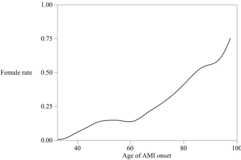
conclusion if we compare two genders without adjusting covariant, especially age. Absolute female proportion in AMI onset of this study is shown in Figure 2. We realize that females are dominant in AMI after age 80.
On the other hand, a female suffering an AMI often receives
less intensive medical therapy and is also less likely to undergo
cardiac catheterization or to receive timely reperfusion 2. Not
surprisingly, that can translate into unfavorable prognoses
for females compared to males 2. In this study, we did not
observe any differences between genders regarding their AMI timeline, such as onset to hospital door or hospital door to reperfusion. Also, treatment strategies, such as rate of emergent cardiac catheterization, cardiac or circulation support system usage, or stent usage, are not different between genders (data not shown). This may affect our result showing that females are not at an increased risk of in-hospital death in this study.
There are several limitations in this study. First, this is an
observational registration study; therefore, we cannot avoid confounding factors. To eliminate as many as possible, we need many registrations, but this study was relatively small. This time, we used PS to eliminate the defect of observational registration. Meta-analysis with large numbers would be one remedy. Second, we did not collect medical treatment data, such as administration of aspirin, anti-platelet drugs other than aspirin, angiotensin-converting enzyme (ACE) inhibitors, angiotensin receptor blockers, or ß-adrenergic blockers. Although evidence-based medical treatment after AMI is
3
0.00 0.25 0.50 0.75 1.00
Female rate
40 60 80 100
Age of AMI onset
References
1. Smith SC Jr, Collins A, Ferrari R, et al. Our time: a call to save prevent-able death from cardiovascular disease (heart disease and stroke). J Am Coll Cardiol 60: 2343-2348, 2012
2. Godoy LC, Abud ICK, Serrano CV Jr, Cardiovascular disease in wom-en: are we aware? Rev Assoc Med Bras 63: 195-196, 2017
3. Greenland P, Reicher-Reiss H, Goldbourt U, Behar S, and the Israeli SPRINT Investigators, In-hospital and 1-year mortality in 1,524 women after myocardial infarction; Comparison with 4,315 men. Circulation
83: 484-491, 1991
4. Pancholy SB, Subash Shantha GP, Patel T, Cheskin LJ, Sex differences in short-term and long-term all-cause mortality among patients with ST-segment elevation myocardial infarction treated by primary percutane-ous intervention; A meta-analysis. JAMA Intern Med 174: 1822-1830, 2014
5. Wang Y, Zhu S, Du R, Zhou J, Chen Y, Zhang Q. Impact of gender on short-term and long-term all-cause mortality in patients with non-ST-segment elevation acute coronary syndromes: A meta-analysis. Intern Emerg Med 2017, DOI 10.1007/s11739-017-1684-y
6. Numasawa Y, Inohara T, Ishii H, et al. Comparison of outcomes of women versus men with non-ST-elevation acute coronary syndromes undergoing percutaneous coronary intervention (from the Japanese Na-tionwide Registry). Am J Cardiol 119: 826-831, 2017
7. Koek HL, de Bruin A, Gast F, et al. Short- and long-term prognosis after acute myocardial infarction in men versus women. Am J Cardiol 98: 993-9, 2006
8. Kannel WB, Feinleib M. Natural history of angina in the Framingham study. Prognosis and survival. Am J Card 29: 154–63, 1972
9. Andreotti F, Marchese N. Women and coronary disease. Heart 94: 108-116, 2008
10. Kristian T, Joseph SA, Allan SJ, et al. Third universal definition of myo-cardial infarction. Circulation 126: 2020-2035, 2015
11. Mozaffarian D, Benjamin EJ, Go AS, et al. American heart association statistics committee and stroke statistics subcommittee. Heart disease and stroke statistics—2015 update: a report from the American Heart Association. Circulation 131: e29-322, 2015
12. Mansur AP, Favarato D. Trends in mortality rate from cardiovascular disease in Brazil, 1980-2012. Arq Bras Cardiol 107: 20-25, 2016 13. Rosenbaum, Paul R.; Rubin, Donald B. The central role of the
propen-sity score in observational studies for causal effects. Biometrika 70: 41–55, 1983
14. Cochran, W. G. The effectiveness of adjustment by subclassification in removing bias in observational studies. Biometrics 24: 295–313, 1968 15. Champney KP, Frederick PD, Bueno H, et al. The joint contribution of
sex, age and type of myocardial infarction on hospital mortality follow-ing acute myocardial infarction. Heart 95: 895-899, 2009
recommended in guidelines, the treatment depends on, and varies with, physicians. Some medications given to patients after the admission might affect in-hospital death, but we cannot test this theory without data. Third, we focused only on gender differences. One article concluded that gender-related differences in short-term mortality of AMI are
age-dependent15. Because the validity of observational registration
may be limited by confounding factors, we performed a propensity analysis focused on genders. PS is an established method to eliminate confounding factors, but it must be focused on one factor, not two or more. Another limitation of PS is that it cannot control for unobserved confounders like many other statistical approaches. And after the PS was matched, we needed to be careful to determine if the matched population was representative of the whole of registry.
In conclusion, we demonstrated in this study that gender
is not a risk factor of in-hospital death from AMI by using
the PS matching and the PS stratification method.
Acknowledgments
This registry was funded in part by Nagasaki Prefectural
Authority of Medical Policy. We appreciate N-AMI Registry