The ribosomal RNA gene repeat is transported to the
nuclear pore complex for the repeat maintenance
Eri Unozawa
Doctor of Philosophy
Department of Genetics
School of Life Science
SOKENDAI (THE GRADUATE UNIVERSITY FOR ADVANCED STUDIES)
2015
Contents
1. Abstract 3~5
2. Introduction 6~10
3. Results 11~21
3.1 Tel1 is involved in recombination repair for DSB caused by Fob1 at the RFB
3.2 The rDNA relocates to the NPC in a Fob1-dependent manner
3.3 Factors for anchoring the HO—induced DSB to the NPC are also required for the transportation of the rDNA to
the NPC
3.4 Condensin recruiting factors to the RFB are required for the transportation of the rDNA to the NPC
3.5 The transportation of the rDNA to the NPC affects rDNA stability
3.6 A histone variant is not involved in the transportation of the rDNA to the NPC
3.7 Ubiquitin ligases are related to the transportation of rDNA to the NPC
3.8 The rDNA is transported not only to the NPC but also to Mps3
2.9 The RFB is not always required for the transportation
4. Discussion 22~31
5. Acknowledgements 32
6. Materials and Methods 33~44
7. References 45~51
8. Tables and Figures 52~
1. Abstract
The broken chromosomal DNAs need to be repaired correctly. The broken ends are
usually repaired by DNA repair systems. If the broken ends are not repaired properly, they may
cause rearrangement of the genome, such as deletion, translocation and amplification. These
rearrangements could lead to diseases including cancer and cellular senescence. Therefore, it is
important for cells to repair the broken ends correctly.
In DNA replication, the replication fork is stalled at a replication fork blocking site. The
stalled replication fork possibly leads to the broken end at the site. The ribosomal RNA gene
repeats (rDNA) in the budding yeast Saccharomyces cerevisiae has a replication fork block site
in which gene amplification recombination is induced. By the recombination, rDNA repeat
number is recovered through the DNA double-strand break (DSB) repair pathway. In this system,
DSB that is induced by Fob1 at the replication fork barrier (RFB) site is repaired by unequal
sister-chromatid recombination. As the result, the some repeats are replicated twice to increase
the repeat number. When the broken end recombines with improper repeating unit, deletional
recombination or reorganization of the repeat may occur and the rDNA becomes unstable.
Therefore, the rDNA is expected to have a system in which the recombination is properly
regulated.
In our laboratory, through screening of genes that reduce rDNA stability, TEL1 was
identified. TEL1 is an ortholog of ATM in mammals. Tel1 has several functions, such as telomere
maintenance, checkpoint control, and DSB repair.
First, to confirm the change of repeat number in the rDNA, Chr.Ⅻ that has the huge
rDNA repeat was analyzed by pulsed-field gel electrophoresis (CHEF). As the result, in the tel1
mutant, the rDNA was highly unstable to compare with that in the wild-type. Next, I investigated
whether the function of Tel1 in the rDNA is dependent on Fob1 or not. When both TEL1 and
FOB1 were deleted, the rDNA was stable. Thereby suggesting that Tel1 is involved in
recombination repair for DSB caused by Fob1 at the RFB.
Tel1 is also known to be involved in the transportation of unrepairable DSB of
non-rDNA to the NPC. However, the significance of the system is not revealed. We speculated
that the rDNA is also isolated near the NPC and prevented recombination with improper copies.
Our research purposes were to uncover whether the rDNA is translocated to the nuclear pore
complex and to identify genes that are related to the translocation. If Tel1 is related to
translocation of the rDNA to the NPC, it will be an important function of Tel1 to maintain the
rDNA.
To know whether the transportation system exists in the rDNA region, I performed ChIP
assay and investigated the localization of the rDNA. As the result, the rDNA is surely located
around the NPC. On the other hand, the pore localization of the rDNA is decreased in the fob1
mutant. In the fob1 mutant, as DSB is not induced, I speculate the Fob1-dependent DSB is
necessary for translocation of the rDNA to the NPC. Moreover, the rDNA association with the
NPC is decreased in the tel1 mutant.
The DSB translocation of non-rDNA region was shown in an artificial system that an
unrepairable DSB is induced by HO endonuclease without any template for repair in the mating
type locus on the Chr . On the other hand, DSB in the rDNA is induced in the natural process.
In the replication of the rDNA, the broken end occurs at the RFB site by blocking the replication
fork machinery. Hence I expected the some factors for the transportation of the rDNA to the NPC
are different from those for the anchoring the artificial DSB to the NPC. I focused on factors that
function in both the rDNA and the nuclear membrane. By ChIP assay, I found that the condensin
recruiting factors to the RFB are necessary for the transportation of the rDNA to the NPC.
As for the biological meaning of the rDNA translocation, I speculate that DSB should be
isolated from other repeats to avoid improper recombination. Moreover, we assume that the
localization is required for the rDNA condensation that is important for chromosome segregation
and repeats maintenance.
2. Introduction
DNA has an important role for inheriting genetic information to the next generation.
Nevertheless, DNA is constantly under thread of change by break. DNA double-strand breaks
(DSBs) have serious effects to genome integrity if they are not repaired properly. Unrepaired
DSBs lead to loss of genetic material, chromosomal duplications / translocations and
carcinogenesis (Adkins et al., 2013). The radiation, such as X-rays or gamma-rays, is direct
external causes to induce DSB. DSB is also occurred by inhibition of replication fork
progression. DSB is commonly repaired by homologous recombination (HR) or non-homologous
end joining (NHEJ). These repair pathways are relatively conserved to higher eukaryotes. It
means that DSB repair is important for life existence.
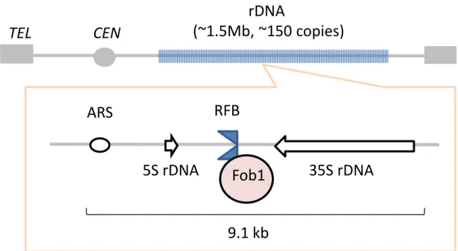
The ribosomal RNA gene cluster (rDNA) in budding yeast is an extreme repeat domain
that is well-studied in eukaryotic cells. In S. cerevisiae, the rDNA is located on chromosome Ⅻ
(Fig. 1). The rDNA exist in nucleolus as approximately 150 tandem-repeats in the wild-type. The
size of one rDNA unit is ~9.1kb and the rDNA occupy 56% of chromosome Ⅻ. There are two
ribosomal RNA genes, 35S rDNA and 5S rDNA in a unit. These genes are transcribed by RNA
polymerase I (pol I) and III (pol III), respectively. The transcripts of 35S rDNA and 5S rDNA are
pre-35S rRNA and 5S rRNA. The pre-35S rRNA is processed into matured 18S, 5.8S and 25S
rRNAs. These rRNAs are essential components of ribosome. There are two inter genic spacers
(IGS1 and IGS2) that are located between 3’-35S rDNA and 3’-5S rDNA and between 5’-5S
rDNA and 5’-35S rDNA, respectively. The replication fork barrier (RFB) and non-coding
promoter (E-pro) are located in the IGS1. Cohesion associated region (CAR) and the replication
origin (rARS) are located in the IGS2.
Because of repetitive structure of the rDNA, it easily forms the abnormal high-order
structure that causes inhibition of DNA replication and induces DSB. In addition, DSB triggers
the reduction of the copy number by popping out the copy and the recombination between the
repeats. Consequently, the rDNA is known to be the most unstable region in the genome
(Kobayashi, 2006). However, there is a system that maintains the rDNA stability, and each
organism regulates the copy number at proper level. In the yeast Saccharomyces cerevisiae, a
gene amplification system involving DNA recombination maintains the rDNA copy number
(Kobayashi et al., 1998) (Fig. 2). First, DSB occurs by the function of Fob1 at the RFB where
inhibits the progression of replication fork in the rDNA. The DSB is induced on the leading
strand of the fork (Burkhalter and Sogo, 2004). In case that this broken end is repaired with
unequal sister-chromatid, some copies are replicated twice and the copy number increases
(Kobayashi, 2006). This system amplifies the rDNA copy number at the rate of ~1 copy per cell
division in the S phase of cell cycle. This process is induced when the copy number is reduced
(Kobayashi et al., 1998). Like this, rDNA copy is recovered by the DSB repair pathway with
sister-chromatid recombination. However, once DSB recombines with improper copies,
deletional recombination of the repeats occurs, and the rDNA becomes unstable. Therefore, to
avoid the risk, the rDNA is expected to have a system, recombination is properly regulated.
When DSBs by DNA damage is not repaired, checkpoint mechanism activates. The
mechanism also activates in the rDNA, however, it occurs independently of homologous
recombination (Mundbjerg et al., 2015). Thus, I speculate that DSB in the rDNA is repaired
immediately and restart of replication for the normal rDNA amplification. However, it is unclear
the detailed mechanism how DSB in the rDNA is repaired.
In general, homologous recombination can randomly occur in the repeat sequences.
Therefore, DSB in the rDNA should have a system to separate the broken ends to avoid such a
random recombination with the other copies. In the previous study, it was shown that DSB
induced artificially on the chromosome III physically interacted with components of the nuclear
pore complex (NPC). This suggests that the nuclear pore is related to DSB repair in addition to
the material transportation between nuclear and cytoplasm though the biological significance is
unknown (Nagai et al., 2008). I speculated that similar separation may occur in the rDNA and
it functions to avoid improper recombination.
In our laboratory, by screening of genes that reduce rDNA stability, TEL1 was
identified. TEL1 is an ortholog of ataxia telangiectasia mutated (ATM) in mammalian cells
(Sabourin and Zakian, 2008). Ataxia telangiectasia (AT) is a multisystem syndrome that results
from the mutation of ATM. In human, the patients have an increased risk for cancer and an
abnormal immune system (Economopoulou et al., 2015). In budding yeast, TEL1 plays an
important role in telomere maintenance, especially in the replication of telomere (Shore and
Bianchi, 2009). Tel1 protein is a member of Serine / Threonine kinase family. Through the kinase
activity, telomerase elongates telomere (Shore and Bianchi, 2009). TEL1 also participates in the
checkpoint signaling response to DNA damage such as DSB (Longhese et al., 2010). It plays an
important role in the first step with MEC1. The checkpoint helps the cell to repair DSB.
Although some functions of TEL1 are known, it is still unclear why the rDNA is unstable by
deletion of TEL1. Recently, the nuclear pore is remarked as repair scaffold and Tel1 is involved
in the transport of unrepairable DSB to the NPC (Nagai et al., 2008).
In this study, I tried to clarify whether DSB in the rDNA is located to the NPC. If it is
the case, what kind of proteins participates in this process? First, in budding yeast, I analyzed the
interaction between rDNA and nuclear pore complex by chromatin immunoprecipitation (ChIP)
assay using anti-NPC antibody in the wild-type and the fob1 mutant in which DSB in the rDNA
doesn’t occur. Next, I identified genes that are required for the localization of the rDNA to the
NPC. Absence of Nup84, Arp5, and Mec1, the interaction between the rDNA and the NPC is
decreased to compare with that of wild-type. These factors need for the anchoring of the
HO-induced DSB to the NPC. Therefore, the rDNA is transported to the NPC by the similar
pathway to the recruitment of the induced-HO DSB to the NPC. Moreover, I analyzed rDNA
stability in these mutants by pulsed field electrophoresis (CHEF) and searched for genes directly
involved in the recruitment of DSB in the rDNA to the NPC. I identified the condensin recruiting
factors as the transportation factors for the rDNA to the NPC.
I speculate that the NPC works as a transient anchoring scaffold to prevent DSB in the
rDNA from recombining with the improper copies. In this study, together with the interaction
between the rDNA and the NPC, I also investigated stability of the rDNA and found that the
separation is required to rDNA maintenance. Moreover, I identified new factors for the
separation of DSB in the rDNA that are not detected in the artificial system to induce DSB by
HO endnuclease.
3. Results
3.1- Tel1 is involved in recombination repair for DSB caused by Fob1 at the RFB
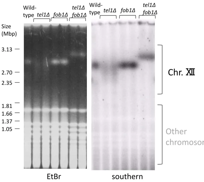
TEL1 was identified through screening of genes that reduce rDNA stability. First we
confirmed whether the phenotype is reproducible. To investigate the change of repeat number in
the rDNA, chromosome Ⅻ (chr.XII) including rDNA, was analyzed by pulsed-field
electrophoresis. Chr.Ⅻ was detected by southern hybridization with rDNA specific probe. In this
analysis, we can judge the rDNA stability by the shape of the band. A sharp band indicates that
the copy number doesn’t change, that is, the rDNA is stable. On the other hand, a broader band
indicates that the copy number frequently changes, that is, the rDNA is unstable. As the result, in
the tel1 mutant, the rDNA was actually unstable to compare with that in the wild-type (Fig. 3).
The band of the fob1 mutant is sharper than that of the wild type, that is, rDNA is stable. This is
because the replication fork is not arrested at the RFB and DSB to cause recombination is not
induced (Kobayashi et al., 1998).
To uncover the function of Tel1 in the rDNA, I analyzed the relationship between Tel1
and Fob1. I investigated whether the rDNA instability in the tel1 mutant is Fob1-dependent or
not. When FOB1 was deleted in the tel1 mutant, the rDNA became stable (Fig. 3). Therefore,
this suggests Tel1 functions in the downstream of Fob1 and it is involved in the recombination
repair for DSB caused by Fob1 at the RFB.
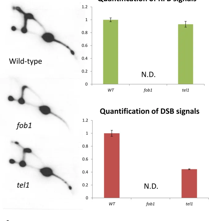
Actually, TEL1 maintains replication fork stability with MEC1 (Doksani et al., 2009).
We assumed that the reduced replication fork stability arrested at the RFB causes change of the
repeat number in the rDNA in the tel1 mutant. To test the possibility, we performed 2D-gel
analysis. This analysis enables to separate replication and recombination intermediates according
to these structures and observe DSB signal. In the tel1 mutant, as the RFB signal is similar to
that of the wild type, the fork stability should be fine. However, the DSB signal was weaker than
that of wild-type (Fig. 4). In general, less DSB induces less instability. In contrast, as the rDNA
is quite unstable in the tel1 mutant, the broken end may be highly resected by abnormally repair
and the amount of DSB was reduced.
3.2- The rDNA relocates to the NPC in a Fob1-dependent manner
Tel1 is required for the transportation of the HO-induced DSB to the NPC (Nagai et al.,
2008). Therefore, I speculated that naturally induced DSB in the rDNA is also transported to the
NPC. To test the idea, I performed ChIP assay using anti-NPC antibody and investigated
localization of the rDNA in wild-type. As a positive control, I used probes for telomere regions
in chr. XII and chr. VI, because telomeres in known to be localized in the nuclear membrane
(Palladino et al., 1993; Gotta et al., 1996). As a negative control, I also used probes in CUP1
gene (data not shown).
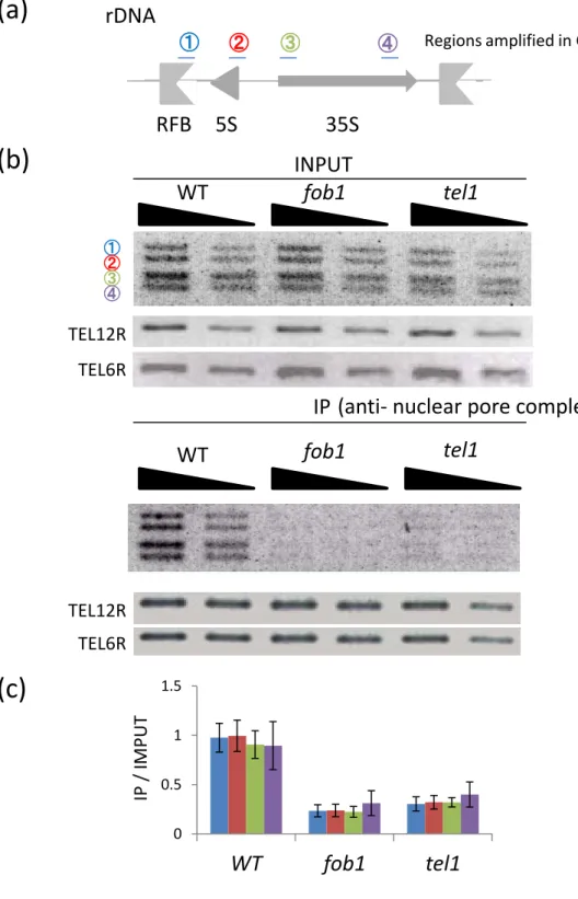
For the ChIP assay I used four probes (RFB, 5S rDNA, 5’-35S rDNA, and 3’-35S
rDNA ; Fig. 5a, blue, red, green, purple). As the result, in the wild-type, the rDNA is surely
located around the nuclear pore complex (Fig. 5b, c). Next, I performed ChIP assay using
anti-NPC antibody in the fob1 mutant. In contrast to the wild-type, interaction of the rDNA to the
NPC was considerably decreased in all four regions in the rDNA (Fig. 5b, c).
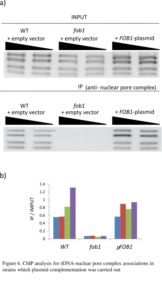
To investigate whether the phenotypes in the fob1 mutant depend on the fob1 mutation,
I performed a plasmid complementation experiment. The plasmid having the intact FOB1 gene
was transformed to the fob1 mutant. The empty vector, not having the intact FOB1 gene, was
also transformed to the wild-type and fob1 mutant. In the wild-type with the empty vector, the
rDNA was transported to the NPC (Fig. 6a, b). When the intact FOB1 gene is transformed in the
fob1 mutant, association between rDNA and NPC recovered (Fig. 6a, b). Therefore, the results
confirm that the Fob1-dependent recruitment of rDNA to the NPC.
In addition, I performed ChIP assay in the tel1 mutant and found that NPC association
with the rDNA was also clearly reduced in all four regions of the rDNA (Fig. 5b, c). Taken
together, the rDNA is transported to the NPC in both Fob1- and Tel- dependent manners.
3.3- Factors for anchoring the HO — induced DSB to the NPC are also required for
transportation of the rDNA to the NPC
In addition to Tel1, there are some factors that are involved in the transportation of the
HO-induced DSB to the NPC and/or inner membrane protein Mps3 (Horigome et al., 2014). To
identify genes that are required for localization of the rDNA to the NPC, I performed the ChIP
assay in various mutants using anti-NPC antibody.
Nup84p is a subunit of the nuclear pore complex, forms the outer ring of nuclear pore
(Strambio-De-Castillia C et al., 2010). The HO-induced DSB is known to be transported to the
Nup84 complex (Nagai et al., 2008). As the result of the ChIP assay using anti-NPC antibody, in
the nup84 mutant, the NPC association with the rDNA was also clearly reduced to compare with
that in the wild-type (Fig. 7a, b).
I also investigated the nuclear pore association in a strain with Nup84 adding FLAG
tag by ChIP assay using anti-FLAG antibody and compare with results of ChIP assay using
anti-NPC antibody. The result of ChIP assay using anti-FLAG antibody was equivalent to that of
ChIP assay using anti-NPC antibody (Fig. 8a, b). The association was also Fob1 dependent.
These results indicate that the rDNA interacts with Nup84 of the NPC, and it is possible that the
interaction is dependent on DSB at the RFB.
The INO80 and Arp5 are members of the chromatin remodeling complex that is
conserved in eukaryotes (Yao et al., 2015). This complex is involved in DNA repair and is
recruited to the phosphorylated histone H2A (Morrison and Shen, 2009). Arp5, one of the
actin-related proteins, also binds to DNA replication origins (Shimada et al., 2008). Moreover,
Arp5 is recruited to the HO-induced DSB by the accumulation of the phosphorylated histone
H2A (van Attikum et al., 2004). In the arp5 mutant, the HO-induced DSB is transported to the
NPC (Horigome et al., 2014). As the result of the ChIP assay using anti-NPC antibody, in the
arp5mutant, the NPC association with the rDNA was clearly reduced to compare with that in the
wild-type (Fig. 9a, b).
After the induction of DSB, yeast histone H2A around the DSB is phosphorylated
(Shroff et al., 2004). The phosphorylated histone H2A is called gamma-H2AX (γ-H2AX) in the
yeast S. cerevisiae (Lee et al., 2014). The serine of 129 (S129) of H2A is a target of
phosphorylation by Mec1 and Tel1, DNA damage checkpoint kinases (Lee et al., 2013). By
altering the S129 of H2A to alanine, the hta1S129A hta2S129A mutant can be constructed. This
mutant is not phosphorylated by Mec1 / Tel1. As the result of the ChIP assay using anti-NPC
antibody, in the hta1S129A hta2S129A non-phosphorylated mutant, the NPC association with the
rDNA was reduced to compare with that of the wild-type (Fig. 10a, b).
Mec1 (ortholog of human ATR) is one of the DNA damage checkpoint kinases, and has
an important role for DNA damage check-point activation (Friedel et al., 2009). Mec1 and Tel1
catalyze the phosphorylation of Rad53 (Pellicioli and Foiani, 2005). Mec1 also phosphorylates
histone H2A with Tel1 in response to DNA damage (Lee et al., 2013). In S. cerevisiae, MEC1 is
an essential gene. However, the lethality is suppressed by deletion of the ribonucleotide
reductase inhibitor Sml1 (Zhao et al., 2000). Therefore, I performed ChIP assay in the sml1 mec1
double mutant. As the result of the ChIP assay using anti-NPC antibody, in the sml1 mec1
mutant, the NPC association with the rDNA was reduced to compare with that of the wild-type
(Fig. 11a, b).
Sir2 is conserved well in eukaryotes and is known to one of long-life associated genes.
In the yeast S. cerevisiae, the sir2 mutant has shorter lifespan than the wild-type. Sir2 functions
as a histone deacetylase (HDAC) that is responsible for gene silencing in telomere, rDNA and
silent mating type loci (Cuperus et al., 2000). Moreover, in the rDNA, Sir2 plays an important
role in maintenance of sister chromatid cohesion to reduce the frequency of unequal sister
chromatid cohesion (Kobayashi et al., 2004). Although the rDNA is unstable in the sir2 mutant,
as the result of the ChIP assay using anti-NPC antibody, the rDNA association with the NPC is
similar to that of wild-type (Fig. 12a, b). Therefore, Sir2, a key factor in rDNA amplification
recombination, is not involved in the recruitment system.
In the nup84 (Fig. 7), arp5 (Fig. 9), hta1S129A hta2S129A (Fig. 10), and mec1 mutants
(Fig. 11), the rDNA association with the NPC was clearly reduced to compare with the wild-type.
On the other hand, in the sir2 mutant (Fig. 12), the rDNA association with the NPC was similar
to that of the wild-type. Consequently, some factors involved in the transportation of the
HO-induced DSB to the NPC and/or Mps3 were also related to the transportation of the rDNA to
the NPC.
3.4- Condensin recruiting factors to the RFB are required for the transportation of the
rDNA to the NPC
As DSB is naturally induced in the rDNA, I speculate there are specific factors
required for this recruitment pathway to the NPC. I focused on proteins that present and function
in both rDNA and nuclear membrane. Tof2, Csm1 and Lrs4 actually present in the both regions.
They are related to condensin recruitment to the RFB site (Johzuka and Horiuchi, 2009). Tof2
binds to Fob1, Csm1 and Lrs4 complex interact with this Tof2, and then condensin is recruited to
the RFB. By the recruitment, the rDNA is condensed and the chromosome can segregate
properly. These three factors, especially, Csm1 and Lrs4 are also known to present on the nuclear
membrane (Huang et al., 2006) and the association is important for rDNA stability (Mekhail et
al.,2008). I performed ChIP assay using anti-NPC antibody in the tof2, csm1 and lrs4 mutant.
In these mutants, rDNA association with the nuclear pore complex was also clearly reduced to
compare with that in the wild-type (Fig. 13a, b). Therefore, these results indicate that the
condensin recruitment factors are also involved in the localization of DSB in the rDNA to the
NPC.
3.5- The transportation of the rDNA to the NPC affects rDNA stability
In the HO-induced DSB experiment, the homologous sequences for the DSB repair are
deleted and the experiment is performed in the G1 phase of cell cycle (Nagai et al., 2008).
Therefore, the DSB repair process is not observed. On the other hand, in case of the rDNA, the
DSB is repaired by usual homologous recombination with the sister-chromatid or other copies.
To test the effect of recruitment for the repair, I investigated rDNA stabilities in the mutants that
have reduced interactions with the nuclear pore complex by pulsed field gel electrophoresis. The
results are shown in Fig.14. As the control, I tested fob1 (Kobayashi et al, 1998) and sir2 mutants
(Kobayashi et al., 2004). In the fob1 mutant the band of chr.XII is sharper and in the sir2 mutant
the band is broader than that in the wild type. These indicate that the rDNA is more stable and
less stable to compare with the wild type, respectively. In the csm1 and lrs4 mutants, the rDNA
was less stable to compare with that in the wild-type as reported (Mekhail et al., 2008). In the
hta1S129A hta2S129A, mec1, tof2 and lrs4 mutants, the rDNA was also less stable to compare
with that in the wild-type (Fig. 14a, b). Moreover, in the nup84, arp5 and csm1 mutants, the
rDNA was slightly unstable though the chromosome is duplicated in the arp5 mutant. Taken
together, these suggest that the localization of DSB in the rDNA to the NPC affects rDNA
stability.
3.6- A histone variant is not involved in the transportation of the rDNA to the NPC
Htz1 is a variant of histone H2A in the yeast S. cerevisiae (Jackson and Gorovsky, 2000).
As DSB is induced in a DNA region, the histone H2A of the DSB site replaces the histone
variant Htz1 (van Attikum et al., 2007). Incorporation of Htz1 to the nucleosome in the DSB site
is mediated by the chromatin remodeling complex SWR1 (Luk et al., 2010, Mizuguchi et al.,
2004). Reversely, the incorporated Htz1 is eliminated by INO80 complex from the DSB site
(Papamichos-Chronakis et al., 2006). In the swr1 and htz1 mutants, the HO-induced DSB is not
transported to the NPC (Horigome et al., 2014).
To investigate whether Htz1 is required for the transportation of the rDNA to the NPC, I
performed ChIP assay using anti-NPC antibody in the mutant. As the result, in the htz1 mutant,
the rDNA association with the NPC is similar to that of wild-type (Fig. 15a, b). Therefore, I
concluded that Htz1 is not involved in the transportation system of the rDNA to the NPC.
3.7- Ubiquitin ligases are related to the transportation of rDNA to the NPC
In the pathway of unrepairable DSB to the NPC, Slx5 and Slx8, members of
SUMO-targeted ubiquitin ligases (STUbLs) have an important role for genome stability (Nagai
et al., 2011). The STUbLs family is conserved from yeast to mammal and SUMO proteins are
enriched at the NPC. These proteins associate with the DSB and the broken end is recruited to
the NPC. In addition, SUMO proteins are also involved in the maintenance of rDNA stability by
reducing recombination with improper copies (Torres-Rosell J et al., 2007).
To investigate whether ubiquitin ligases are required for the transportation of rDNA, I
performed ChIP assay using anti-NPC antibody in the slx5 and slx8 mutants. As the result, in the
slx5and slx8 mutants, the rDNA association with the NPC is slightly decreased to compare with
that of wild-type (Fig. 15a, b). Therefore, contribution of SUMO proteins to the transportation of
rDNA to the NPC might be minor.
3.8- The rDNA is transported not only to the NPC but also to Mps3
Mps3 is the inner membrane protein that is required for the recruitment of the
HO-induced DSB to the nuclear membrane (Oza et al., 2009). In S.cerevisiae, Mps3 is an
essential protein (Jaspersen et al., 2002) for establishment of Spindle Pole Body (SPB) that
corresponds to the Centriol in mammal (Nishikawa et al., 2003). Moreover, Mps3 is required for
localization of telomere with nuclear membrane (Antoniacci et al., 2007; Bupp et al., 2007).
To test whether Mps3 is required for the transportation of rDNA to the NPC, I
performed ChIP assay using anti-NPC antibody in the mps3 mutant. In this mutant, only
N-terminal acidic domain of Mps3 is deleted to maintain viability of the cell. This mps3 mutation
declines telomere localization on the nuclear membrane in S phase but maintains the distribution
of nuclear pore on the nuclear membrane (Oza et al., 2009). As the result of the ChIP assay using
anti-NPC antibody, in the mps3 mutant, the NPC association with the rDNA was similar to that
of the wild-type (Fig. 16a, b).
Then, I performed ChIP assay in the fob1 mutant with Mps3-FLAG using anti-FLAG
antibody and compare with results of ChIP assay using anti-NPC antibody. As the result, the
association of the rDNA with Mps3 is reduced as in the fob1 mutant (Fig. 17a, b). Therefore, the
rDNA is recruited to Mps3 in a Fob1 dependent manner, too.
3.9- The RFB is not always required for the transportation
Tof1 is a subunit of the replication-pausing checkpoint complex (Tof1-Mrc1-Csm3), and
Rrm3 is a helicase in the rDNA replication. Tof1p is required to maintain the fork stability and
also fork arrest at the RFB. In the 2D gel analysis, the RFB spots in wild-type are almost
abolished in the tof1 mutant (Bastia et al., 2006). In addition, in the tof1 mutant, the rDNA is
unstable (Saka et al., submitted). On the other hand, in the absence of the Rrm3p helicase, there
was a slight enhancement of replication fork arrest at the RFB sites in comparison with the
wild-type (Bastia et al., 2006). As these mutants affect on the fork block activity, we investigated
the recruitment of the rDNA to the NPC by ChIP assay in these mutants. As the result of the
ChIP assay using anti-NPC antibody, both in the tof1 and rrm3 mutants, the rDNA association
with the NPC is similar to that of wild-type (Fig.16a, b). As DSB is expected to be occurred in
the tof1 mutant, the RFB is not always required for the transportation to the NPC. In this case,
the transportation may be similar to the HO-induced one.
4. Discussion
In this study, I found the rDNA is transported to the NPC and Mps3 in S. cerevisiae. As
the rDNA is spontaneously and highly recombinogenic region, this transportation should have
important physiological functions to maintain rDNA stability. Actually, mutation in the pathway
reduced rDNA stability. The results in this study suggest that DSB in the rDNA is repaired at the
nuclear pore complex. In addition, I identified that Tel1 and some other genes are required for
the recruitment of DSB in the rDNA to nuclear pore complex. In HU-treated cells, Ino80 is
recruited to the RFB site in the rDNA (Shimada et al., 2008). However, it was unclear whether
Tel1 and some other genes are actually recruited to the rDNA region. Some condensin
recruitment genes are included in the genes for transportation of rDNA. In addition, for the
transportation to the NPC, the inner membrane protein, Mps3 wasn’t required, however, the
rDNA is also associated to Mps3 in a Fob1-dependent manner. As the conclusion, I will present a
model as follow.
DSB is induced at the RFB site in the rDNA by the function of Fob1. Mec1/Tel1
phosphorylated histone H2A of the DSB and Ino80 complex is recruited to the phosphorylated
histone H2A. Then the rDNA is transported to the NPC and/or Mps3 (Fig. 18). I assume that the
condensin recruiting factors might transport the rDNA to the NPC and/or Mps3. This
transportation system prevents DSB from recombining with improper copies and ensures the
normal chromosome segregation. Therefore, I assume that this transportation system maintains
rDNA stability and regulates the senescence of mother cell.
4.1- rDNA association to the NPC
I performed ChIP assay to investigate the localization of DSB in the rDNA. As the result,
DSB in the rDNA is surely associated with the nuclear pore complex. It is better that this
phenomenon is confirmed by other methods except ChIP assay, for example, with microscopy.
Actually, the transportation of HO-induced DSB was observed by microscopy (Nagai et al.,
2008). Moreover, an I-SceI-induced DSB in the rDNA relocates to the extranucleolar site for
repair (Torres-Rosell et al., 2007). While, it is known that the rDNA repeat presents in the
nucleolus through the cell cycle (Miyazaki and Kobayashi, 2011). In the wild-type, the rDNA
copy number is about 150. Among them, only a few copies are broken (Zou and Rothstein, Cell).
I speculate Therefore, it could be difficult to observe the direct interaction between DSB in the
rDNA and the nuclear pore because most of rDNA copy is still located in the nucleolus.
I performed ChIP assay by using anti-Nuclear Pore Complex Proteins antibody. This
antibody recognizes the proteins in the NPC that contain phenylalanine-glycine (FG) repeats.
NPC proteins containing this repeats are especially called FG Nups and are well conserved
through species. As the NPC is a huge protein complex, the antibody precipitates the whole
complex with the associating DNA. However, as in the nup84 mutant the rDNA association to
the NPC was much reduced, Nup84 is mainly associated with rDNA.
As for unrepairable HO-induced DSB, DSB association with nuclear pore complex is
observed even 9.6kb away from the HO cut site (Nagai et al., 2008). As a unit of rDNA is about
9.1kb (Kobayashi, 2006; Miyazaki and Kobayashi, 2011), it is not so surprising that all over the
rDNA region is associated with the nuclear pore complex. Therefore, I speculate that DSB
specifically associates with Nup84 and other part of rDNA may non-specifically interact with
other components of NPC.
4.2- What is the role of Tel1 in rDNA stability rDNA?
This study uncovered that Tel1 is required for the transportation of the rDNA to the
NPC. In addition, the ChIP assay using anti-NPC antibody uncovered that the phosphorylated
histone H2A is also involved in the transportation. Therefore, these suggest that Tel1 functions to
phosphorylate the histone H2A of DSB in the rDNA. In mammalian cells, the phosphorylated
histone H2AX (γ-H2AX) is a specific DSB marker (Turinetto and Giachino, 2015). In the yeast
S. cerevisiae, throughout about 5kb around a break site, γ-H2A is formed (Shroff et al., 2004).
In case that Tel1 is not functioning, the rDNA is not transported to the NPC and DSB in the
rDNA might be not repaired correctly. Therefore, the phosphorylated histone H2A by Mec1/Tel1
is possibly the trigger of the transportation of the rDNA to the NPC.
Tel1 maintains replication fork stability with Mec1 (Doksani et al., 2009; Lopes et al.,
2001). In addition, Mec1 and Tel1 prevent the fork collapse (Branzei and Foiani, 2010). As in the
mec1 and tel1 mutants, the interaction between the rDNA and the NPC is decreased compare to
that of wild-type, I first assumed that the fork collapse causes change of the repeat number in the
rDNA in these mutants. However, in Fig.4, as the RFB signal was not reduced in the tel1 mutant,
the fork at the RFB is not collapsed. Therefore, the tel1 mutation doesn’t affect the fork stability.
Instead, the mutation may reduce recombination repair after DSB.
In mammalian cells, DSB in the nucleolus causes ATM-dependent silencing of the
transcription by RNA polymerase (Pol ) (Kruhlak et al., 2007). In addition, by
ATM-dependent silencing of the rDNA transcription, DSB in the rDNA is located at the
nucleolar periphery, and is recognized by DNA damage response factors (Harding et al., 2015).
If Tel1 dysfunctions on the rDNA in S. cerevisiae, DSB in the rDNA might be not recognized by
DNA damage response factors and it might be not repaired correctly. As the result, the broken
end is recombined with improper copies. I assume Tel1 acts on the transportation of the rDNA to
the NPC and the relocation is required for the proper repair.
4.3- Is the interaction actually dependent on DSB in the rDNA?
I showed that the rDNA interaction with NPC is “FOB1” dependent. However, there is
no evidence that the interaction is “DSB” dependent. I speculate that rDNA behaves as the
HO-induced DSB dose. It is possible that the arrested fork by the function of Fob1 or Fob1
association to the RFB itself is necessary for the transportation. As the result of the ChIP assay
using anti-NPC antibody in the tof1 mutant and the rrm3 mutant, the rDNA association with the
NPC was similar to that of wild-type (Fig.16). If stalled fork is transported to the NPC, the rDNA
association with the NPC should decrease in the tof1 mutant and increase in the rrm3 mutant
compare to that of wild-type. As well as Rrm3, the Pif1 helicase is associated with rDNA, has an
important role for rDNA replication (Ivessa et al., 2000). In the pif1 mutant, rDNA breakage and
level of rDNA circles decreased compare to that of wild-type. To check the involvement of Pif1
to the transportation, we need to perform the ChIP assay in the pif1 mutant. In addition, Tof1 is
required for fork-stabilizing (Voineagu et al., 2008). The replication fork destabilizes in the tof1
mutant, and DSB is most likely induced in the region except the RFB. Therefore, we need to
perform the 2D gel analysis in the tof1 mutant, and investigate the signal pattern. Moreover, to
investigate whether DSB increases and the rDNA is transported to the NPC, we need to perform
the ChIP assay in the fob1 tof1 double mutant.
4.4- Why is rDNA not so unstable in the transportation-related factors mutants?
As the result of CHEF and the southern hybridization, in the hta1S129A hta2S129A, tel1,
and mec1 mutants, the rDNA was much less stable to compare with that in the wild-type (Fig.
14). Moreover, in the nup84 mutant, the rDNA was slightly unstable. One possible idea is that
the relocation itself is not related to the DSB repair. On the way to the NPC, most of DSB is
repaired. For example, in the nup84 mutant, rDNA is not relocated to the NPC. But the
Tel1-dependent transportation is induced and the DSB is repaired outside of the nucleolus with
proper sister chromatid. Therefore, rDNA is not so unstable. During the repair process, a very
few DSB is not repaired properly and relocated at the NPC in a Nup84 dependent manner. In
other words, in the tel1 mutant, as neither repair nor translocation occurs, the rDNA becomes
unstable. On the contrary, in the nup84 mutant, as repair occurs but relocation doesn’t occur, the
rDNA is less unstable. In fact, the HO-induced DSB is unrepairable and relocated to the NPC
(Nagai et al., 2008). I assume the transportation of DSB to NPC is the system for isolation of the
unrepaired DSB from other DNA to avoid recombination with improper copies.
4.5- Is condensin involved in the transportation of the rDNA to the NPC?
Condensin regulates the rDNA compaction. In addition, condensin is important for DSB
repair and the mutants are sensitive to the UV (Sheedy et al., 2005). Condensin is thought to
promote the equal sister-chromatid recombination by attachment of sister-chromatids (Ide et al.,
2010). This study indicates that condensin recruiting factors to the RFB are involved in the
transportation of the rDNA to the NPC. Therefore, condensin is also possibly involved in the
transportation. I assume that condensin recruiting factors act as the transporter of the rDNA to
the NPC, and condensin condenses the rDNA around the NPC to facilitate the chromosome
segregation. To investigate the direct involvement of condensin to the transportation, we need to
perform the ChIP assay in the condensin mutant.
4.6- Dynamics of the rDNA in the nucleolus with regards to this transportation system
The condensin recruiting factors, Csm1 and Lrs4 (Mekhail et al., 2008) interact with the
CLIP (chromosome linkage INM protein) complex that anchor the rDNA to the nuclear envelope
to stay the rDNA in the nucleolus. The complex keeps the rDNA away from DNA recombinases
(Rad52 etc) in the nucleoplasm and maintains rDNA stability (Mekhail and Moazed, 2010). In
general, the rDNA exists in the nucleolus throughout cell cycle (Miyazaki and Kobayashi, 2011)
in which there isn’t DNA repair factor in the nucleolus (Torres-Rosell et al., 2007). Therefore,
the rDNA has to go out the nucleolus for repair. Actually, in the rad52 mutant, the rDNA copy
number did not recover in the strain having approximately 80-copies (Kobayashi et al., 2004).
This study suggests ubiquitin ligases are involved in the transportation of the rDNA to
the NPC though the contribution is minor. In the slx5 mutant, and especially in the slx8 mutant,
the rDNA association with the NPC was a little decreased to compare with that of wild-type (Fig.
15). Slx5 and Slx8 have a key role for genome stability (Zhang et al., 2006). They are a
heterodimeric complex in the yeast S. cerevisiae (Cook et al., 2009), The Slx5-Slx8 complex
reflects on the sumoylation of DNA repair factors containing Rad52 (Burgess et al., 2007). In
addition, in the slx8 mutant, the rDNA recombination is increased in a Rad52 dependent manner
(Eckert-Boulet and Lisby, 2009). The frequency of nucleolar Rad52 foci is also increased in the
slx8mutant (Burgess et al., 2007). Therefore, the Slx5-Slx8 complex has a role for the regulation
of the rDNA recombination. As for the transportation of the rDNA to the NPC, the complex is
not so critical.
4.7- What is the role of Mps3 in the transportation of the rDNA to the NPC?
In the yeast S. cerevisiae, Mps3 has a role for establishment of spindle pole body (SPB)
(Jaspersen et al., 2002). The HO-induced DSB is transported to Mps3 in an INO80 complex
dependent manner (Oza et al., 2009 and Horigome et al., 2014). My results suggest that the
transportation of the rDNA to the NPC possibly requires INO80 complex. In this study, I did not
perform the ChIP assay in the ino80 mutant (non-essential gene). One of the subunit of INO80
complex, Ino80 has a role as ATPase for chromatin remodeling (Seeber et al., 2013).
Actin-related proteins, such Arp5 and Arp8, are required not only for the recruitment of INO80
complex to chromatin but also for the ATPase activity of INO80 complex (Osakabe et al., 2014).
In addition, in the htz1 mutant, the rDNA association with the NPC was similar to that of
wild-type (Fig. 15). Htz1 is necessary for the transportation of the HO-induced DSB to Mps3
(Horigome et al., 2014). Therefore, the incorporation of Htz1 by SWR1 chromatin remodeling
complex is possibly required for the transportation of the rDNA to Mps3, not to the NPC.
SPB and NPC link on the nuclear envelope (Jaspersen and Ghosh, 2012). The SPB is
important for chromosome segregation. Centromeres of chromosomes are tethered to the SPB
(Taddei et al., 2010). This tethering permits to segregate chromosomes. My results indicate that
the transportation of the rDNA to the NPC requires condensin recruiting factors. Consequently,
by the cooperation of Mps3 and condensin recruiters, the rDNA is normally condensed and
chromosome might be easy to segregate.
4.8- Association of the transportation of the rDNA to the NPC for aging
After the induction of unrepairable DSB, it takes for 2~4h to transport the DSB to the
NPC (Nagai et al., 2008). On the other hand, DSB in the rDNA could occur frequently by
blocking replication fork at the RFBs, and the broken end is quickly repaired in one way or
another. In this study, I identified genes that are involved in the recruitment of the rDNA to the
NPC. However, it is unclear when and which these factors act on the recruitment pathway.
Moreover, although there are approximately 200 NPCs on the nuclear membrane in the yeast
S.cerevisiae, it is unknown which NPCs on the nuclear membrane interacts with the rDNA.
The condition of rDNA (the copy number and stability) affects the function of the cell.
Notably, it is known that cellular senescence (replicative senescence) is affected by rDNA
stability (Kobayashi, 2008). In the budding yeast S. cerevisiae, whenever a cell (mother cell)
produces a new cell (daughter cell), the mother cell becomes older, and dies finally. On the other
hand, the newborn daughter cell doesn’t inherit the aging phenotypes of the mother cell and
achieves the rejuvenation. To rejuvenate the daughter cell in the cell division, the mechanism of
the mother cell aging is uncovering. The previous study proposes the hypothesis that the
instability of the rDNA is a cause of the aging (Kobayashi, 2006). In this hypothesis, according
to cell divisions, the mother cell specific rDNA instability is induced (Ganley et al., 2009).
However, the mechanism to distinguish between the mother cell and the daughter cell is still
unknown. In this current study, it uncovers that condensin recruiting factors needs for the
transportation of the rDNA to the NPC. Condensin functions the unity of the rDNA. Dysfunction
of condensin causes non-disjunction of chromosomes. Therefore, I speculate that the unequal
distribution of the rDNA between mother cell and daughter cell contributes to the differentiation
of both cells and the transportation links the rDNA stability and aging. I expect that the NPC and
condensin become a key to solve the cooperation between these mechanisms.
5. Acknowledgements
I’m grateful thank to Prof. Takehiko Kobayashi for constant guidance during my PhD study. I
thank Dr. Kenji Shimada (Friedrich Miescher Institute for Biomedical Research), Prof. Masahiko
Harata (Tohoku University), Prof. Masato Kanemaki, and Assistant Prof. Chihiro Horigome for
providing us various yeast strains and plasmids in this study. I also thank Prof. Hiroyuki Araki,
Assistant Prof. Chihiro Horigome and Assistant Prof. Mariko Sasaki for advice and discussion
throughout my study. Moreover, I appreciate to Assistant Prof. Tetsushi Iida, Assistant Prof.
Yuhuko Akamatsu, Kimiko Saka, and the all progress committee members for supporting my
PhD study.
6. Material and Methods
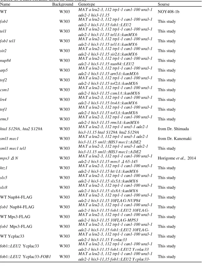
Yeast strains, plasmids, and growth conditions
Yeast strains used in this study were derived from NOY408-1b (W303 derivative).
Unless indicated, strains were grown at 30 ˚ C in YPD medium. YPD (yeast
extract-peptone-dextrose) is rich medium for the normal culture, YP-Galactose (yeast
extract-peptone-galactose) is for induction of GAL promoter and synthetic complete (SC)
medium lacking the appropriate amino acids (Sherman et al., 1986) is for the gene maker
selection. To control the FOB1 expression under the GAL7 promoter, cells were pre-cultured in
medium containing 2% (w/v) raffinose as a sole carbon source until induction. Induction of
FOB1 was triggered by adding galactose solution to the culture (2% [w/v].) Plasmids were
maintained in Escherichia coli DH5α strain. Yeast strains and plasmids used in this study are
listed in Table 1, 2.
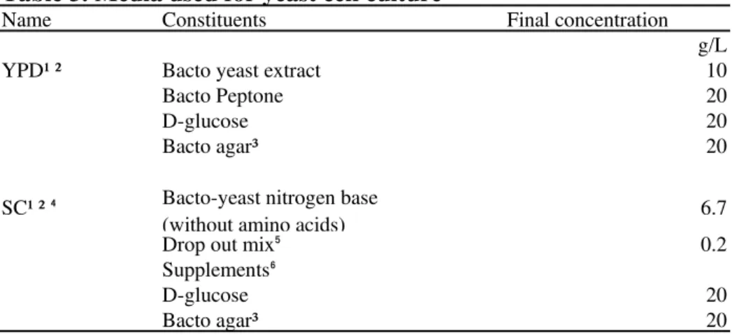
Medium used for yeast cell culture is listed in Table 3. They were prepared as described
(Dan Burke, 2000) with some modification. If necessary, G418 (Sigma) and 5-Fluoroorotic acid
(5-FOA; Wako) were added to the medium with the concentration shown in Table 3.
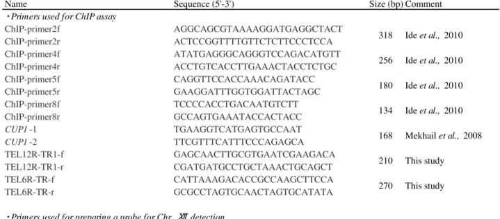
PCR primers
PCR primers used in this study are listed in Table 4. Primers were stored in 50 ㎕ TE
buffer at -20˚C freezer.
Yeast genetic transformation
Yeast genetic transformation was performed by using Frozen-EZ Yeast Transformation
Kit (Zymo Research Corporation) according to the instruction of manufacturer. Yeast cells
were cultured in appropriate liquid medium (10ml) until mid-log phase (O.D. ~1.0, 600nm) and
collected by centrifugation at 10,000 rpm for 1 min. Cells were washed with 0.5 ml of EZ
solution 1 and repelleted. After supernatant was discarded, ~1× cells were suspended into 50 ㎕
of EZ solution 2 for one transformation reaction. 5 ㎕ of DNA solution (~200 ng/㎕) was mixed
with the cell suspension, 500 ㎕ of EZ solution 3 was added and suspended gently. The mixture
was incubated in 30 ˚C for at least 45 min with vigorously mixing every 15 min. Cell mixture
was pelleted by centrifugation at 10,000 rpm for 1 min, and spread onto an appropriate plate
medium. When a drug resistance marker was used for selection, cells were cultured in
non-selective liquid medium for at least 2 h before spreading.
Plasmid construction
The galactose-inducible FOB1 plasmid, YCplac33-GALFOB1, was constructed as
follow. ~ 3kb fragment that contains galactose-inducible FOB1 cassette was excised from
YCpGALFOB1 by BamH / Sal digestion and sub-cloned into these sites of YCplac33.
DNA labeling with radioactive dCTP
Radio labeled DNA probes for Southern hybridization was obtained as follow. To label
DNA fragments with [α-] dCTP, High Prime (Roche diagnostic) was used according to the
instruction of manufacturer. Before the labeling, the template DNA (probe, 50 ng) in dH₂O was
boiled for 10 min and immediately chilled on ice. The template DNA was mixed and 5 ㎕ of [α-]
dCTP, then incubated for at least 10 min at 37˚ C for labeling. After the reaction was finished,
labeled DNA was purified by using NICK columns (GE). For denaturing, the purified DNA was
boiled for 5 min, immediately chilled on ice for at least 1 min and then used for hybridization.
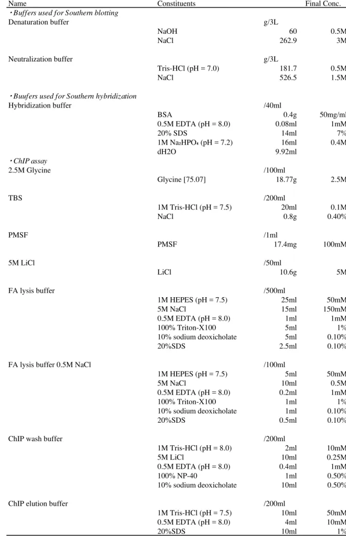
Southern blotting and hybridization
DNA transfer from agarose gel to nylon membrane was performed as described
previously (Sambrook and Russell, 2001). After electrophoresis, the DNA was depurinated in 0.2
N HCl, denatured in Denaturation buffer (see Table 5), and neutralized in Neutralization buffer
(see Table 5) for 20 minutes, respectively. Next, the DNA was transferred to Nylon membrane
(Hybond N+ , GE) in 20×SSC by capillary transfer for at least 15 h. After the membrane was
washed with 6×SSC, DNA was cross-linked to the membrane before the hybridization with 120
mJ of UV (254 nm) irradiation by Stratalinker (Stratagene).
The membrane was pre-hybridized in 40 ml Hybridization buffer (see Table 5) at 65˚C
for 30 min, followed by hybridization in 40 ml of Hybridization buffer containing heat-denatured
probe at 65˚C for overnight in a roller bottle. The membrane was washed with 2×SSC, 2% SDS
for 30 min at 65˚C and in 0.2×SSC, 0.2% SDS for 30 min at 65˚C. Next, the membrane was
briefly rinsed with 0.2×SSC, 0.2% SDS at room temperature. Then, the membrane was exposed
to the Imaging plate (GE) for a day. The signals were detected by Typhoon FLA 9000 (GE) and
analyzed by Image Quant (GE).
Pulsed field (CHEF: Countour- clamped homogenous electric field) electrophoresis
Samples for pulsed-field (CHEF) electrophoresis were prepared as described previously
(Kobayashi et al., 2001) using ~1.0× cells per one plug. The sample plug was cut in half and
used for electrophoresis.
Electrophoresis was performed in a 0.8% agarose gel with 0.5 × Tris-borate-EDTA
(TBE) buffer, using CHEF-MAPPER (Bio-Rad). For Fig. 1, the conditions were a 300-900 sec
pulse time and 100V for 68 hours at 14˚C in a 0.8% agarose gel.
Two-dimensional (2D) gel electrophoresis
To detect replication and recombination intermediates and DSB spot by 2D gel
electrophoresis (Ide and Kobayashi, 2010), DNA was prepared from cells growing in YPD, and
DNA was isolated and embedded in plugs (Ide et al., 2010). Yeast cells were cultured in YPD
medium until mid-log phase (O.D. 0.8, 600nm). After the incubation on ice, 10% sodium azide
(final conc. 0.1%) was added to the sample. The sample cells were collected by centrifugation at
3,500 rpm for 5 min at 4˚C. After supernatant was discarded, the cells were suspended into 10
ml ice-cold sorbitol solution with sodium azide for cell wash. The cells were collected by
centrifugation at 3,500 rpm for 5 min at 4 ˚ C, then supernatant was discarded. This sorbitol
solution with sodium azide wash was performed twice. There are ~1.0× cells in each plug.
) Digestion with restriction enzyme
The plugs were treated with Bgl . Before the digestion of the restriction enzyme, the
plugs were put in 1.5 ml tubes, 1 ml TE buffer (pH 8.0) was added to the tubes for wash. After
the incubation for 30 min at room temperature, supernatant was discarded. This incubation was
performed twice. After that, 0.5 ml 1 × reaction buffer was added to the tube. After the
incubation for 30 min at room temperature, the buffer was discarded. This incubation was
performed twice. DNA in the plugs were digested with Bgl for 4h at 37˚C. The reaction was
carried out in 200 ㎕ 1×reaction buffer with 150 units of Bgl .
) The first dimension gel electrophoresis
The plugs were set in 0.4% agarose gel (200 ml 1×TBE, 0.8g SeeKem LE agarose), the
first dimension electrophoresis was performed at 32V/cm for 12~13h at room temperature. After
the electrophoresis, the gel was stained in 300 ml 1 × TBE containing 0.5 μ g/ml ethidium
bromide for 30 min at room temperature. After the staining, the electrophoresis band patterns
were checked. By this first dimension electrophoresis, DNA in the plugs was separated with size.
) The second dimension electrophoresis
The gel (lane) containing the objective size was excised from the first dimension gel,
turned it 90˚, and put on the second dimension gel tray. 1.2 % agalose solution (200 ml 1×TBE,
2.4g SeeKem LE agarose, 6.0 ㎕ 10mg/ml ethidium bromide at ~55˚C) was pored to the tray,
and the gel was hardened for 20min at room temperature. The second dimension electrophoresis
was performed at 132V/cm for 4.5h at 4˚C.
) Signal detection
After the check of the electrophoresis band patterns, the DNA was transferred to the
membrane. The membrane-bound DNA was hybridized with a radiorabeled probe. The rDNA
was detected with an rDNA specific probe. After wash, the membrane was exposed to the
Imaging plate (GE) for a week. The signal was detected by Typhoon FLA 9000 (GE) and were
analyzed by Image Quant (GE).
DNA sequencing
DNA sequencing was performed by using BigDye Ⓡ Terminator v3.1 Cycle
Sequencing Kits (Applied Biosystems) and 3130xl Genetic Analyzer (Applied Biosystems)
according to the instruction of manufacturer.
Western blotting
Yeast whole cell extracts were prepared by the TCA method (Ide et al., 2010). Proteins
were fractionated by SDS-polyacrylamide gel electrophoresis (PAGE), transferred to a PVDF
membrane (Millipore) and subjected to Western blotting analysis as described previously (Ide et
al.,2010). For detection of a related family of nuclear pore complex (NPC) proteins, anti-nuclear
pore complex proteins antibody Mab414 (Abcam) or anti-FLAG antibody, F2 were used.
Chromatin immunoprecipitation
ChIP analysis was performed based on the method described previously (Aparicio et al.,
2004). Buffers used in this assay are shown in Table 5. Yeast cells were cultured in appropriate
liquid medium until mid-log phase (O.D. 0.6~0.8, 600nm).
) Cross-link protein-DNA complexes in vivo
Cells were fixed in 0.55ml 37% formaldehyde for 20 min. After that, 3ml 2.5M Glycine
was added to stop the cross-link reaction for 5 min. The cells were collected by centrifugation at
3500 rpm for 3 min at 4˚C. After supernatant was discarded, the cells were suspended into 5ml
of ice-cold TBS (see Table 5) solution for cell wash. The sample cells were collected by
centrifugation at 3,500 rpm for 3 min at 4˚C, then the supernatant was discarded. This TBS wash
was performed twice. Next, the cells were suspended into 5ml of ice-cold FA lysis buffer (see
Table 5) for cell wash. The cells were collected by centrifugation at 3,500 rpm for 3 min at 4˚C,
then the supernatant was discarded. This FA lysis buffer wash was performed three times.
) Lyse cells and isolate chromatin
After cell wash, the sample cells were suspended into 0.5ml of ice-cold FA lysis buffer /
2mM PMSF. These samples were transferred to 2.0ml screw cap microfuge tubes (Sarstedt). This
tube was pre-added 1.7g Glass beads, acid washed 425-600um (SIGMA). These tubes were
closed by screw caps tightly, and mixed by inversion. By using Multi-Beads Shocker (YASUI
KIKAI), the beads in the tubes were stirred and the cells were crashed in the condition (interval
[ON 30 sec: OFF 60 sec]×16).
) Isolate lysate
The bottom of the tube was punched a hole by a needle and the tube was inserted to a
size larger tube. The samples were collected by centrifugation at 3,500 rpm for 3 min at 4˚C.
The collected sample was transferred to a 1.5ml Eppendorf tube. The sample was collected by
centrifugation at 15,000 rpm for 15 min at 4˚C, then the supernatant was discarded.
) Shear DNA
The samples were suspended into 0.5ml of ice-cold FA lysis buffer. The suspension
sample was transferred to the tubes for sonication by Bioruptor (Cosmo Bio). The tube was set to
the machine, and DNA sheared in the condition (interval [ON 30sec: OFF 30sec]×9) to reduce
the average size of DNA fragment to ~500bp. The sonicated samples were transferred to new
eppendorf tubes, and collected by centrifugation at 15,000 rpm for 30 min at 4˚C. The
supernatants were transferred to new eppendorf tubes, stored -80˚C deep freezer. This was used
as the whole cell extract (WCE) for ChIP assay.
) Check chromatin fragment size
50 ㎕ WCE was added 50 ㎕ ChIP elution buffer (see Table 5). Next, 4 ㎕ 20mg/ml
proteinase K (Merck) in PBS was added to the 100 ㎕ sample, and it was incubated for 2h at 37˚
C and for 6h at 65˚C. Proteins were removed by phenol chloroform, and DNA was precipitated
by ethanol. After 70% ethanol wash, dried fragments were suspended into 4 ㎕ TE buffer (pH
8.0). The 2 ㎕ suspension was added 1 ㎕ dH₂O and 1 ㎕ 10×loading buffer (TaKaRa). These
samples were applied to 1.5% agarose gel, and performed electrophoresis with 100bp DNA
ladder marker (New England Biolabs, NEB) for 18min at 135V.
) Immunoprecipitate
1 ㎕ anti-nuclear pore complex (NPC) protein antibody (Mab414, abcam) (1,000mg/ml)
was suspended to 10 ㎕ Dynabeads Protein G . Total 11 ㎕ beads and antibody was tapped for
mix, and spun down. After the incubation on ice for 30 min, the tube was spun down, and set to
the magnet holder. After the beads were attracted by the magnet, the supernatant was discarded
and the tube was removed for the holder. 22 ㎕ 5mg/ml BSA in PBS was added to the tube and
mixed. The tube was set to the magnet holder. After the beads were attracted by the magnet, the
supernatant was discarded. This beads wash step was repeated total three times. After final
supernatant was discarded, 33 ㎕ 5mg/ml BSA in FA Lysis buffer was added to the tube, tapped
for mix, and spun down. The tube was rotated for 1h at 4˚C. After the tube was spun down, the
tube was set to the magnet holder. After the beads were attracted by the magnet, the supernatant
was discarded and 110 ㎕ ice-cold FA Lysis buffer was added, tapped for mix, and spun down.
After the tube was spun down, the tube was set to the magnet holder. After the beads were
attracted by the magnet, the supernatant was discarded and 11 ㎕ ice-cold FA Lysis buffer was
added, tapped for mix, spun down and put on ice. 240 ㎕ WCE was added to the new tube, and
the 11 ㎕ beads-antibody solution was suspended. The tube was rotated for 90 min at 4˚C. After
the rotation, the tube was spun down. Then, the tube was set to the magnet holder. After the
beads were attracted by the magnet, the supernatant was discarded.
) Wash beads
300 ㎕ ice-cold FA Lysis buffer was added to the tube, tapped for mix, and incubated
for 3 min at room temperature. After the incubation, the tube was set to the magnet holder. After
the beads were attracted by the magnet, the supernatant was discarded. The wash by ice-cold FA
Lysis buffer was repeated again. Next, 300 ㎕ FA Lysis buffer / NaCl (see Table 5) was added to
the tube, tapped for mix, and incubated for 3min at room temperature. After the incubation, the
tube was set to magnet holder. After the beads were attracted by the magnet, the supernatant was
discarded. The wash by FA Lysis buffer / NaCl was repeated again. Then, 300 ㎕ ChIP wash
buffer (see Table 5) was added to the tube, tapped for mix, and incubated for 3 min at room
temperature. After the incubation, the tube was set to the magnet holder. After the beads were
attracted by the magnet, the supernatant was discarded. The wash by ChIP wash buffer was
repeated again. Finally, 300 ㎕ TE buffer (pH 7.5) was added to the tube, tapped for mix, and
incubated for 3 min at room temperature. After the incubation, the tube was set to magnet holder.
After the beads were attracted by the magnet, the supernatant was discarded. The wash by TE
buffer was repeated again.
) Elute protein from beads
50 ㎕ ChIP elution buffer was added to the tube, tapped for dissolution, and spun down.
After mild tapping, the tube was incubated for 10 min at 65˚C. Then the tube was tapped mildly,
and spun down. The tube was set to the magnet holder. After the beads were attracted by the
magnet, the supernatant was used for reverse cross-link to purify DNA.
) Reverse cross-link and purify DNA
40 ㎕ TE buffer (pH 7.5) and 10 ㎕ 20 mg/ml proteinase K in TBS were added to the
supernatant in a new tube. Total 100 ㎕ solution was incubated for 2 h at 37˚C and for 6 h at 65˚
C. This sample was treated as immunoprecipitated (IP) sample. In addition, 50 ㎕ ChIP elution
buffer, 40 ㎕ TE buffer (pH 7.5) and 10 ㎕ 20 mg/ml proteinase K in TBS were added to the
whole cell extract (input). This solution was also incubated for 2 h at 37˚C and for 6 h at 65˚C.
After the incubation, 8 ㎕ 5M LiCl was added to the tube. DNA was extracted by phenol
chloroform and precipitated by ethanol. After 70% ethanol wash, dried pellet was suspended into
30 ㎕ TE buffer (pH 8.0).
) Quantitative PCR and agarose gel electrophoresis
The input and IP samples were analyzed by quantitative PCR (qPCR). To confirm that
PCR reaction is in the linear range, input and IP samples were serially two-fold diluted and the
PCR products were separated on 2.0 % agarose gels and stained with ethidium bromide. The
values are given as a percentage of immunoprecipitates (IP/input). Four regions in the rDNA
were analyzed by qPCR. Primer sequences were described previously (Ide et al., 2010) and
shown in Table 4.
7. References
Adkins NL, Niu H, Sung P, Peterson CL. (2013): Nucleosome dynamics regulates DNA processing. Nat Struct Mol Biol. 20:836-842
Antoniacci LM, Kenna MA, Skibbens RV. (2007): The nuclear envelope and spindle pole body-associated Mps3 protein bind telomere regulators and function in telomere clustering. Cell Cycle.6:75-79
Aparicio O, Geisberg J, Struhl K. (2005): Chromatin immunoprecipitation for determining the association of proteins with specific genomic sequences in vivo. Current Protocols in Cell Biology17.7.1-17.7.23
Bairwa NK, Mohanty BK, Stamenova R, Curcio MJ, Bastia D. (2011): The intra-S phase checkpoint protein Tof1 collaborates with the helicase Rrm3 and F-box protein Dia2 to maintain genome stability in Saccharomyces cerevisiae. J Biol Chem. 286:2445-2454
Branzei D and Foiani M. (2010): Maintaining genome stability at the replication fork. Nat Rev Mol Cell Biol.11:208-219
Bupp JM, Martin AE, Stensrud ES, Jaspersen SL. (2007): Telomere anchoring at the nuclear periphery requires the budding yeast Sad1-UNC-84 domain protein Mps3. J Cell Biol. 179:845-854
Burgess RC, Rahman S, Lisby M, Rothstein R Zhao X. (2007): The Slx5-Slx8 complex affects sumoylation of DNA repair proteins and negatively regulates recombination. Mol Cell Biol.27:6153-6162
Burkhalter MD and Sogo JM. (2004): rDNA enhancer affects replication initiation and mitotic recombination: Fob1 mediates nucleolytic processing independently of replication. Mol Cell. 15:409-421
Cook CE, Hochstrasser M, Kerscher O. (2009): The SUMO-targeted ubiquitin ligase subunit Slx5 resides in nuclear foci and at sites of DNA breaks. Cell Cycle. 8:1080-1089
Cuperus G, Shafaatian R, Shore D. (2000): Locus specificity determinants in the
Dan Burke D D and Tim Stearns (2000): Methods in Yeast genetics, ED. 2000. COLD SPRING HARBOR LABORATORY PRESS.
Doksani Y, Bermejo R, Fiorani S, Haber J E, Foiani M. (2009): Replication dynamics, dormant origin firing, and terminal fork integrity after double-strand break formation. Cell. 137:247-258
Doksani Y, Bermejo R, Fiorani S, Haber JE, Foiani M. (2009): Replicon dynamics, dormant origin firing, and terminal fork integrity after double-strand break formation. Cell. 137:247-258
Eckert-Boulet N and Lisby M. (2009): Regulation of rDNA stability by sumoylation. DNA repair.8:507-516
Economopoulou P, Dimitriadis G, Psyrri A. (2015): Beyond BRCA: new herediary breast cancer susceptibility genes. Cancer Treat Rev. 41:1-8
Friedel AM, Pike BL, Gasser SM. (2009): ATR/Mec1: coordinating fork stability and repair. Curr Opin Cell Biol.21:237-244
Ganley AR, Ide S, Saka K, Kobayashi T. (2009): The effect of replication initiation on gene amplification in the rDNA and its relationship to aging. Mol Cell. 35:683-693
Ganley ARD, Ide S, Saka K, Kobayashi T. (2009): The effect of replication initiation on gene amplification in the rDNA and its relationship to aging. Mol Cel.l 35:683-693
Gotta M, Laroche T, Formenton A, Maillet L, Scherthan H, Gasser SM. (1996): The clustering of telomeres and colocalization with Rap1, Sir3, and Sir4 proteins in wild-type Saccharomyces cerevisiae. J. Cell Biol.143:1349-1363
Horigome C, Oma Y, Konishi T, Schmid R, Marcomini I, Hauer MH, Dion V, Harata M, Gasser SM. (2014): SWR1 and INO80 chromatin remodelers contribute to DNA double-strand break perinuclear anchorage site choice. Mol Cell. 55:626-639
Huang J, Brito IL, Villen J, Gygi SP, Amon A, Moazed D. (2006): Inhibition of homologous recombination by a cohesion-associated clamp complex recruited to the rDNA recombination enhancer. Genes Dev. 20:2887-2901
Ide S and Kobayashi T. (2010): Analysis of DNA replication in Saccahromyces cerevisiae by
22.14.1-22.14.12
Ide S, Miyazaki T, Maki H, Kobayashi T. (2010): Abundance of ribosomal RNA gene copies maintains genome integrity. Science 327:693-696
Ivessa AS, Zhou JQ, Zakian VA. (2000): The Saccharomyces Pif1p DNA helicase and the highly related Rrm3p have opposite effects on replication fork progression in ribosomal DNA. Cell.100:479-489
Jackson JD and Gorovsky MA. (2000): Histone H2A.Z has a conserved function that is distinct from that of the major H2A sequence variants. Nucleic Acids Res. 28:3811-3816
Jaspersen SL and Ghosh S. (2012): Nuclear envelope insertion of spindle pole bodies and nuclear pore complexes. Nucleus. 3:226-236
Jaspersen SL, Giddings TH Jr, Winey M. (2002): Mps3p is a novel component of the yeast spindle pole body that interacts with the yeast centrin homologue Cdc13p. J Cell Biol. 159:945-956
Johzuka K and Horiuchi T. (2009): The cis element and factors required for condensin recruitment to chromosomes. Mol Cell. 34:26-35
Kobayashi T, Heck DJ, Nomura M, Horiuchi T. (1998): Expansion and contraction of ribosomal DNA repeats in Saccharomyces cerevisiae: requruitment of replication fork blocking (Fob1) protein and the role of RNA polymerase . Genes Dev. 12:3821-3830
Kobayashi T, Nomura M, Horiuchi T. (2001): Identification of DNA cis elements essential for expansion of ribosomal DNA repeats in Saccharomyces cerevisiae. Mol Cell Biol. 21:136-147
Kobayashi T, Horiuchi T, Tongaonkar P, Vu L, Nomura M. (2004): SIR2 regulates
recombination between different rDNA repeats, but not recombination within individual rRNA genes in yeast. Cell 117:441-453
Kobayashi T. (2006): Strategies to maintain the stability of the ribosomal RNA gene repeats –Collaboration of recombination, cohesion, and condensation- Genes Genet. Syst. 81:155-161.
Kruhlak M, Crouch EE, Orlov M, Montańo C, Gorski SA, Nussenzweig A, Misteli T, Phair RD, Casellas R. (2007): The ATM repair pathway inhibits RNA polymerase transcription in