t ype l oc us i n t he gr een s eaw
eed U
l va par t i t a
著者
Yam
az aki Tom
okaz u, I c hi har a Kens uke, Suz uki
Ryogo, O
s hi m
a Kens hi r o, M
i yam
ur a Shi ni c hi ,
Kuw
ano Kaz uyos hi , Toyoda At s us hi , Suz uki
Yut aka, Sugano Sum
i o, H
at t or i M
as ahi r a, Kaw
ano
Shi geyuki
j our nal or
publ i c at i on t i t l e
Sc i ent i f i c Repor t s
vol um
e
7
page r ange
11679
year
2017- 09
権利
( C) The Aut hor ( s ) 2017
Thi s ar t i c l e i s l i c ens ed under a Cr eat i ve
Com
m
ons At t r i but i on 4. 0 I nt er nat i onal Li c ens e,
w
hi c h per m
i t s us e, s har i ng, adapt at i on,
di s t r i but i on and r epr oduc t i on i n any m
edi um
or
f or m
at , as l ong as you gi ve appr opr i at e c r edi t
t o t he or i gi nal aut hor ( s ) and t he s our c e,
pr ovi de a l i nk t o t he Cr eat i ve Com
m
ons
l i c ens e, and i ndi c at e i f c hanges w
er e m
ade.
The i m
ages or ot her t hi r d par t y m
at er i al i n
t hi s ar t i c l e ar e i nc l uded i n t he ar t i c l e’
s
Cr eat i ve Com
m
ons l i c ens e, unl es s i ndi c at ed
ot her w
i s e i n a c r edi t l i ne t o t he m
at er i al . I f
m
at er i al i s not i nc l uded i n t he ar t i c l e’
s
Cr eat i ve Com
m
ons l i c ens e and your i nt ended us e
i s not per m
i t t ed by s t at ut or y r egul at i on or
exc eeds t he per m
i t t ed us e, you w
i l l need t o
obt ai n per m
i s s i on di r ec t l y f r om
t he c opyr i ght
hol der . To vi ew
a c opy of t hi s l i c ens e, vi s i t
ht t p: / / c r eat i vec om
m
ons . or g/ l i c ens es / by/ 4. 0/ .
U
RL
ht t p: / / hdl . handl e. net / 2241/ 00148428
doi: 10.1038/s41598-017-11677-0
Cr eat i ve Commons : 表示
Genomic structure and evolution of
the mating type locus in the green
seaweed Ulva partita
tomokazu Yamazaki
1, Kensuke Ichihara
1, Ryogo suzuki
1, Kenshiro oshima
2, shinichi
Miyamura
3, Kazuyoshi Kuwano
4, Atsushi toyoda
5, Yutaka suzuki
2, sumio sugano
2,
Masahira Hattori
2,6& shigeyuki Kawano
1the evolution of sex chromosomes and mating loci in organisms with UV systems of sex/mating type determination in haploid phases via genes on UV chromosomes is not well understood. We report the structure of the mating type (Mt) locus and its evolutionary history in the green seaweed Ulva partita, which is a multicellular organism with an isomorphic haploid-diploid life cycle and mating type determination in the haploid phase. Comprehensive comparison of a total of 12.0 and 16.6 Gb of genomic next-generation sequencing data for mt− and mt+ strains identiied highly rearranged MT loci of 1.0 and 1.5 Mb in size and containing 46 and 67 genes, respectively, including 23 gametologs. Molecular evolutionary analyses suggested that the Mt loci diverged over a prolonged period in the individual mating types after their establishment in an ancestor. A gene encoding an RWP-RK domain-containing protein was found in the mt− MT locus but was not an ortholog of the chlorophycean mating type determination gene MID. taken together, our results suggest that the genomic structure and its evolutionary history in the U. partita Mt locus are similar to those on other UV chromosomes and that the MT locus genes are quite diferent from those of Chlorophyceae.
Sexual reproduction systems in eukaryotes can be divided into two types, in terms of determining sex/mating type in the haploid phase (UV systems) or in the diploid phase (XY/ZW systems)1. In the XY/ZW systems of
mammals, insects, and plants, the structures of XY/ZW chromosomes and their evolution correspond reasonably well with predictions based on population genetics theory, whereby the suppressed recombination of the two chromosomes results in degeneration through Muller’s ratchet, background selection, the Hill-Robertson efect with weak selection, and the “hitchhiking” of deleterious alleles along with favorable mutations2,3. hese
theoret-ical predictions are constructed under the postulate that both sex chromosomes (XY or ZW) are heterozygous in the diploid phase and that they are distributed separately into the gametes (egg and sperm) via meiosis. In this case, deleterious mutations in an allelic gene on a sex chromosome, referred to as a gametolog, are masked by the counterpart gene on the other sex chromosome, resulting in sex chromosome degeneration driven by the above population genetic mechanisms of gene ixation. Sex chromosomes undergo stepwise degeneration, such as a size decrease, gene loss, accumulation of transposable elements, and decrease in codon bias, at diferent evolutionary times, resulting in “evolutionary strata”1,4,5. Several recent studies about plant Y chromosomes suggest that
puri-fying selection inluences their degeneration6. On the other hand, this postulate is not applicable to organisms
with UV systems in which mutations in both sex chromosomes, named UV chromosomes, are not sheltered, because they have no allelic counterparts in the dominant haploid phase, leading to the expectation of diferent evolutionary patterns for UV chromosomes7. Recent simulations of UV systems suggest that the degeneration
of sex chromosomes due to the accumulation of deleterious mutations by reduced recombination at mating type (MT) loci or sex-determining regions (SDRs) should be slower than in diploid determination systems because of
1Department of integrated Biosciences, Graduate School of frontier Sciences, University of tokyo, Kashiwa, chiba,
Japan. 2Department of Medical Genome Sciences, Graduate School of frontier Sciences, University of tokyo, Kashiwa, chiba, Japan. 3faculty of Life and environmental Sciences, University of tsukuba, tsukuba, ibaraki, Japan. 4Graduate School of fisheries and environmental Sciences, nagasaki University, nagasaki, Japan. 5center for
information Biology, national institute of Genetics, Shizuoka, Japan. 6Graduate School of Advanced Science and engineering, Waseda University, tokyo, Japan. tomokazu Yamazaki, Kensuke ichihara and Ryogo Suzuki contributed equally to this work. correspondence and requests for materials should be addressed to S.K. (email: kawano@k.u-tokyo.ac.jp)
Received: 7 November 2016
Accepted: 29 August 2017
Published: xx xx xxxx
the absence of masking of these mutations in the haploid phase; their diferentiation would be driven by balancing selection, which involves maintenance of allelic genes in a population, to a greater extent than in XY/ZW sys-tems8. However, there have been very few empirical studies of the structure and evolution of UV chromosomes.
In the green plant lineage, the genomic sequences of MT loci and SDRs on UV chromosomes have been reported in four species: the unicellular green alga Chlamydomonas (Chlorophyta), the colonial green alga Gonium (Chlorophyta), the multicellular alga Volvox (Chlorophyta), and the liverwort Marchantia
(Marchantiophyta)9–11. he three green algal MT loci and SDRs have been compared along with the evolution
of multicellularity and oogamy because these algae evolved from an ancestral unicellular green alga, similar to
Chlamydomonas, into Volvox with multicellularity and oogamy since least 200 million years ago (Mya)12. he
sizes of Volvox male and female SDRs are over 1.0 and 1.5 Mb at the distal ends of the UV chromosomes and contain 70 and 80 genes, respectively, and they are larger than those of the of Chlamydomonas and Gonium MT loci, which are 200–300 kb and 360–500 kb and contain 40–41 genes and 24 genes, respectively9,10. Many genes of
the Volvox SDR are the same as those located inside and outside of the Chlamydomonas MT locus, suggesting that expansion of the MT locus involves the surrounding genes9. hese green algal MT loci/SDRs show high degrees
of gene rearrangement9,10,13. Other well-studied green lineages include bryophyte species, speciically the
liver-wort Marchantia (Marchantiophyta) and the moss Ceratodon (Bryophyta). Marchantia has accumulated repeats in sex chromosomes, and gametologs are exposed to purifying selection11,14,15. Although the genomic sequences
of Ceratodon have not been reported, population genetics and molecular evolutionary approaches indicate that non-recombination of SDRs exposes gametologs16. he genomic sequences of the MT loci have been reported
in several species outside the green lineages. he brown alga Ectocarpus has a non-recombining SDR in which gametologs are exposed17. In fungi, the ilamentous self-fertilized ascomycete Neurospora tetrasperma and the
anther-smut fungus Microbotryum lychnidis-dioicae have UV chromosomes called “mating type chromosomes”; both the former and latter show an early stage of MT locus degeneration with two inversions via transposable elements over a 1.2–5.3 Mb region and a highly divergent MT locus with rearrangement over18–20. In both cases,
degeneration signals, such as transposable element accumulation and relaxed codon bias, are found, but there are no clear evolutionary strata. hese studies of the MT locus/SDR sequences provided both similar and contrast-ing indcontrast-ings (Fig. 1). he similar results included low levels of MT locus/SDR recombination, rearrangement of gametolog locations, exposure of most gametologs to purifying selection, low gene density, and gene loss. he contrasting results were a size range of 200 kb to 4 Mb, lack of clear strata except in Ceratodon, lack of relaxed codon usage bias in Chlamydomonas (but this has not been estimated in some species), and lack of accumulation of some transposable elements. he rules governing the generation of these diferences are not yet clear.
Green seaweeds of the Ulvophyceae are multicellular and grow in coastal areas worldwide21,22. Ulva partita is
a species of the Ulvophyceae and shows representative features of the life cycle of this order (Fig. 1). he species exhibits a typical haploid-diploid life cycle with alternating haploid and diploid phases, and the gametes have two mating types, mt− and mt+23,24. Our previous study indicated a diference between the mating types, as evidenced
by the arrangement of a putative mating structure involved in the fusion of gamete cells and an eyespot required for the recognition of photons; the putative mating structure and eyespot are arranged on opposite sides in mt−
gametes and on the same side in mt+ gametes25. he asymmetry between the mating structure and the eye spot is
observed even in the isogamous green alga Chlamydomonas reinhardtii26. hus, U. partita develops a multicellular
body and produces gametes with the determination of mating types in the haploid phase. Ulva species are anisog-amous but not ooganisog-amous. U. partita develops isomorphic gametophytes and sporophytes with a thallus (leaf-like) shape, and their somatic cells diferentiate into bilagellate gametes and tetralagellate zoospores, respectively23,24.
Compared with the other previously analyzed organisms with UV systems, U. partita may provide several insights into the drivers of MT locus/SDR evolution in terms of the life cycle. Isomorphism between gametophytes and sporophytes is expected to restrict the functions of the MT locus genes because they must function equally in the haploid gametophyte, haploid gamete, diploid sporophyte, and haploid zoospore. Natural populations of Ulva
species show no dominance of haploid or diploid phases and no sexual bias between seasons, suggesting that isomorphism and sexuality do not afect itness in either phase27. his is distinct from other organisms. For
exam-ple, the two mosses develop extremely heteromorphic gametophytes and sporophytes or egg, sperm, and spores, and not all SDR genes are necessarily required for both phases, resulting in evolutionary relaxation of selective pressure on particular genes. Ulva genetically determines mating type ater meiosis by harboring individual UV chromosomes in gametophytes, and it may acquire a transcriptional regulation system between mating types at the gamete stage or during gametogenesis.
Chlorophyta contains several classes; the major classes are Prasinophyceae, Trebouxiophyceae, Chlorophyceae, and Ulvophyceae28. In all Chlorophyta, the only known sex- or mating-determining gene is the Chlamydomonas MID (Minus dominance), encoding a putative transcription factor containing an RWP-RK domain, including a leucine zipper-like motif29,30. A MID ortholog has been found in the Volvox SDR, and its genetic manipulation
results in the transformation of sex, from female to male or from male to female. However, the expression level of this gene is constant during spermatogenesis in males, suggesting that this gene does not play a role in sex deter-mination but instead has a male-speciic function in the diferentiation of male vegetative cells into sperm31. MID
is highly conserved in the Chlorophyceae lineage, but it is unclear whether other green algal lineages also possess this gene. With regard to green algal evolution, it is of interest to examine the conservation of MID among the distinct taxonomic classes Chlorophyceae and Ulvophyceae.
the evolution of the MT locus. We also investigated the orthologs among the U. partita MT locus, the MT locus in Chlamydomonas, and SDRs in Volvox.
Results
structure of the Mt locus in the green seaweed
Ulva partita
.
he PacBio long reads (1.7 × 10⁶ readsand 12.0 Gb for mt− and 2.7 × 10⁶ reads and 16.6 Gb for mt+) from the genomes of both mating types were
assem-bled into scafolds. Although comparison of the scafolds with unassemassem-bled PacBio long reads revealed mating type-speciic (MTS) PacBio long reads, these reads were distributed over many scafolds (Supplementary Tables 1
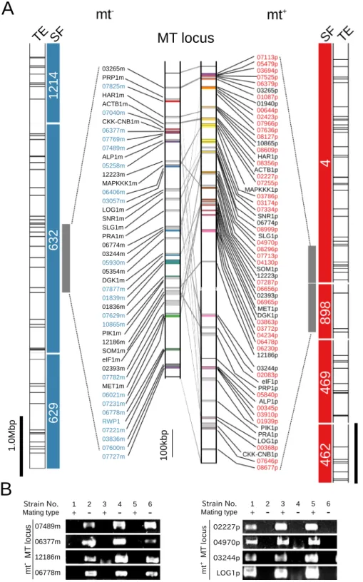
and 2; Supplementary Fig. 1). To select the MTS PacBio long reads located within a particular narrow region, the ratio of the sum of the lengths of 5–15 successive MTS PacBio long reads on the same scafold per genomic length to that of the distal positions of the successive reads was determined (Supplementary Fig. 2). For reads located close together in a narrow region, the ratio reached 1 (see Supplementary Text for detailed analysis). his analysis identiied a scafold (# 632) containing a region that was highly divergent between the two mating types (Supplementary Fig. 3). In addition, the mapping results of the Illumina short reads from the two mating type genomes and RNA-sequencing (RNA-seq) reads derived from gametes and gametophytes were mapped, and the gene models predicted by the RNA-seq assemblies are shown in Supplementary Fig. 3. he highly adjacent MTS region was located in the middle of mt− scafold 632 over ~1.0 Mb (designated as the mt− MT locus), and this
region was particularly well mapped using short-read nucleotide sequences generated from the mt−, but not the
mt+, genome and RNA-seq reads (Supplementary Fig. 3, 4th and 5th lanes). In addition, two highly adjacent MTS
regions (mt+ MT locus) were identiied for mt+ (scafolds 4 and 898; Supplementary Fig. 2D–F, Supplementary
Figs 4 and 5). hese regions had lower gene density than the surrounding regions (8.2 and 5.8 genes/100 kb for the mt− MT locus and the mt+ MT locus, and 10.6 and 9.8 genes/100 kb for the regions around these loci,
respectively).
Using mt− scafold 632 and mt+ scafolds 4 and 898, homologous scafolds for the opposite mating type
genome were identiied based on a reciprocal homology search of scafolds, which revealed that all had com-plementary scaffolds, and the two mt+ scaffolds were estimated to be a single fragmented mt+ MT locus
(Supplementary Fig. 6). he mt− and mt+ MT loci, together with the surrounding complementary regions
iden-tiied in the lanking scafolds, extended for 7.19 and 7.33 Mb, respectively (Fig. 2A).
As no genome of Ulva relatives has yet been analyzed, there are no training data for gene prediction based on genome sequencing data. hus, for precise prediction of genes based on expression, sets of RNA-seq assemblies from gametes and gametophytes of the individual mating types were assembled and mapped on the scafolds in and around the MT locus. he sets of RNA-seq assemblies were gathered based on homology, and then deined as genes. hese analyses indicated that the mt− MT locus and mt+ MT locus contained 46 and 67 mRNA-coding
loci, respectively; several of the loci were assumed to generate splicing variants (Supplementary Tables 4 and 5). Comparisons of the genes in the mt− MT locus and the mt+ MT locus by reciprocal BLASTX analysis showed
that 23 genes were shared by the two regions (Supplementary Table 6). hese genes were deined as gametologs and were used to compare the genomic structures of the two regions; the results indicated that the mt− and mt+
MT loci were highly rearranged and contained many MTS reads (Fig. 2A; Supplementary Figs 3–5).
In XY and ZW systems, particular transposable elements accumulate in the sex chromosomes32. No such
accu-mulation of transposable elements has been detected in the MT loci of Chlamydomonas and Gonium, but it has been found in Volvox or Ectocarpus, Marchantia9,11,17. Transposable elements were predicted based on homology
with known transposable elements and comparison of the genome with itself. he results showed that transpos-able elements were present at the MT locus but were not more highly accumulated than in neighboring regions (mt−, MT locus: 0.32 ± 0.56/100 kb; neighboring region: 0.57 ± 0.84/100 kb; mt+, MT locus: 0.61 ± 0.85/100 kb;
neighboring region: 0.62 ± 0.81/100 kb) (Supplementary Fig. 7).
U. partita has no genetic marker for estimating homologous recombination. hus, a genomic PCR analysis was performed using four MT locus genes in both mating types for the two genome-sequenced strains and four other strains (mt−, MGEC-3 and 5; mt+, MGEC-4 and 6) isolated from diferent areas along the Japanese coast
(Supplementary Table 6). All examined genes were linked to the mating types of the individual isolates (Fig. 2B), suggesting that these two regions contain the characteristics expected of an MT locus.
Finally, to examine the linkage between mating type and the identiied MT locus, the mt− strain, which was
a diferent isolate than the one for which the genome was sequenced, was crossed with the mt+ strain, and the
linkage between the mating types of their progeny and the unique gene markers of the MT locus was examined (Supplementary Table 7). A total of 10 of 16 progeny were mated with an mt+ tester strain, and 7 of 16 progeny
in the moss, with the other organisms harboring sex-determining regions (SDRs) in the UV chromosomes. he green seaweed Ulva partita has a life cycle with even domination of haploid and diploid phases, but it is not oogamous, in which gametes diferentiate into eggs and sperms. he bilagellate gametes are anisogamous, apart from their size and ultrastructure. U. partita develops isomorphic gametophytes with a tubular thallus, and somatic cells diferentiate into gametes24. Gametes of opposite mating types fuse, and a zygote develops into a
sporophyte that is identical to a gametophyte in terms of morphology. he sporophyte somatic cells diferentiate into zoospores that have four lagellae and are slightly larger than the gametes. he gametes have two mating types, mt−
and mt+
, deined by the inheritance of chloroplast DNA from the mt+
gamete to the zygote23,24. hus,
Figure 2. Genomic structures of the mating type (MT) locus in the green seaweed Ulva partita. (A) Scafolds, transposable elements surrounding the MT locus, the MT locus in the green seaweed U. partita. SF, the MT locus scafolds and neighboring scafolds in both the mt+ and mt− strains are shown. Blue and red bars indicate mt−
and mt+ scafolds, respectively. Numbers are scafold numbers. Transposable elements (TEs) of the same type are
indicated by the same colors. he predicted MT locus genes were mapped on both genomes, and the positions of the 23 gametologs were compared between the mt+ and mt− strains. he colored vertical bars indicate individual
mating type-speciic genes. Light gray vertical bars indicate mating gametologs. mt−, mating type minus; mt+,
mating type plus. (B) Genomic PCR of MT locus genes for distinct wild-type strains. he presence of the U.
partita MT locus genes in the MGEC-2 (mt−) and MGEC-1 (mt+) strains was conirmed in four other strains
were mated with an mt−
tester strain. Eight of the mt−
progeny harbored only mt−
MT locus markers, but two of them harbored both types of MT locus marker. In addition, six of the mt+
progeny harbored only mt+
MT locus markers.
Evolution of gametologs in the
U
.
partita
Mt locus.
To analyze the evolutionary history of the gametologs in the MT locus, the homologous sequences of an MT locus gene encoding proliferation-associated protein 1, PAR1, and a gene encoding G-strand telomere-binding protein 1, GTBP1, in a region neighboring the MT locus, were isolated from species related to U. partita. Molecular phylogenies were then reconstructed (Fig. 3A and Supplementary Table 7). In all species examined, two types of homologous sequence were identi-ied from the two distinct mating types, and the phylogenetic tree showed that the genes could be classiidenti-ied into two clades (Fig. 3A). In addition, these two clades were associated with the previously determined mating typesFigure 3. Evolution of the gametologs at the mating type (MT) locus. Molecular phylogenies of a gametolog in and a gene outside the MT locus. (A) Genes orthologous to an MT locus gametolog (encoding proliferation-associated protein 1, PAR1) from other species of the order Ulvales and (B) those of a gene outside the MT locus (encoding G-strand telomere-binding protein 1, GTBP1). he mating type of each strain of the species was determined previously (see Supplementary Table 7) and is indicated ater the species name. Bootstrap values were obtained from analyses of 100 pseudoreplicates and are shown close to the branches (>50). he numbers
above the scale bar indicate nucleotide substitutions per site. (CF) Synonymous (dS) and non-synonymous (dN) substitution rates of gametologs between the mt+
and mt−
strains of green algae. (C) dS and dN values for
C. reinhardtii. (D) dS and dN values for Volvox carteri. (E) dS and dN values for Ulva partita. (F) dN/dS ratios of three algae. Up, U. partita. Cr, C. reinhardtii. Vc, V. carteri. MS, model selection method. MYN, modiied YN method. Lines show speciic thresholds of the dN/dS ratio. To make the dN/dS ratio of a gametolog clearly understandable, useful thresholds of this ratio (1, 0.2, 0.1, and 0.05) are indicated as follows: continuous line, 1, dashed line, 0.2, dotted line, 0.1, and dashed and dotted line, 0.05. (G) dS values and (H) dN values of the gametologs and genes around the MT locus. dN and dS values of the gametologs and the genes around the MT locus were calculated and plotted according to their positions in the mt−
(Supplementary Table 7). In contrast, the neighboring-region genes in each species were almost identical and were not classiied into diferent clades in the molecular phylogeny (Fig. 3B). hese data suggest that the investi-gated gametolog existed in the MT locus when this locus was established and evolved independently within the MT loci of the individual mating types.
Next, to determine the type of selective pressure exerted on the gametologs ater their divergence, the nucleo-tide substitution rates at synonymous and non-synonymous sites (dS and dN, respectively) were estimated. hey were also compared with those for genes at the known MT locus of Chlamydomonas and in the SDR of Volvox9,13
(Supplementary Tables 8 and 9). Mean distances between individual genes on the plot were also calculated as an index to compare the divergence of MT locus genes (Supplementary Tables 10–12). Among 23 genes with several mRNA variants deined as gametologs by BLASTX analysis, two (06550 m/01365p and 12186 m/06628p) did not have coding sequences (CDSs) that could be aligned between gametologs; thus, the CDSs of the other 21 game-tologs were aligned.
Both maximum-likelihood and approximate methods showed that the synonymous substitution rates for the gametologs in U. partita were considerably higher than the non-synonymous rates, and the non-synonymous rate/synonymous rate (dN/dS) ratios were <1 for all genes except one (means were 0.16 ± 0.36 for the approx-imate method and 0.16 ± 0.40 for the maximum-likelihood method), suggesting that these genes have been exposed to negative selective pressure and that their functions are highly restricted (Fig. 3E,F and Supplementary Table 13).
Mean dS values were much higher in U. partita and Volvox than in Chlamydomonas, and those of U. partita
were higher than those of Volvox (Supplementary Fig. 8 and Supplementary Table 13). In addition, the dN means of U. partita and Volvox were higher than that of Chlamydomonas, but the diference between U. partita and
Volvox was small.
Means of all distances between the two dots for each estimation method were calculated as an index of scattering (Supplementary Tables 10–13). If the mean distance is short, the dN/dS ratios would be expected to be homogeneous. he mean distances for U. partita in the two estimation methods were 1.43 ± 1.19 and 1.59 ± 1.23 (Supplementary Table 10). All mean distances were lower in Chlamydomonas than in U. partita, while some of the dN/dS ratios were slightly higher (0.17 ± 0.18 for the approximate method and 0.17 ± 0.17 for the maximum-likelihood method) and were plotted in a small area (mean distances were 0.03 ± 0.02 for the approx-imate method and 0.03 ± 0.02 for the maximum-likelihood method; Fig. 3C,F; Supplementary Table 11). Mean distances in Volvox were similar to those of U. partita (1.20 ± 0.92 for the approximate method and 1.10 ± 0.98 for the maximum-likelihood method), but comparison of the standard deviations showed that the divergence of their ratios was similar to that of Chlamydomonas (0.21 ± 0.22 for the approximate method and 0.22 ± 0.24 for the maximum-likelihood method; Fig. 3D,F; Supplementary Table 12). he molecular phylogeny and nucleotide sub-stitution rates suggest that the U. partita gametologs were present at a common MT locus before the divergence of the relatives and that this region experienced a prolonged period ater separation.
he synonymous and nonsynonymous substitution rates of the genes around the MT locus were estimated by the two methods. From the scafold data around the MT loci of mt− and mt+, the CDSs of the genes were
extracted and their associations were determined using BLASTN. Ater estimation of the synonymous and nonsynonymous substitution rates of 119 genes by the two methods, both values were plotted according to the mt− positions of the MT locus (Fig. 3G,H). he data showed that almost all synonymous and non-synonymous
substitution rates of the genes around the MT locus were near zero or zero; additionally, the synonymous sub-stitution rates were higher than those of the MT locus, and the non-synonymous subsub-stitution rates were slightly higher than those of the MT locus.
It has been reported that relaxed codon usage bias occurs with reduced recombination in sex chromosomes33,34.
Furthermore, the codon usage in CDSs obtained from all mRNA data and the codon usage for the MT locus genes were compared with those of other autosomal locus genes (Supplementary Table 14). he codon usage patterns of all of the autosomal genes and the MT locus genes did not appear to difer (Supplementary Fig. 9), and comparisons between them and those of mt− and mt+ autosomal locus genes showed high correlations (Poisson’s
correlation, 0.98; p-value, 2.2 × 10⁻¹⁶). However, the codon usage of half of the MT locus genes in both mating
types difered signiicantly from that of the autosomal genes, and these included the gametologs, which showed a low dN/dS ratio (Supplementary Table 14).
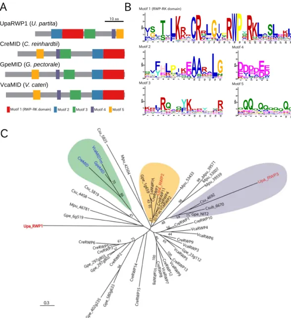
Motifs and molecular phylogeny of RWP-RK domain-containing proteins in the MT locus
and autosomes.
We investigated whether there were orthologs among the U. partita MT locus genes, theChlamydomonas MT locus, and the Volvox SDR. Although no gene was clearly shared among the MT loci and SDR in the three species, very weak homology with MID was found in a gene only in the U. partita mt− MT
locus, named UpaRWP1. To assess the relationship between MID and UpaRWP1, a BLAST analysis was per-formed using all Chlamydomonas RWP genes as queries against the entire U. partita mRNA database from the assembly of RNA-seq data. Two of the autosomal RWPs (UpaRWP2 and UpaRWP3) were identiied and mapped to locations other than the MT locus. In addition, genes encoding proteins containing the RWP-RK domain in Chlorophyta were collected from the annotated genes from the genomes of ive species: Chlamydomonas rein-hardtii, Volvox carteri, Gonium pectorale, Coccomyxa subellipsoidea, and Micromonas pusilla. Although these genes encode proteins containing a single RWP-RK domain, the protein lengths are very diverse.
Figure 4. Conserved motifs and molecular phylogeny of U. partita RWP1, Volvocales MIDs, and RWP-RK domain-containing proteins in green algae. (A) Schematic of the protein structure of U. partita RWP1. Among proteins containing the RWP-RK domain in Chlorophyta, conserved motifs were identiied by using MEME, and the motifs of U. partita RWP1 and Volvocales MIDs are shown by distinctly colored boxes, relecting their positions and lengths. Gray bars show the total lengths of the individual proteins. In particular, Motif 1 contains RWPxRK sequences. Bar shows 10 amino acids. (B) Sequence logos of individual motifs. Information contents of individual amino acids at a position in each motif are visualized by the height of the capital letters designating amino acid residues. he y-axis represents the bit value, as information content, of which the maximum is log220 ≈ 4.32. he x-axis represents the position of an amino acid residue in each motif. (C) Molecular phylogeny of the RWP-RK domain-containing proteins in Chlorophyta. From the deduced amino acid sequences of the proteins containing the RWP-RK domain in the genomes of Ulva partita, Volvox carteri, C. reinhardtii, Gonium pectorale,
Coccomyxa subellipsoidea, and Micromonas pusilla, the amino acid sequences of motifs conserved in all proteins were combined in individual proteins, and the aligned combined sequences were used for the construction of an unrooted molecular phylogeny using the maximum-likelihood method. U. partita RWP1 (UpaRWP1) in the MT locus is shown in red. he highlighted genes indicated in green, orange, and purple are classiied into clades containing Volvocales MIDs (blue), one of the U. partita autosomal RWP genes (UpaRWP2; accession ID, DN37992), and the other of the U. partita autosomal RWP genes (UpaRWP3; accession ID, DN130398), respectively. he preixes before the gene symbols are abbreviations of the species names: Upa, U. partita, Cre,
A molecular phylogenetic tree was constructed using all ive motifs of UpaRWP1, two U. partita autosomal RWPs, MIDs, and other Chlorophyta RWPs (Fig. 4C). he three MIDs were classiied into a clade with high statistical support, whereas UpaRWP1 was classiied into a diferent clade from that containing the MIDs, albeit with low statistical support (Fig. 4C). In addition, an autosomal U. partita RWP (UpaRWP2) was classiied into a clade containing Chlamydomonas RWP11 (CreRWP11) with high statistical support and few amino acid sub-stitutions35. he other (UparRWP2) was classiied into a clade containing NIT2, which is a regulator of nitrate
assimilation36. hus, UpaRWP1 difered not only from MIDs but also from the autosomal RWPs, suggesting that
this gene is not an ortholog among the three species and was acquired independently in the U. partita MT locus and the two other species.
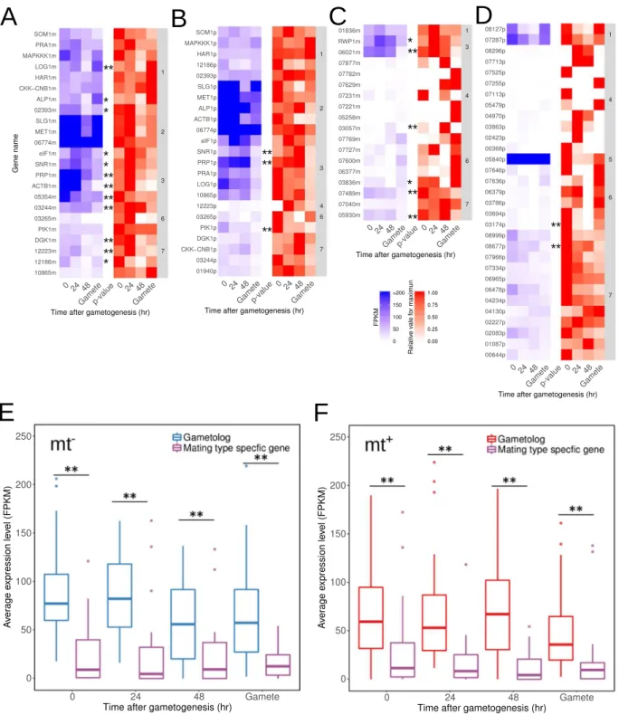
Expression of MT locus genes in gametogenesis.
Mating type-speciic genes are expected to provide genetic diferences to opposite mating types ater meiosis, and the expression of the MT locus genes may provide diferentiation of gametophytes and gametes between mating types. On the other hand, there are no diferences in gametophytes between mating types in U. partita, but there are some diferences in gametes with asymme-try of mating structure and eye spot and mt−-speciic fusion machinery25,37. To analyze the expression levels ofthe U. partita MT locus genes, RNA-seq data during gametogenesis from the two mating types with biological replications were mapped on the genome, and the expression levels of the MT locus genes were estimated. he expression levels of all splicing variants are shown with the results of one-way ANOVA for four time points and the Dunnett test for multiple comparisons between gametophytes before induction and at various time points ater gametogenesis (Supplementary Table 15). Ater removing splicing variants with low expression and cluster-ing these data, the expression data of gametologs and matcluster-ing type-speciic genes as well as the relative expression changes were plotted separately (Fig. 5A–D). he expression data showed that most of the genes, including both the unique genes and the gametologs, were expressed constantly during gametogenesis in both mating types. In addition, the expression levels of gametologs were much higher than those of the mating type-speciic genes (Fig. 5E,F).
Statistical analyses showed that the expression levels of 18 and 6 genes changed signiicantly during game-togenesis in mt− and mt+, respectively. Among the 18 mt− MT locus genes, the two gametologs LOG1m and
ALP1m were upregulated in gametes; two gametologs (DGK1 and 12223m) and two mating type-speciic genes (07489m and 05930m) were classified into cluster No. 7 and, except for 12223m, were downregulated dur-ing gametogenesis; six gametologs (elF1m, SNR1m, PRP1m, ACTB1m, 05354m, and 23244m) and two mat-ing type-speciic genes (RWP1 and 06021m) were co-expressed (cluster No. 3), and, except for 05354m, their expression levels increased at 24 h ater gametogenesis and decreased to the pre-gametogenesis level in gametes (Fig. 5A,C). Of the mating type-speciic genes at the mt− MT locus, the expression level of 03057m was increased
at 48 h ater gametogenesis and then decreased to zero in gametes; the others were downregulated in gametes. Among the 6 mt+ MT locus genes, two gametologs (SNR1p and PRP1p) were classiied into co-expression
clus-ter (#3, see above); one other gametolog (Pik1p) and two mating type-speciic genes (03154p and 08677p) were downregulated (Fig. 5B,D). In summary, among the genes signiicantly upregulated in mt−, thirteen mt− genes
and two mt+ genes were signiicantly upregulated during gametogenesis or in gametes, and the others were
downregulated.
Discussion
Comparison of the genomic structures of the
Ulva partita
Mt locus with those of other
organ-isms.
In this study, we used a third-generation sequencing technology with a single-molecule sequencing method to identify the putative mating locus in the genome of the green macroalga Ulva partita. he size of theU. partita MT locus (~1.0–1.5 Mb; Fig. 1 and 2) resembles that of the SDR of Volvox, which is a UV system with a dominating haploid phase in a life cycle showing phasic heteromorphism (sporophytes do not develop, and mei-osis occurs in the zygote) and gamete dimorphism (eggs and sperm), and the brown alga Ectocarpus, which is also a UV system with a haploid-diploid life cycle with phasic heteromorphism and gamete dimorphism (motile and immotile gametes in males and females, respectively)9,17. hese sizes are smaller than those in two fungal MT loci
(Neurospora and Microbotryum) and larger than those in unicellular and colonial green algae (Chlamydomonas
and Gonium)9,10,18–20. In addition, the gametolog location rearrangements in the individual mating types
resem-ble not only the SDRs of Volvox and Ectocarpus but also the MT loci of all others sequenced to date. herefore, genomic rearrangement in the MT loci and SDRs is a common phenomenon in haploid organisms. Accumulation of transposable elements and low gene content are found in the MT loci and SDRs of Volvox, Ectocarpus, and
Microbotryum but not of Chlamydomonas, Gonium, or Neurospora. Note that the Neurospora locus is thought to be young and therefore to show less accumulation of transposable elements18. he Ulva MT locus showed lower
gene content but not a high level of transposable element accumulation. he low gene content suggests chro-mosomal degeneration with gene loss, while the low level of transposable element accumulation may relect the shortage of transposable element data for Ulvophyceae.
Chlamydomonas and Gonium are unicellular and colonial, respectively, with few cells in their gametophytes, and meiosis occurs in diploid spores38. his is the clearest diference from other organisms except the two fungi Neurospora and Microbotryum. On the other hand, the fungi Neurospora and Microbotryum exhibit automictic reproduction, which is a mating system involving a meiotic tetrad20,39. Such automictic reproduction is predicted
to favor successive linkage to a set of mating type genes that experience deleterious and recessive mutations40,41.
have not yet been reported11. When available, they will likely provide some insight into the evolution of the MT
locus/SDR in organisms in which the sex/mating type is determined in the haploid stages.
Figure 5. Expression changes in the mating type (MT) locus during gametogenesis. (A) Gametologs of mt−
. (B) Gametologs of mt+
. (C) Mating type-speciic genes of mt−
. (D) Mating type-speciic genes of mt+
. he blue heat map shows the average fragments per kilobase of exon per million fragments (FPKM) values from three biological replicate for gametologs and mating type-speciic genes in each mating type during gametogenesis. High FPKM values (>200) are shown in the same color as values of 200. he red heat map shows relative values
for maxima of FPKM values. he numbers in the right column are the numbers of clusters calculated by the k-means method. Results of one-way ANOVA for individual genes are shown on the right side of the heat maps (**p < 0.01, *p < 0.05). (E),(F) Average FPKM and values normalized to the beta-tubulin gene. Box-whisker
Although the MT locus genes were tightly linked to the mating types of the U. partita isolates, inheritance patterns from a sporophyte to gametophytes were somewhat unusual. Several progenies had the both mating type-speciic genes, suggesting that they are diploid. However, these progenies mated with the mt+ tester strain.
We now hypothesized that this is an apomixis-like phenomenon found in several organisms and will report on this in more detail in the future. In addition, the inding that the gametes of the diploid progenies mated with the mating type plus gametes is similar to an observation in Chlamydomonas, in which diploid gametes artiicially generated by using auxotrophic mutants exhibited the mt− phenotype42. his suggests that the MT locus gene(s)
of U. partita may determine the mating type.
Evolution of the
U
.
partita
Mt locus.
A comparison of the dS and dN values of the gametologs in the U.partita MT locus showed that the dS values were signiicantly higher than the dN values (Fig. 3). Although they are dependent on the substitution model and generation time, dS values are underestimated when they are greater than ~243. In total, 15 dS values for the approximate method and 13 for the maximum-likelihood method were >2, indicating that nucleotide substitution at a given site may have occurred several times and that substitutions at some sites are saturated. hus, the possibility that the dS values estimated from this data set are not accurate cannot be ruled out. In contrast, with the exception of one gene, all dN values were <1. his suggests that the dN/ dS ratios for all gametologs, except one, are <1, although dS may be over- or underestimated, and that the genes have been exposed to purifying selection with a functional constraint.
Volvox is considered to have diverged from a unicellular ancestor, similar to Chlamydomonas, at least 200 Mya12. Comparison of the MT locus/SDR and neighboring autosomal genes between Chlamydomonas and Volvox
suggested that the expansion from an ancestral MT locus to an SDR involving neighboring autosomal genes occurred with cooption of gene functions12. Although the timing of the establishment of the U. partita MT locus
is currently unclear, the molecular phylogeny of a gametolog among U. partita relatives was associated with mating type, and the U. partita gametologs showed high proportions of synonymous substitutions (Fig. 3). here are no fossil samples available to calibrate the molecular clock of the order Ulvales, including U. partita, and it is therefore diicult to determine the timing of the establishment of the Ulva MT locus. However, the diversity of gametologs and the molecular phylogeny within Ulvales suggest that this locus was established at least at the ori-gin of the examined species and has experienced a long period of evolution. he low diversity of the dN/dS ratio in the U. partita gametologs suggests that no such expansion during evolution of the Volvox SDR has occurred for a prolonged period, because newer gametologs would be expected to have lower dN and dS values than those of older genes if there had been expansion involving the addition of autosomal genes adjacent to the MT locus9;
alternatively, rapid gene losses may have been occurring, and this may be related to a larger number of mating type-speciic genes than are present in other green linage organisms. his is similar to the SDR of the UV chro-mosome in Ectocarpus, which was estimated to have been established more than 70 Mya17.
Evolutionary relationship between
Chlamydomonas MID
and an Mt locus gene encoding
an RWP-RK domain.
In the Chlorophyta, a gene determining mating type has been identiied only inChlamydomonas, namely, Minus Dominance (MID), which is located at the mt− MT locus29. his gene encodes
a putative transcription factor containing an RWP-RK domain, which includes a leucine zipper-like motif29,30.
Although MID homologs have been found across the Volvocales and the Volvox MID (VcaMID) is located on the SDR of the V chromosome in males, genetic manipulation data and the constitutive expression of VcaMID in males during both vegetative and sexual stages suggest that this gene does not play a role in sex determination but instead has a sex-speciic function in the diferentiation of male vegetative cells into sperm9,31,44. One RWP-RK
domain-containing gene was found only at the mt− MT locus of U. partita and was named RWP1 (Fig. 4). Genes
containing the RWP-RK domain are present in the genomes of various plants, including green algae, and their orthologs in Arabidopsis play roles in the development of eggs and embryos45–49. Although RWP1 is a potential
determinant of mating type, transcriptome analysis showed that it is expressed even in gametophytes, with a slight increase at an early time point, and decreases to the initial level in gametes, suggesting that this gene is related to mating type diferentiation at the transcriptional level (Fig. 5C). his is similar to the case of Volvox MID9. If RWP1 is a master gene for mating type determination, future studies should address whether post-transcriptional or post-translational regulation occurs during gametogenesis and, if so, which mechanisms underlie this process.
Degeneration of
U
.
partita
Mt locus genes.
Expression levels of most of the MT locus genes were constant during gametogenesis in both mating types, and those of gametologs were much higher than those of mating type-speciic genes (Fig. 5E,F). Ectocarpus sp. show much lower transcript abundance in haplotype mating type-speciic SDR genes, and this may relect degradation of the promoter and cis-regulatory sequences of these SDR genes17. his corresponds to the mating type-speciic genes of U. partita. Although comparisons amongspecies closely related to U. partita are required, these low levels of mating type-speciic gene expressions may indicate their degeneration via mutations not only in protein-coding sequences but also in promoter regions. Degeneration is supported by the presence of degeneration signals such as relaxed codon usage bias in both gametologs and mating type-speciic genes (Supplementary Table 14).
expression levels at the early stage of gametogenesis. hese genes encoded orthologs of actin (ACTB1m), alin-like protein (ALP1m), small nuclear ribonucleoprotein polypeptide G (SNR1m), proliferation-associated protein 1 (PRP1m), and eukaryotic initiation factor (eIF1m) that are expected to be involved in important cellular func-tions, and their allelic genes, except eIF1m and PRP1m, did not show changes in expression levels, suggesting that these genes may modulate diferentiation of gametes via transcriptional regulation. On the other hand, other mat-ing type-speciic genes (RWP1 and 06021m) were upregulated, and still others were downregulated; most of them, except RWP1, were shown to encode proteins with no homology to known proteins, making it diicult to predict their function(s) during gametogenesis. Our group and another team have developed a system to introduce and transiently express a transgene using a polyethylene glycol method. his method, as well as the application of other methods, including RNA interference and genome-editing technologies, will provide further information regarding the molecular functions of the MT locus and its constituent genes50,51.
In conclusion, we identiied a locus linked to mating type in the green macroalga U. partita with an isomor-phic haploid-diploid life cycle and without oogamy. his locus was highly rearranged and exhibited suppressed recombination for a prolonged period. In addition, the U. partita MT locus has features similar to UV chro-mosomes. Although the U. partita mt− MT locus harbored a gene encoding a protein containing an RWP-RK
domain, RWP1, which is found in the Chlamydomonas mating type determination gene MID, this gene is not an ortholog of MID. During gametogenesis, the expression level of RWP1 increased once and then decreased in gametes as much as in gametophytes.
Materials and Methods
Algal materials and culture conditions.
Algal strains. he pairs of mt−(MGEC-2) and mt+
(MGEC-1) strains of Ulva partita used were collected from the coast of Japan (Supplementary Table 7)23–25,52. We recently
renamed this species from Ulva compressa to U. partita based on its molecular phylogeny and morphology35. he Ulva strains were obtained from culture collections at Kochi University, and their mating types were determined previously based on gamete sizes53–55. he strains are maintained in the culture collection of Nagasaki University
(Nagasaki, Japan).
Culture conditions. Laboratory cultivation and induction of gametogenesis were performed as described pre-viously23,56. Briely, thalloid gametophytes were grown at 16 °C under 150 µmol photons m−2 s−1 light under a
10 h:14 h light (L):dark (D) cycle in artiicial seawater for 28 days. hen, 2 days ater the induction of gametogen-esis by rinsing several times and transferring to long-day conditions at 23 °C under 150 µmol photons m−2 s−1
light under a 14 h:10 h L:D cycle in seawater, migrating gametes were released from the gametophytes. Positive phototaxis was used to collect the gametes. his alga was cultured with symbiotic bacteria because inappropriate development occurs in the absence of symbiotic bacteria.
Genome and RNA-sequencing.
DNA isolation. To remove bacterial DNA contamination prior to genome sequencing, gametic cells were gathered by illumination using a natural white luorescent light that induced positive phototaxis. Gametes of both mating types with a fresh weight (FW) of approximately 1.5 g were collected. he collected gametic cells were frozen in liquid nitrogen, ground, and subjected to genomic DNA isolation using a Plant Maxi Kit (QIAGEN, Venlo, he Netherlands).RNA extraction. Gametic cells were collected by the same method as used to gather cells for RNA isolation. Gametophytic thalli were collected at 0, 24, and 48 h ater the induction of gametogenesis, and three replicates of each mating type were included. Total RNA was extracted from 50 mg of gametic cells and gametophyte thalli using an RNeasy Plant Mini Kit (QIAGEN) according to the manufacturer’s protocol. Contaminating DNA was removed using RNase-Free DNase I (QIAGEN).
DNA and RNA-sequencing. Genomic sequences of U. partita MGEC-1 and MGEC-2 were determined using PacBio single-molecule real-time sequencing (long reads) and Illumina MiSeq for paired-end (PE) short reads. Briely, for PacBio sequencing, a library was constructed using the PacBio DNA Template Prep Kit 2.0 (Paciic Biosciences, CA, USA) according to the manufacturer’s protocol. For Illumina sequencing, a library was con-structed using the TruSeq- DNA LT Sample Prep Kit (Illumina, CA, USA). he PacBio sequencing and selection of reads of more than 500 bp provided 1.7 M reads of 12.0 Gb and 2.7 M reads of 16.6 Gb from the mt− and mt+
genomes, respectively. he average lengths of mt− and mt+ PacBio reads were 7182 and 6136 bp, respectively.
he Illumina sequencing generated 271 M reads of 100 bp (totaling 27.1 Gb) and 252 M reads of 100 bp (totaling 25.2 Gb) from the mt− and mt+ genomes, respectively. Sequences in the obtained long reads were corrected by
mapping the short reads and comparing individual sites, and the corrected long reads were assembled into scaf-folds using HGAP3 sotware (Paciic Biosciences, CA, USA). Finally, BLASTN analysis was performed using the scafolds as query sequences against the RefSeq microbial genome database (http://www.ncbi.nlm.nih.gov/
refseq/) with a threshold e-value of 1 × 10⁻³⁰ to exclude contaminating sequences from the symbiotic bacteria.
For mt− and mt+ genomes, the inal numbers of scafolds ater removing bacterial genome contamination were
851 and 1385, and the total lengths of scafolds were 110.2 and 116.7 Mb. hese sequences were used for later analyses. Ater assembly, the Illumina short reads were mapped onto the assembled sequences of both mating types. he proportions of properly paired reads were 99.8% and 99.5%, respectively. In addition, the proportions for mt− scafold 632 and mt+ scafolds 4 and 898 were 99.2%, 99.1%, and 99.4%, respectively.
was sequenced using an Illumina HiSeq 2500 instrument. A summary of the reads obtained is shown in Supplementary Table 1.
Identiication of mating type-speciic long genomic sequence reads. he long reads and the scafolds were used to identify mating type-speciic genomic regions. First, the corrected long reads from mt− were mapped to the
scafolds from mt+ by BLAST search57 using the criteria of nucleotide sequences >3 kb matched with 97% identity
and a gap length of less than 100 bp, and then the unmapped long reads were obtained. For the individual mating types, RNA-seq data derived from the gametes and the gametophytes before gametogenesis were assembled into isotigs using Newbler (Roche Applied Science, Penzberg, Germany) with default parameters, and these isotigs were used as gene models. he isotigs of a mating type were mapped on the scafolds of the same mating type. he mt− unmapped long reads on the above mt+ long scafolds were used as query sequences against a database
gen-erated from the mt− scafolds using BLASTN, and a pool of long reads with e-values of >1 × 10−100 was selected.
From the selected long reads, MTS reads were deined using the following criteria: 1) identity with a region over-lapping the read was less than 80%, 2) the read contained the gene model(s), and 3) length >1 kb. An equivalent analysis of the mt+ long reads was also performed. Finally, 241 and 320 sites were identiied for mt− and mt+,
respectively. A summary of the MTS reads is shown in Supplementary Table 2.
Identiication of MTS genomic scafolds. To identify genomic regions in which successively mapped MTS reads were present, “moving sums” were calculated. he length of a read among the MTS reads on a scafold and lengths of n − 1 reads toward the 3′ end from the irst read were summed (l, vertical axis in Supplementary Fig. 3) and
deined as the “moving sum” unit. From a read at the 5′ end of a scafold to its 3′ end, the same calculations of
moving sums were performed. In addition, the distance between the position at the 5′ end of the irst read of a
moving sum and the position at the 3′ end of its last read, which were n reads, was calculated (L, horizontal axis
in Supplementary Fig. 3). All moving sums for n = 5, 10, and 15 were calculated. he l/L ratio was used as an
index of the extent to which the MTS reads were successive on a scafold that is part of the U. partita genome. If the moving sum is completely successive, the ratio will be >1. MTSs that met the following criteria were
sub-jected to further analysis: (1) l/L was >0.1, (2) l was >50 kb in n = 15, and (3) L was >1 Mb. Scafold 632 from
mt−
and Scafold 4 and Scafold 898 from mt+
were identiied as containing highly successive MTS reads (HMTS scafolds).
Comparison of MTS genomic scafolds from mt−
and mt+
genomes. he data from short reads, MTS reads, Culinks gene models (not the same as the models from isotigs: see RNA-seq analysis for the generation of these gene models), and RNA-seq reads from gametes and gametophytes were mapped onto the scafolds and visualized using the GBrowse genome browser58. he HMTS scafolds were used as query sequences against the
database for the opposite mating type using LAST (long-sequence alignment sotware) with default parameters59,
and their counterpart scafolds were identiied. his process was performed reciprocally, and three scafolds for mt− (#632, #629, and #1214) and four scafolds for mt+ (#4, #898, #462, and #469) were identiied. hese scafolds
were aligned and visualized as a dot plot using a script in the LAST sotware with default parameters. Finally, the mt− and mt+ HMTS scafolds were estimated as complementary scafolds, and the sums of the HMTS scafolds
and adjacent scafolds for mt− and mt+ were 7.19 and 7.33 Mb, respectively.
Comparison and visualization of the MT locus. he regions containing HMTS reads were ~1.0 and ~1.5 Mb, respectively. RNA-seq data of gametes and gametophytes in the individual mating types were merged and bled using Newbler, and CDSs were generated automatically. hese isotigs, were mapped on the genomic assem-blies of both mating types, and genes contained in these regions were identiied from the isotig gene models. In total, 84 and 95 genes with splicing variants were identiied for mt−
and mt+
, respectively (Supplementary Tables 3 and 4). Clustering analyses using the DNACLUST package60 were performed for the genes in the HMTS
genomic regions in mt−
and mt+
; ater manual correction of the clusters, they were classiied into 46 and 67 clus-ters that were deined as splicing variants transcribed from individual loci. hese genes were compared by using reciprocal BLASTX analyses61 with e-values of >1 × 10⁻³, and 23 were identiied as gametologs (Supplementary
Table 5). Representative genes were selected and their positional data in the scafolds were visualized using the ggplot2 (1.0.0) and ggbio (1.14.0)62 packages in R (3.1.3). he resulting data were modiied using drawing
sot-ware. he identiied regions in the mt−
and mt+
scafolds were termed the MT loci. he nucleotide sequences of the MT loci were submitted to the DNA Data Bank of Japan (DDBJ; accession numbers: LC091542 for the mt+
MT locus; and LC091540 and LC091540 for mt−
MT loci in scafolds 4 and 898, respectively). he nucleotide sequences of the MT locus genes for RNA-seq analysis were submitted to DDBJ, and the accession numbers are shown in Supplementary Tables 3 and 4.
Segregation analysis. From MGEC-5 (mt−) and MGEC-2 (mt−) gametophytes, gametes were induced and
mixed. hen, mated zygotes were gathered together by negative phototaxis. Ater cultivation for 3 weeks, some sporophytes were transferred to MGEC-5 or MGEC-2 culture lasks, from which zoospores were induced by the same method as for the gametogenesis. Microscopy was used to determine whether the zoospores had four lagella, which are diferent from gametes with two lagella. Zoospores that showed exactly four lagella were cul-tured in 1-L lasks for 3 weeks. Approximately 100 small gametophytes of MGEC-5/MGEC-2 progeny were trans-ferred into respective 1-L lasks and cultured for 3 weeks. Before checking the mating types, small pieces of thalli were collected, frozen in liquid nitrogen and stored at −80 °C until the extraction of genomic DNA. Gametes were induced from approximately 50 healthily developed gametophytes. MGEC-2 (mt−) and MGEC-1 (mt+) were used
by negative phototaxis and the observation of zygotes. Finally, the mating types of a total of 16 MGEC-5/MGEC-2 progeny were determined.
Identification of repeats. To identify repetitive sequences, we used RepeatMasker (ver. 4.05) and RepBase (20140131) with RepeatMasker in polymorphism mode (Open-4.0. 2013–2015, http://www.repeatmasker.org). From all mt−
genome assemblies, in total, 92 kb of repeats containing 1,126 transposable elements, 223 small RNAs, and one satellite were identiied. For all mt+
genome assemblies, in total, 106 kb of repeats containing 1,147 transposable elements, 173 small RNAs, and one simple repeat were identiied. he data of the mt−
scafolds (#632, #629, and #1214) and the mt+
scafolds (#4, #898, #462, and #469) were extracted and visualized using R/ ggbio.
Molecular evolution analysis.
Phylogenetic analysis. To construct phylogenetic trees, an MT locus gene (PRA1) and a gene (GTBP1) in the lanking region of the MT locus (homologous genes) were isolated from Ulva spp. (Supplementary Table 7). he CDS regions of the two genes from the examined species were amplified using degenerate primers (mt−PRA1m/mt+
PRA1f, 5′-TTCATTGCYGTTCAAGCTACWAC-3′
and 5′-AACAAGCTCWCCRTCTTTCTCCCA-3′; G-strand telomere-binding protein 1 (GTBP1),
5′-TGGCGCACATCATGGCAAGATT-3′ and 5′-CAGCCCCACTGATCGAGCTTCAC-3′). he PCR program
for PRA1 and GTBP1 consisted of one initial denaturation step for 2 min at 94 °C, followed by 45 cycles of dena-turation for 30 s at 94 °C, annealing for 30 s at 50 °C, and extension for 40 s at 68 °C. Sequences were aligned using Muscle in MEGA663. Model tests for each analysis were performed using KAKUSAN 4.064. he best it models
for maximum-likelihood analysis were GTR + G for mt−
PRA1m/mt+
PRA1p and J2 + G for GTBP1, based on
the Akaike information criterion (AIC). Phylogenetic analyses were performed using the maximum-likelihood method in TREEFINDER65. Bootstrap values66 were obtained from analyses of 100 pseudoreplicates. he
nucle-otide sequences of the genes homologous to mt−
PRA1m/mt+
PRA1p and GTBP1 were submitted to DDBJ, and the accession numbers are shown in Supplementary Table 7.
Using the C. reinhardtii RWP-RK domain-containing proteins deined by Chardin et al.45 as queries, RWP-RK
domain-containing proteins were retrieved by BLAST analysis from data sets in Phytozome 11 (
http://phyto-zome.jgi.doe.gov/pz/portal.html) for C. reinhardtii (ver. 5.5), Volvox carteri (ver. 2.1), Coccomyxa subellipsoidea
(ver. 2.0), and Micromonas pusilla (ver. 3.0), and from NCBI for Gonium pectorale38. MIDs for Chlamydomonas
and Volvox were retrieved from NCBI9,13. U. partita autosomal genes encoding an RWP-RK domain-containing
protein were identiied with BLASTX using all Chlamydomonas RWP-RP domain-containing proteins as que-ries, similar to the description above, against the RNA-seq assembly database. he resulting data set served as input for a conserved motif analysis performed using MEME (http://meme.sdsc.edu/meme/meme.html), and five conserved motifs were identified. The five motifs were combined and used for molecular phylogenetic analyses. Phylogenetic analyses were performed using the maximum-likelihood method in MEGA6. Model tests for the analysis were also performed using MEGA6. he best-it model, based on AIC, for the RWP-RK domain-containing proteins was JTT + G. Bootstrap values were obtained from analyses of 100 pseudoreplicates. Calculation of synonymous and non-synonymous substitution rates. From mRNA assembly data, CDSs of game-tologs were extracted, and the deduced amino acid sequences were checked manually with BLAST analyses. If the amino acid sequences were not similar between gametologs, the full-length assembly sequences were analyzed with ORF inder (http://www.ncbi.nlm.nih.gov/gorf/gorf.html) and an appropriate frame was identi-ied. Ater manual checking, two gametologs were found to contain no CDSs that could form pairs with their counterparts. Pairs of CDSs for the individual gametologs were aligned using the ParaAT package67, and the
alignments obtained were used to calculate the synonymous and non-synonymous substitution rates using the maximum-likelihood and approximation methods with nucleotide substitution models in the KaKs_Calculator 2.0 package68,69. For comparison with the substitution rate in U. partita, CDSs of gametologs in C. reinhardtii and V. carteri9,13 were obtained from the NCBI database (Supplementary Table 8). he data were plotted using ggplot2
for R.
Calculation of codon usage. CDSs of all mRNAs of mt− and mt+ were extracted using a custom-made Perl script,
and codons in individual CDSs were counted using the cusp command of EMBOSS (ver. 6.6.0.0). he results were merged using a custom-made Python script. From these data, the MT locus genes and autosomal genes were separated, and Pearson’s product-moment correlations and p-values for the sums of individual codons of all autosomal genes and the MT locus genes were determined using the R default “cor” command. he correlation between all autosomal mt− and mt+ genes was 0.98 (p = 2.2 × 10⁻¹⁶).
Ampliication of MT locus genes. DNA was extracted from six U. partita strains using the CicaGeneus DNA Extraction Reagent DNA kit (Kanto Chemical, Tokyo, Japan). he mating types are given in Supplementary Table 7. To amplify DNA fragments of individual genes, the Kapa Taq PCR kit (Kapa Biosystems) was used in accordance with the manufacturer’s protocol. he PCR program for the ampliication of each gene consisted of an initial denaturation step of 3 min at 95 °C, followed by 45 cycles of denaturation for 15 s at 95 °C, annealing for 15 s at 60 °C, and extension for 90 s at 72 °C. Primer sets are shown in Supplementary Table 15. he ampliied DNA fragments were separated by electrophoresis and visualized by ethidium bromide staining.
RNA-seq analysis. Triplicate RNA-seq data from gametophytes, gametophytes ater the induction of gametogen-esis (24 and 48 h), and gametes from mt− and mt+ were obtained using an Illumina HiSeq. 2500. To compare the