Dual role of 20‑hydroxyecdysone in the
apoptosis of the anterior silk gland of the silkworm,Bombyx mori
著者 寺島 潤
year 2001‑03‑22
URL http://hdl.handle.net/2297/30606
博士論文
Dual r o l e o f 20‑hydroxyecdysone i n t h e a p o p t o s i s o f t h e a n t e r i o r s i l k g l a n d o f t h e s i l k ¥ ν o r n l
,B0 l 1 1byx
lHOri金沢大学大学院自然科学研究科 生命科学専攻 遺伝情報学講座
学籍番号
9623032315
氏 名 寺 島 潤
主 任 指 導 教 官 棲 井 勝
Dual role of 20-hydroxyecdysone in the apoptosis of the anterior silk gland of the silkworm, Bomb.y.\ mortl
Jun Terashima
Contents
Generalintroduction ' ' ' ' ' ' ' ' ' ' ' ' ' ' ' ' ' '2
Chapterl ''''''''''''''''''4
20E, a triggering factor of apoptosis in the anterior .silk ...oland of the silkwoi'm, Bomb>;.v i7iori
Chapter ll ' ' ' ' ' ' ' ' ' ' ' ' ' ' ' ' ' ' 17
Analysjs of abrupt progression of apoptosis triggered by protein synthesis
inhibitor, cycloheximide
Cyhapter HI ' ' ' ' ' ' ' ' ' ' ' ' ' ' ' ' ' '22
Increase in intracellular concentration of cAMP by 20E
General conclusion ' ' ' ' ' ' ' ' ' ' ' ' ' ' ' ' ' '29 Acknowledgement ' ' ' ' ' ' ' ' ' ' ' ' • ' ' ' ' •31
References ' ' ' ' ' ' ' ' ' ' ' ' • ' ' ' • •32
Tables and Figures ' ' ' ' ' ' ' ' ' ' ' ' ' ' ' ' ' Attached
1
General introduction
The holometabolous insect, Bombyx mori belongs to Lepidoptera, and its larvae undergo metamorphosis into pupae then into moths. At the pupal metamorphosis, most of larval specific tissues degenerate. The degeneration is known as programmed cell death (PCD), and recent studies showed that most PCD is
apoptosis. Bombyx silk gland is a larval specific tissue, and it is consisted of threeparts, anterior, mid and posterior silk gland. Silk proteins are produced in the middle and spun out as silk thread after passing through an anterior silk gland, An anterior silk gland is a mere duct surrounded by a single layer of about 300 large flattened
cells and lined with a thick cuticular intima at the lumen. After completion of
spinning of cocoon, the silk glands begin to degenerate.I studied the PCD of anterior silk glands of Bombyx Jast instar larvae in
respect of 20-hydroxyecdysone requirements in vivo and in vitro. The glands in vivo began to exhibit signs of PCD 2 days after gut purge and completed the PCD by 48 h after pupation. In vitro, 20E precociously induced the PCD, which took l20 or 144 hfor completion, An oligo-nucleosomal ladder pattern was observed in DNA extracted at the end of PCD, and chromatin condensation occurred as revealed by acridine orange staining. These results indicate that the PCD of anterior silk gland is
apoptosis that is induced by a steroid hormone.ct-Amanitin, which is mRNA synthesis inhibitor, and cycloheximide and
emetine, both of which are protein synthesis inhibitors, inhibited the apoptosis if added simultaneously with 20E, but did not inhibit the progressing of apoptosis whenadded to the culture 8 and 18 h after 20E challenge, respectively. These results
indicate that 20E-stimulated transcription and protein synthesis for apoptosis arecompleted in 8 and 18 h, respectively. Nevertheless, wjthdrawal of 20E from the
medium in the period of l8-42 h after 20E challenge suppressed the full apoptosis,showing that 20E must be present in vitro after completion of gene expression required for the full apoptosis. This led an assumption that 20E effects on the progression of apoptosis are mediated by two distinct processes -- one through
nuclear hormone receptors, and the other independent from de novo gene expression.In the latter, dibutyryl cAMP was capable of substituting for 20E, and 20E induced
an increase in intercellular cAMP concentrations under an appropriate in vitro
condition. The increase was observed within one minute after 20E challenge. These results let us to propose that the later effect of 20E is mediated by a membrane receptor. The central dogma of a steroid signaling shows that ecdysteroids act
through nuclear receptors that are hormone-regulated transcriptional factors. On the other hand, studies in the latest decade argue that steroid hormone may also exert their effects without gene expression. Apoptosis of anterior silk gland takes 6 days to be completed. An addition of cycloheximide or emetine at an appropriate time after 20E stimulation shortened the period for completion of apoptosis, These indicate thatinhibiting protein synthesis from a long-life mRNA encoding apoptosis inhibitory protein(s) might bring about the accelerated progression of apoptosis by
cycloheximide. Probably, the inhibitory protein suppresses apoptosis accelerators.
The progression of 20E-induced apoptosis is slower than the apoptosis of
other tissues or glands, which makes it easy to analyze each step of apoptosis. In the present thesis, 1 used this advantage to describe each step of morphological change of apoptosis in anterior silk gland and examined the signaling pathway of 20E. In the signaling pathway, I have proposed a new model for steroid action on apoptosis. 20Eis one of steroid hormones. 20E is not only a triggering factor acting as a
transcriptional factor to activate death genes but also the factor that leads activation
of signal transducing pathway culminating in execution of death. 20E is one of
steroid hormones. The present thesis is thus the first report that concretely describes the dual mode of action of the same steroid hormone in the progression of apoptosis.Beside gene activation, 20E may activate caspase cascade through cAMP. Then the signaling pathway induces chromatin condensation and DNA fragmentation.
The progression of 20E-induced apoptosis is slower than the apoptosis of
other tissues or g]ands, which makes it easy to analyze each steps of apoptosis at molecular level. In the present thesis, I used this advantage in research and haveproposed a new model for the mode of action of an insect steroid hormone, ecdysone.
3
20E,
Chapter I
a triggering factor of apoptosis in the anterior silk gland of
the silkworm, Bombyx mori
Introduction
Programmed cell death (PCD) commonly occurs in tissues and cells during
animal development. PCD removes the tadpole's tail and gills (Kerr et al. I974) andis involved in hemocyte differentiation (Hortsh et al. 1998) and sperm maturation (Wong et al. 1994). In insects, imaginal tissues and organs are newly formed from
primordial cells and imaginal discs while larval tissues degenerate in pupa and pupaltissues are lost in adult. For example, PCD in holometabolous insects removes intersegmental muscles (Lockshjn and Williams 1965), motoneurons (Truman and
Schwartz 1982), prothoracic glands (Dai and Gilbert 1997) and silk glands (Chinzei1975).
Typically, PCD occurs in a temporal window of opportunity following a
molt. Intersegmental muscles, which facilitate adult eclosion, degenerate in the firstfew days of adult life (Lockshin and Williams 1965; Schwartz and Truman 1982;
Schwartz 1992). The motoneurons innervating these muscles undergo PCD concurrently (Truman 1983). Similarly, prothoracic glands degenerate following of
the ecdysteroids that stimulate adult differentiation (Ozeki 1966; Dai and Gilbert 1997). PCD of those glands or tissues 'are confirmed mostly as apoptosis by detection
of DNA fragmentation or chromatin condensation.
Various blood-born factors trigger apoptosis through binding with membrane receptor followed by intercellular receptor-mediated pathways (Ashkenai and Dixit 1998). In addition, steroid and thyroid hormones, whose actions are mediated by cytosolic receptor proteins, are known to induce apoptosis (Thompson
1994). Glucocorticoid induces the apoptosis of lymphoid cells through a nuclear receptor complex (Caron-Leslie et al. 1991). Ecdysteroid is an insect steroid
hormone and its mode of action is similar to other steroid hormones (Thummel 1995;Chambers et al. 1996). Ecdysteroids are implicated in the regulation of apoptosis in several larval tissues (Lockshin and Williams 1965; Ozeki 1968). The mode of action of steroid hormones in the apoptcsis is little known (Thompson 1994). Accordingly, elucidation of the role of steroids in the induction of apoptosis remains an important issue for understanding both invertebrate and vertebrate development.
Chinzei (1975) demonstrated an involvement of 20-hydroxyecdysone (20E)
in the apoptosis of anterior silk gland of Bombyx mori and Streichert et al. (1997)4
showed a similar role of 20E for motoneuron apoptosis in the tobacco hornworm,
Manduca sexta. In Bombyx, silk gland is the largest tissue in last instar larvae and degenerates entirely after completion of cocoon spinning. Silk gland consists of an anterior, a middle and a posterior division (Akai 1983). Anterior silk gland is a mere duct surrounded by a single layer of approximately 300 large, flat cells and lined with a thick cuticular intima at the internal suiface (Akai 1983; Korayem 1998). The cellular simplicity of the gland appears to facilitate examination of apoptosis at cellular and molecular levels.In chapter I, I described the in vitro progression of apoptosis. Ecdysteroid titer was determined from the day of gut purge until pupation in conjunction of the progression of apoptosis. The apoptosis of the anterior silk gland was reproduced in vitro by culturing the glands with 20E and the entire progression was divided into
seven stages according to the gland morphology. The time-period for completion of
transcription and translation after 20E challenge were determined using ct-amanitin and cycloheximide, respectively. In this period, genes required for the apoptosis may be transcribed followed by proteins synthesis for apoptosis. After the translation,DNA fragmentation and chromatin condensation occurred as demonstrated by
acridine orange staining are induced.
Materials and Methods
Animals
Larvae of the silkworm, Bombyx mori (racial hybrid Gunpo x Shugyoku),
were reared on an artificial diet (Silkmate II, Nihon Nosan Kogyo, Yokohama) under a 12L: 12D photoperiodic regime at 25 OC (Sakurai 1984). Last instar larvae exhibited gut purge in the scotophase of day 6 or 7 of the instar and the day of gut purge wasdesignated as day O (GP O) (Sakarai et al. 1998). Pupal age was counted in hours
from the time of pupal molting.Hormones and Chemicals
20E (Sigma, Saint Louis, MO) was dissolved in ethanol and stored at -20 OC
until use. When added to the culture medium, an aliquot of the stock solution was
evaporated, dissolved in distilled water and added to the medium to desired concentrations. a-Amanitin (Sigma) and cycloheximide (Sigma) were dissolved in
distilled water at a concentration of 1 and 10 mglm;, respectively, and stored at -20 OC When used, an aliquot of each stock solution was added to the culture medium to desired concentrations.In vitro cLtllblre ofanterior silk gland
Anterior silk glands were obtained from gut purged larvae during the photophase of day O unless otherwise indicated. The glands were rinsed with MGM- 450 modified medium (Nariki et al. 1988) and separately cultured in O.3 ml of the medium at 25 OC. They were observed every 24 h under a microscope.
DNA extraction an.d electrophoresis
Anterior silk glands were homogenized in homogenizing buffer (100 mM Tris-HCI (pH. 8.0), 400 mM NaCl, 40 mM EDTA, 29o sodium dodecylsulfate (SDS)).
DNA was extracted using a modified phenol extraction method (Sambrook et al.
1989) and l "g aliquots were electrophoresed on a 39o NuSieve GTG agarose (Bio Products, Rockland, ME): agarose S (Nippon gene, Toyama) (3 : l) gel followed by
visualization with ethidium bromide.Staining with acridine orange
Acridine orange staining was conducted according to Abrams et al. (1993)
with minor modifications. After being washed with insect Ringer's solution (130 mMNaCl, 4.7 mM KCI, 1.9 mM CaC12), anterior silk glands were incubated in phosphate-buffered saline (PBS:137 mM NaCl, 2.7 mM KCI, 8.1 mM Na2HP04,
I.47 mM KH2P04) containing acridine orange (100 ptglml) at 25 OC in the dark for 15 mjn. Glands were examined with a fluorescence microscope using a UV
excitation filter (BX•-50, Olympus, Tokyo).
Mea.sblrement ofhemolymph ecdysteroid concentration
Hemolymph was collected individually from larvae, and 50 "l aliquots were
added with 450 ptl methanol. After centrifugation at 10,OOO xg for 5 min, aliquot ofthe supernatant was dried in vacuo at room temperature, and the residue was
6
dissolved in water and to subject to ecdysteroid radioimmunoassay (RIA) (Sakurai et
al. 1998). Cross-reactivity of the antibody to ecdysone and 20E was 1:5 (Yokoyama
et al. I996).
ResuRts
Alpoptosis qfanterior silk gland in vivo
Morphological changes of anterior silk glands after gut purge were
examined (Fig. 1). Day O and day 1 glands (a and b) were similar to those before gutpurge (not shown). The cells were clear and arranged in an orderiy line, the cell boundaries were distinguishable, and no intercellular spaces were observed. Day 2 cells (c) were somewhat opaque. Cell boundaries were clearly distinguishable (d),
indicating that the cells may be detached from each other. On day 3 (e), the cellswere smaller than those one day before and rounded, and the nuclei appeared to be condensed.
In the glands of O h pupae, the nuclear condensation progressed (b, and the
outer suiface of the gland became irregular due to the shrinkage. At 12 h after
pupation (g), the cell boundaries were obvious, and the cells appeared to be
dissociated. The outer surface of the g}and, probably surrounded by basement
membrane, was not clearly visible until 24 h after pupation. By this time, the cells were discrete dark bodies that were completely separated from one another. By 36 h, several dark bodies were clustered together (i) but not fused O. At 42 h, the darkbodies were fused in to a line (k). Occasionally empty parts surrounded by plasma
membrane were observed (m). The dark bodies disappeared by 48 h after pupation,
but a tube-like structure remained in the pupal body (1). The outer surface structure of the tube was similar to that of day O glands and the inner thick line was probably cuticular intima (n). The translucent, not transparent (compare with k) material in the space between these surfaces is the remnant of the dark body's contents. At this time, anterior end of the glands was always detached from the spinnerets and the anterior portions of the glands were partially or completely missing.Changes in hemol.ymph ecd.ysteroid ti,ter
Changes in hemolymph ecdysteroid titer were determined from 12 h before
the scotophase of day O to 10 h of day 2. As shown in Figure 2, the ecdysteroid titerincreased from the beginning of photophase prior to gut purge and attained a peak
titer of 22 nglml at O h (beginning of scotophase) of day O, then decreased to about10 ng!n)1 8 h later. The titer increased again to a peak at 14 h of day 1 (362 Å} 10 nglml), followed by a decline, and then an increase to 745 Å} 349 nglml at 6 h of day
2. The highest titer during the prepupal period was reported to be more than 1500
ng/ml found in late photophase of day 2 (Kiguchi et al. 1985).lnduction ofapoptosis by 20E in vitro
Figure 3 shows an in vitro progression of apoptosis in day O glands that
were cultured with O.5 ptg!ml 20E. In order to express quantitatively the degree of apoptosis. the glands were scored from 1 to 6 according to their morphologies (Table1). Day O glands cultured in 20E-free medium did not exhibit any morphological
changes in culture up to l92 h.Dose-response ofday 0 glands for 20E
Day O anterior silk glands were cultured with varying concentrations of 20E
for 144 h. The degree of apoptosis increased in a dose-dependent manner between
O.1 and O.5 ptglml. The EDso for induction of apoptosis with a score of 3 was O.12 ptg/ml (Fig. 4A) which is close to the peak titer in early scotophase of day 1 (Fig. 2).The progression of apoptosis in vitro was analyzed for 4 concentrations of
20E (Fig. 4B). At O. I ptglml, the period with no morphological changes, or the latentperiod, was 96 h. By 144 h, the score had only reached 1.8. At the intermediate concentrations of O.25 and O.125 ptglml, the latent periods were 80 and 96 h,
respectively. At O.5 ptg!ml, the score remained at O for the first 24 h of culture, and then it increased reaching 6 at 144 h. In each case, once the latent period was over, the apoptosis scores increased at the same rate, indicating that 20E concentration affected the length of the latent period.DNA fra,g,mentation and chromatin condensation
Day O glands were cultured with O.5 ptglml 20E, and DNA was
extracted8
from the glands with apoptosis scores 2, 4 and 6. The oligonticleosomal ladder
pattern that is typical of apoptosis was observed for the glands that exhibited score 4 and more (Fig. 5, lanes 3 and 4). A similar ladder pattern was observed in the DNA from the glands (lane 5) that showed the in vivo morphology (g in Fig. 2: 12 h after pupation) similar to score 5 glands in vitro.To further examine the correlation between the progression of apoptosis and the timing of DNA fragmentation. I applied acridine orange staining to the glands of
various stages in vivo (Fig. 6) and in virro (Fig. 7). The nuclei were extremely branched in the glands of larvae one day before gut purge and of GP O day (GP O)
laryae. The GP l and GP2 gland cells were substantially similar to GPO gland cells.There was no sign of chromatin condensation in those glands. In GP 3 glands,
chromatin condensation was observed in some gland cells but not in all. The nucleishrunk and appeared to be compacted in comparison with GP O gland cells. In the
condensed nuclei, there were many bright blots, like pomegranate seeds, that may bebrought about by chromatin condensation, showing that GP 3 gland cells were
typical for chromatin condensation.When GP O glands were cultured with 20E in vitro, the nuclear shapes underwent the changes similar to the in vivo changes as described above. Nuclear
morphologies of score O, 1 and 2 gland cells were quite similar to those of GPO and GP 2 gland cells. In score 3 cells, chromatin started to condense critically and was not observe for fine branches. The cells of score 4 glands exhibited typical chromatincondensation, same as the GP 3 gland cells. Accordingly, there seems to be correlation between chromatin condensation and nuclear condensation as judged by light-microscope observations. The cells of score 6 glands contained many bright dots of various sizes that may be brought about by nuc}ear fragmentation. Such
bright dots, however, were not observed in vivo.ln vivo ('rirical periodfor induction of apoptosis
Since the rise in ecdysteroid titer after gut purge (Fig. 2) was supposed to trigger the apoptosis of anterior silk glands, I determined the critical period when
giands were stimulated sufficiently by ecdysteroids in vivo by dissecting glands
every 24 h after gut purge and culturing them in 20E-free medium for 168 h (Fig. 9).No sing of apoptosis appeared in O (GP O) and 24 h (GP 1) glands. At 48 h (GP 2),
the score of freshly dissected glands was 1. When cultured in 20E-free medium, 2 of 10 glands showed apoptosis score of 2, but the remaining glands stayed at a score of I. The scores of freshly dissected glands at 72 h ranged from 1 to 3. 0f 10 glands, 2
glands had a score of 1, 2 glands had a score of 2 and 2 glands had a score of 3.
When those glands were cultured in 20E-free medium, one gland attained score 4, and nine completed the apoptosis by 168 h of culture. Accordingly, anterior silk glands appear to be sufficiently stimulated by hemolymph ecdysteroids for
completion of apoptosis between 48 and 72 h after gut purge.Changes in responsiveness ofthe anterior silk gland to 20E
As shown in Figure 8, the glands obtained after gut purge completed
apoptosis when cultured with O.5 "glml 20E. By contrast, the glands obtained beforethe scotophase of gut purge never underwent apoptosis even when cultured at much higher concentrations of 20E such as 10 ptg/ml (data not shown), indicating that anterior silk glands became competent to respond to 20E during the scotophase of gut purge. To determine the time when the glands became competent to respond to 20E, anterior silk glands were dissected out every 3 h after the beginning of the
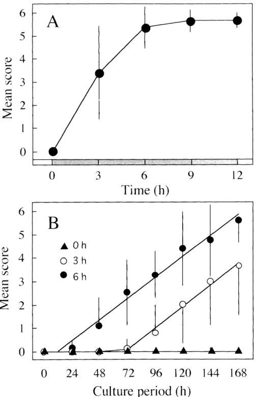
scotophase and cultured with O.5 paglml 20E. All glands acquired the competence to respond to 20E during the first 6 h of the scotophase (Fig. 9A).Progression of apoptosis in vitro was monitored for glands dissected at O, 3
and 6 h after the beginning of the scotophase (Fig. 9B). The score for the glands explanted at 6 h began to increase within 24 h after exposure to 20E and continuously increased to a mean score of 5.6 at 168 h. By contrast, the glands
dissected at 3 h exhibited morphological changes that began 72 h after exposure to 20E. Coefficients of the linear functions for 3 and 6 h glands were O.93 and O.92, respectively, showing that their apoptosis progressed at the same rate once the glands began to exhibit morphological changes.lnhibition ofapoptosis by a-amanitin and cycloheximide
When ct-amanitin (l ptg/ml) was added to the culture of day O anterior silk glands with O.5 ptglml 20E, 23 of 39 glands survived by the end of the 144 h culture.
Fourteen of the 23 glands completed the apoptosis. However when the concentration of a-amanitin was raised to 10 "glml, none of the survivors showed any sign of
IO
apoptosis (Table 2). Since a few glands underwent necrosis without exhibiting the
morphology typical of apoptosis (see Fig. 3), it was possible that the suppression ofapoptosis by a-amanitin was not brought about by the inhibition of RNA polymerase
II but by cytotoxic effects. In order to address this issue, day O glands were cultured with ct-amanitin (10 ptglml) and 20E (O.5 ptg/ml) for 24 h, then transferred them to culture medium containing only 20E (n=10). Under these culture conditions, all the glands completed the apoptosis after having exhibited every stage of apoptosis. In addition, the progression of apoptosis was delayed by about 24 h compared to that ofthe glands cultured with 20E alone for the entire culture period (Fig. 4B). This
indicates that the inhibition of apoptosis by ot-amanitin was not due to its toxic effectbut to the inhibition of RNA synthesis. Thus, 20E--mediated apoptosis may be accompanied by de novo synthesis of poly (A) RNA.
To estimate the time required for sufficient transcription to cause the full
apoptosis, a-amanitin (10 ptglml) was added at varying times after exposure of day O
anterior silk glands to 20E. ct-Amanitin completely inhibited the apoptosis when
added within 6 h of 20E stimulation but exerted Iittle effect when added at 8 h or later. When cultured at a 10-hold higher concentration of ct-amanitin (100 ptglml) 18 h after exposure to 20E (Fig. 10), the glands completed the apoptosis. Accordingly,mRNA synthesis necessary for full apoptosis may be completed within 8 h of
exposure to 20E.To determine the requirement of de novo protein synthesis for apoptosis, cycloheximide was added simultaneously with 20E A concentration of 1 ptglml did
not suppress the apoptosis at all (Table 3). At 50 paglml (i=iO.2 mM), apoptosis did not occur, but the glands showed necrosis which was observed 72 h after exposure tocycloheximide and 20E. Surprisingly, the glands in the presence of only cycloheximide alone did not show any sign of necrosis. When emetine, an another effective protein synthesis inhibitor (Ito et al. 1985), was examined at two concentrations (O.5 and 1 mM), it completely suppressed the apoptosis in the presence of 20E and no glands showed the sign of necrosis (data not shown),
indicating that de novo protein syr,thesis is required for progression of apoptosis.
Differential addition of cycloheximide (50 ptglml) to the culture medium
showed that the inhibitory effect of cycloheximide was gradually reduced over a 6 h- period from hours 12 to 18 after exposures to 20E (Fig. 11). At and after 15 h, nogland exhibited necrosis. These results indicated that protein synthesis for full apoptosis was completed within l8 h of exposure to 20E. It should be noted that when cycloheximide was added at and after 12 h of the culture with 20E, the
apoptosis abruptly progressed from score O to 5 but never attained score 6. No glandsshowed intermediate scores when observed every hour between 48 and 96 h after an
addition of cycloheximide at 18 h of the culture with 20E (Fig. 16),Discussion
Apoptosis Qfanterior silk gland triggered directly hy 20E
In this chapter, it was confirmed that ecdysteroids acted directly on anterior
silk glands to trigger apoptosis as had been indicated by Chinzei (1975). The
sequential progression of apoptosis sequence was reproduced in vitro with exposure of day O glands to O.5 ptglml 20E. The involvement of 20E in the onset of apoptosisof insect tissues is demonstrated in tissues such as intersegmental muscles and motoneurons by in vivo observation (Lockshin and Williams 1965; Schwartz and
Truman l982) or in vitro culture with 20E (Streichert et al. 1997). Although these observations and in vitro manipulations provide strong evidence for a direct action of 20E on tissues, the tissues in those studies consisted of several types of cells. This left up the possibility of intercellular signaling that triggered the apoptosis. Anterior silk gland is consisted of a single type of cells lined with a thin basement membrane(Akai 1984; Korayem 1998). This enabled us to exclude such an intracellular signaling except for an autocrine or paracline pathway between the same types of cells. Accordingly, we concluded that the death of the anterior silk gland was
triggered directly by 20E.Anterior silk gland is stimulated to undergo full apoptosis by the rise of
hemolymph ecdysteroids in the photophase of day 2. Although, hemolymph
ecdysteroid titer increases during the preceding scotophase to O.75 ptglml that is sufficient to induce the larval--to•-pupal transformation (Fukuda l944), this rise is not sufficient to trigger full apoptosis in vivo. Another peak titer of approximately l.5 ptglml in late day 2 (Kiguchi 1983) may trigger the apoptosis. This indicates that the degeneration of tissues requires stronger steroid stimulation than that needed for
l2
tissue diffe:'entiation.
During the scotophase of day 2, the ecdysteroid titer reached to O.75 ptg/ml that was higher than the 20E concentration (O.5 "glml) needed to induce apoptosis in day O glands in vitro. However, this in vivo titer is not sufficient to induce the glands to die. A possible interpretation of the apparent disagreement between in vivo and in vin'o results could lie in the ecdysteroid composition in the hemolymph. Ecdysone is
usually less active than 20E (Applebaum and Gilbert l972), and the major
ecdysteroid in early prepupal period could be primarily ecdysone rather than 20E as in early pupal period (Wai'ren and Gilbert 1986). In Manduca, however, the ratio of
ecdysone and to 20E in hemolymph is 1:5 (Bollenbacher et al. 1981). If this were
the case in Bombyx, hemolymph concentration of 20E would still be greater than O.5ptg!ml. This would argue against a simple composition effect. Alternatively, an
accumulation of covert effects could explain the above. In vivo, the peak titer of O.75 ptglml on day 2 photophase is transient, and the effects accumulated by 48 h after gut purge must be far less than the accumulated covert effects in vitro.Anterior silk gland acquires its competence to respond to 20E during the first 6h of the scotophase of gut purge. While 6 hours seems very short when
compared with other ecdysteroid-dependent phenomena such as pupal commitment,
this short period may be due to the fact that the anterior silk gland consisted of quiteuniform cells. In pupal commitment of epidermal cells in M. sexta, for example, loss of sensitivity to juvenile hormone takes approximately 14 h (Riddiford 1978). This rather long period may be resulted from the different sensitivity of those cells to 20E
since area of pupal cuticle formed in an in vivo commitment assay increases in proportion to the length of exposure to 20E in vitro (Riddiford 1978). Juvenile
hormone application to the last instars larvae of the wax moth, Galleria mellonella causes a composite cuticle with larval and adult characteristics, an indication that developmental events associated svith pupal metamorphosis occur individually at thecellular level, probably independently from each other (Willis et al. I982).
Accordingly, it is likely that developmental events may take a longer time-period to
be accomplished in a tissue consisting of multiple types of cells with different
sensitivities to hormones. By contrast, the gland consists of a single cell type (Akai1983), which may be the cause for that progression of apoptosis of individual cells within a single anterior silk gland is well synchronized.
ReclLtirement ofgene expressionfor completion ofapoptosis
As summarized in Figure 12, the in vitro progression of apoptosis of
anterior silk gland is divided into two phases. The first phase (I) is the initial l8 h- period ofthe culture with 20E This phase is sub-divided to I-1 and I-2. In Phase I-1,
20E activates transcription of genes (Tsuzuki et al. 2001). Transcription is accompiished in the first 8 h-period (Phase I-1) and is probably mediated by the nuclear hormone receptors, ecdysonelUltraspiracle receptor complex (EcRIUSP).
Phase l-2 is the period during which protein synthesis is completed, and the cells rapidly acquire the ability to undergo cell death to a score of 5. The second phase (II) is the period after 18 h of the culture with 20E. During this period, the progression of apoptosis is observable for morphological changes.
An addition of a-amanitin at the beginning of gland culture with 20E resulted in a significant fraction of glands undergoing necrosis as opposed to apoptosis. Since the a--amanitin concentration used (10 ptg/ml) was extraordinary
high for inhibition of RNA polymerase II, it might also suppress RNA polymerase III.In addition, a delayed addition of a-amanitin would induce necrosis since it could be
supposed to suppress mRNA syntheses for house keeping proteins. However, ct- amanitin does not suppress apoptosis when added at 8 h and thereafter. This shows
that the glands do not undergo necrosis but apoptosis once the gene expression has been completed. Alternatively, it was possible that the above result occurred because ct-amanitin did not completely inhibit m-RNA synthesis due to its poor penetration into gland cells. Even at higher concentrations (up 100 ptglml), ct-amanitin failed tosuppress apoptosis. Consequently, we may conclude that gene expression necessary
for apoptosis is completed in the fi rst 8 h after exposure to 20E (phase I-l).Progression ofapoptosis ofthe anterior silk gland
Apoptosis is generally completed through 5 steps, i,e. determination,
triggerring, killing (execution), engulfment and degradation by phagocytes (Ellis et al. 1991 : Steller l995; Hengartner 2000). Anterior silk gland acquires its competence to respond to 20E during the first 6 h of the scotophase in which gut purge occurs (GP O). If tissues responsiveness to 20E determines the timing of pupal commitmentas demonstrated in Manduca epidermis (Riddiford l985), anterior silk gland may be
l4
committed to die during the scotophase of GP O. However, the present data is not
sufficient to conclude the timing of the decision to die for the gland. Though thetiming of death commitment was unsure, we observed the entire process of the
execution of cell death. Therefore, one of the roles of 20E is to trigger the onset of death process itself.In virro morphological changes accompanying the progression of apoptosis
are different from in vivo progression, especially in the later stages of apoptosis. The in vitro changes in day O silk gland from score O to 4, which was triggered by 20E, was similar to the first hajf of the in vivo changes. However, the second half of the in vivo progression, that was observed 24 h after pupation, was not reproduced i.n vitro.In an in vilro culture, granules of various sizes, probably brought about by nuclear fragmentation (see Fig. 7g), appeared in score 6 glands, whereas such granules did
not appear at the end of apoptosis (48 h of pupation) in vivo, One of the most
prominent differences between the in vivo and in vitro conditions is the dynamics of20E concentration. Hemolymph ecdysteroid concentration increases with sharp fluctuations in vivo while the glands exposed to continuous 20E in vitro. In Drosophila salivary gland, the mid and late prepupal puffs do not occur if the wandering stage gland is exposed to continuous 20E (Ashburner and Richards 1976).
Rather inseilion of a culture in hormone-free medium is necessary to reproduce the in vivo progression of apoptosis in the gland. I tried to introduce a hormone-free period in the culture of anterior silk glands with 20E, but such culture conditions did not reproduce similar morphological changes to those seen in vivo. In contrast with the brief exposure to 20E required for the apoptosis of Drosophila salivary gland, Bomb.v.y silk gland undergoes apoptosis with continuous exposure to 20E. The reason why the morphological changes of the late stage in vivo were not observed in vitro remains obscure.
Silk gland is homologous to insect labial gland. The larval labial glands of
the giant silk moth, Antheraea polyphemus (Selman and Kafatos 1974) and M. sexta
(Hakim and Kafatos 1976) are transformed into salivary organs during the pupal
period. Nuclei change their shape during the transformation in the latter species butnuclei do not disappear (Hakim and Kafatos 1976). In Bombyx, anterior silk gland
nuclei apparently disappear from the cells in vivo, and DNA fragmentation occurs
both in i'ivo and in vitro. These results show that the anterior silk gland does notremain in the pupal body nor is used for any structure like the Iabial gland in adult.
DNA of Bombyx silk gland is not completely digested in the apoptosis, and large DNA fragments are translocated to fat body (Chinzei 1975). Similarly digested
contents in the tube-like structure after completion of apoptosis in vivo may be taken up by tissues such as fat body and recycled during adult differentiation.In this chapter, I have described the requirements of 20E for triggering the apoptosis of Bombyx anterior silk gland and the progression of apoptosis in vivo and in vitro. Shoitly after gut purge, 20E probably stimulates transcription and translation of criticai factors which are necessary for the entire progression of apoptosis to
OCCU1'.
16
Chapter II
Analysis of abrupt progression of apoptosis triggered by protein
synthesis jnhibitor, cycloheximide
Introduction
The processes of PCD or apoptosis can be divided into 5 steps, i.e.
determination, triggering, killing (execution), engulfment, and degradation (Ellis et al.
1991; Steller 1995; Hengartner 2000). The step of execution is associated with
morphological changes of tissue or gland. The execution phase is thus the `active'phase in the entire process of apoptosis, following the death commitment The hallmark morphologic features of apoptosis, i.e. membrane blebbing, chromatin condensation and DNA fragmentation characterize the phase. Most studies of the execution phase have been focused on the molecular mechanisms culminating in
DNA fragmentation (Li et al. 1997; Liu et al. 1997; Enari et al. 1998). It is known inmammalian cells that caspase family peptides, Bcl superfamily peptides and their homologues are all important factors to trigger the execution of apoptosis (Meier
2000). The homologues is known or, at least, considered to be involved in apoptosis in various species and their mode of action are similar to that in mammalian cells.Bcl supeifamily peptides are known as a regulatory factor of apoptosis. Bcl
superfamily consists of two types of molecules, one is inhibitor of apoptosis such asBcl-2 and Bcl-XL, and another is accelerator of apoptosis such as BAX and BAD (McDonnell 1996). Zha and colleagues (1996) reported an interaction between BAD
and Bcl-XL that is regulated by phosphorylation of BAD. These indicate that de novo gene expression is not sufficient for inducing the execution of apoptosis, for which accelerator must be liberated from the effects of inhibitors.Apoptosis of anterior silk gland takes 5 or 6 days to be completed. As described in Chapter 1, when a protein synthesis inhibitor, CHX or emetine was added to the culture of the glands with 20E at the time when ct-amanitin did not suppress the progression of apoptosis, CHX stimulated apoptosis rather than
suppressing it. The progression of apoptosis induced by CHX is different from that ofthe glands cultured with continuous 20E. Normally, apoptosis in the anterior silk
gland progresses step by step from stage O to 6. In those glands, membrane blebbing and nuclear condensation take 96 h or 120 h to occur. In the case of addition of CHX however. apoptosis abruptly progressed from stage 2 to 5 with skipping stages 3 and 4. In addition, it took only 72 h for attaining score 5 if CHX is added 12 h after the challenge with 20E (see Fig. 9 in Chapter I). These results indicated an involvement17
of inhibitor in the apoptosis of anterior silk gland,
mammalian cells. Accordingly, effects of CHX in
further examined in the present chapter.similar to that in the apoptosis of the progression of apoptosis were
Materials and Methods
Animal.s'
Larvae of the silkworm and rearing conditions were the same as described
in Chapter I.Hormones and Chemicals
Hormone and chemicals used in this Chapter is the same as described in
Chapter l.In vitro ('ulture ofanterior silk gland
Culture conditions were the same as described in Chapter I.
Results
Abrupt progression ofapoptosis by inhibition ofprotein synthesis
Abrupt progression of apoptosis was induced by CHX added to the medium at 24 h, 48 h or 72 h of the culture with 20E (Fig. 13). It is noteworthy that an
addition of CHX at 48 h and 72 h induced the abrupt progression to stage 5 or 6 in 24 h. In any case examined, the apoptosis jumped up to stage 5 or 6 with skipping the process that was seen in normally progression. The abrupt progression of apoptosis was also induced by emetine (1 mM), a protein synthesis inhibitor(Fig.14).
Morpho/ogical changes in the abrupt progression
When CHX was added 24 h from initiation of culture with 20E, the stages
of apoptosis progressed from primary stages (O, 1 or 2) to stage 5 within a 24 h-
period between 48 and 72 h of the culture with 20E. To examine whether the abrupt
progression occurs in a stepwise manner or by skipping stages up to 5, the glands were observed every 1 h from 48 h to 72 h (Fig. 15). The morphological changes in the abrupt progression were different from that in normal progression. Gap space
appeared in cell-cell boundary as typical morphology of stage 3, Then, cells shrunk and the row of cells became irregular (data not shown).Abrupr progression in the absence of20E.
It was examined if the abrupt progression induced by CHX is 20E-
dependent events, the glands were cultured with 20E for 24 h and transferred to 20E-
free medium into which CHX was added (Fig. 16). The abrupt progression of
apoptosif occurred irrespective of the presence of 20E in the culture with CHX. This indicates that the abrupt progression of apoptosis was independent of 20E action.20E rec/Liirementfor abrupt progression
When anterior silk glands were cultured for 24 h with 20E, then transferred
to a hormone free medium containing CHX, CHX induced abrupt progression (Fig.
17). But, the substitution of CHX for 20E at l8 h did not induce the abrupt progression (Fig. 17). It was examined if the abrupt progressions needed 20E stimulation for further 6 h from 18 to 24 h, the 20E-free period of 6 or 12 h was inserted between the end of 20E challenge and an addition of CHX (Fig. 18). An
abrupt progression was induced in those culture conditions. Individual glands did not synchronously exhibit the abrupt progression. It took 24-72 h for all the glands to attain score 5, which is the reason for the large S.D. values for 24-96 h of cultureperiod (Fig. 18). This indicates that intracellular conditions of the glands are
changing in this period.Discussion
Abrblpt /?rogression showsfeature morphology
In the apoptosis of anterior silk gland induced continuous 20E (normal condition), membrane blebbing and nuclear condensation are clearly observed.
Blebbin..o. appear in stage 4 glands, and nuclear/chromatin condensation appears in
19
stage 4 or 5 glands (Fig. 3 and 7). In normal condition, it takes 96 and 120 h to exhibit blebbing and nuclearlchromatin condensation, respectively, while CHX
added at 24 h of the culture with 20E accelerates their appearance by approximately24 h for blebbing and 48 h for the condensation (Fig. 13). The abrupt progression does not follow a stepwise manner. When CHX is added to culture medium in phase II, cell condensation and nuclear condensation occurred at the same time of the
occurrence of cell blebbing (Fig. 17).The morphological changes associating with apoptosis of mammalian cells
have been studied with reference to cytoskelta] proteins. As the most cells enter the execution phase, they release extracellular matrix (ECM) attachment and reorganizefocal adhesions (FA), adopting a more "rounded" morpho]ogy. The blebbing stage
begins with myosin II-dependent contraction of the actin ring followed by a period of sustained, dynamic plasma membrane protrusion and retraction. It continues until thecell finally enters the condensation stage, which is characterized by nuclear and
chromatin condensations into small apoptotic bodies or into a single, shrunken ball.(Millis et al. I999). These morphologies, releasing matrix, membrane, nuclear and chromatin condensation are manifestations of execution phase. Blebbing dose not
occur in some cells lacking caspase 3 (Janicke et al. 1998; Zheng et al. 1998); cells appear to release but not bleb, suggesting a cellular checkpoint exists between release and blebbing.It is of interest to compare the morphological changes of the silk glands with
those as mentioned above. Blebbing in the silk glands occurred at stage 3 while
condensation was first observed in stage 4 glands. Caspase 3 inhibitor did not
suppress the glands to attain stage 4, indicating that caspase 3 may not be involved in blebbing and nuclear condensation in the silk glands. This indicates that there may be a cellular checkpoint between stage 2 and the stage with condensation although it is not clear what stage 2 indicates. In the anterior silk gland, the stage accompaniedwith condensation and that with formation of apoptotic bodies are clearly
distinguishable in vitro while in mammalian cells, the condensation stage is not
separable from the stage of apoptotic body formation. This indicates that the silk gland provides advantage to the study of molecules involved in the progression from nuclear condensation stage to apoptotic body one.Abrupt pi"ogression caLised by protein synthesi.s inhibitor
Bcl-2 family proteins includes inhibitors of apoptosis. Many of these
proteins interact with each other and with other proteins through a complex network of homo--heterodimers (Sato et al. 1994; Sedlak et al. 1995). It is suggested that such system is used as "rate-limiting" system to progression of apoptosis.Inhibition of protein synthesis by CHX and emetine accelerated the progression of apoptosis in the anterior silk gland (Fig. 13 and 14). The abrupt
progression of apoptosis may be explained as follows. The genes required for the full apoptosis have been transcribed and translated in the period of phase II. Accordingly,the mRNA whose translation is suppressed by CHX must not be the mRNA that is induced by 20E. The fact that inhibition of protein synthesis induces abrupt progression shows that the mRNA in question must be for inhibitor protein(s). The
inhibitor protein of apoptosis may regulate the progression rate of apoptosis, which may cause the gradual progression of apoptosis through stage 2 to 5. The following is only a matter of speculation. If the above explanation is the case, life of inhibitormRNA may be long while the ]ife of inhibitor protein may be around 24 h or less
since abrupt progression occurred with a quiescent period of 24 h after an addition ofCHX. The mRNA amount may decrease gradually along with the progression of apoptosis. Accordingly, the CHX effect was observed when added between 12 and 72 h of the culture with 20E. It is highly possib]e that such inhibitors are the Bcl
superfamily proteins and other associated proteins as found in mammalian cells.
20E reqLtirementfor abrupt progression ofalpoptosis
To complete apoptosis need 20E stimulation during 18 h to synthesize
accelerator protein(s). Abrupt progression of apoptosis is induced by inhibition of protein synthesis on and after l8 h after 20E challenge (Fig. 11 and 13). But, even ifprotein synthesis is inhibited on and after 18 h without 20E during 18-24 h, the abrupt progression is not induced (Fig. 17), and inhibition of protein synthesis
induces the abrupt progression with or without 20E on and after 24 h (Fig. 16). These results indicate that abrupt progression of apoptosis in anterior silk gland needs 20E stimulation during 24 h. 20E induces protein synthesis to progress apoptosis during the first l8 h, and then 20E acts not through gene expression in fo"owing 6 h during18-24 h. But, the abrupt progression occurs in the culture that 20E-free period is
21
inseited during l8-24 h or 18-30 h without CHX, and then the glands are cultured
with CHX on and after 24 h (Fig. 18). These results indicate that there are two
pathways for apoptosis progression, which are independent or dependent for 20E
stimulation in phase I-3 (Fig. 19). The pathway of dependent for 20E is not through gene expression. This pathway is principal way to progress apoptosis in anterior silkglands (described in chapter III). The pathway of independent for 20E is through
protein synthesis. This pathway of independent for 20E may act to rescue in the case without 20E on and after 18 h and maintain condition on standby for progression of apoptosls.Chapter III
Increase in intracellular concentration of cAMP by 20E
Introduction
Hydrophilic signal molp.cular such as peptides and amines are unable to permeate through plasma membrane, and the first step of signal transductin is
mediated by membrane-bound receptors. By contrast, lipophilic steroid hormones get though the membrane and are associated with intracellular receptors. Ligand bindingleads homo- or hetero-dimerization of the receptors, and the dimmer acts as
transcriptional regulators. In cellular response to steroid hormones, the responsebecomes overt through two steps: immediate induction of small number of specific genes occurs within thirty minutes after hormone challenge, which is known as the
primary response. The products of those genes are transcriptional factors and activateother genes that are involved in a delayed, secondary response. Thus a simple
hormonal trigger causes complex changes in the pattern of gene expression.On the other hand, steroid hormone is known to exert its effects that are not mediated by nuclear receptors. In mammals, progesterone inhibits oxytocin signaiing
by binding to the membrane-bound oxytocin receptor (Grazzini l998). In the rainbow trout, 0ncorhynchus mykiss, 17ct, 20P-dihydroxy-4 pregnen-3-one binds a
receptor on the extracel]ular surface of oocytes to induce formation of maturationpromoting factor (MPF) that triggers meiotic maturation of oocytes (Nagahama
l997). The induction of MPF is mediated by signal transducing pathway that involves MoslMAPK (Yamashita 2000).
Ecdysteroid is an insect steroid hormone. Ecdysone is a hormone secreted from prothoracic glands and 20-hydroxyecdysone (20E), a product of oxidation of
ecdysone, is the active form in insects. The generally accepted model of the action of ecdysone to activate gene transcription was first proposed by Karlson (Karlson 1968;Karlson and Sekeris l966) and later sophisticated by Ashburner (Ashuburner and
Richard 1976), as referred to as Ashburner's model. In the anterior silk glands ofBomby.x' mori, 20E activates several genes (Tsuzuki et al. 2001), which are
considered to be tightly involved in the apoptosis of silk glands though elucidation of the gene functions awaits future studies. As mentioned in Chapter I, the expression of genes, required for completion of apoptosis are completed in 8 for transcription andl8 h for translation after challenge with 20E. If Ashburner's model is applicable to
the 20E-induced apoptosis, 20E challenge for 8 h must be sufficient for the full
apoptosis since 20E suppresses expression of late genes after completion of early gene expression. Nevertheless, withdrawal of 20E during a period between 8 and 42
hours of the in vitro culture with 20E interfered the progression of apoptosis, and the extent of inhibition depended on the time of the withdrawal, This suggested that 20Eaction in this period would not be accompanied by gene expression but mediated by
a membrane-bound receptor followed by a signal transduction pathway. If this is thecase, a second messenger analogue must substitute for 20E for the progression of apoptosis. In order to address this issue, I replaced 20E with several second
messengers and found that cyclic AMP could be the candidate.Materials and Methods
Aniinals
Larvae of the silkworm and rearing conditions were the same as describe in Chapter I.
Hormones and Chemicals
20E (Sigma, Saint Louis, MO) was dissolved in ethanol and stored at
---200C until use. When added to the culture medium, an aliquot of the stock solution
was evaporated, dissolved in distilled water and added to the medium to a desired concentration. Dibutyryl-cyclic AMP (dbcAMP; Sigma), caspase-3 inhibitor (CPP32/Apopain inhibitor; CALBIOCHEM, La Jolla, CA) and caspase-1 inhibitor
(ICE inhibitor I; CALBIOCHEM) were dissolved in distilled water at a concentration of 10 mM and stored at -200C. Phorbol ester (Sigma) was dissolved in distilled waterat a concentration of 1mglml and stored at 40C Calcium ionophore (A23187; Sigma) was dissolved in dimethyl sulfoxide (DMSO) at a concentration of lmM, and stored at -200C 3-Isobutyl-1-methylxanthine (IBMX; Sigma) and caffeine (Han-i Chemical,
Kyoto, Japan) were dissolved in distilled water at a concentration of 10 mM and 10mglml, respectively, and stored at -200C When used, an aliquot of each stock
solution was added to the culture medium to a desired concentration.ln vitro ('ultblre
24
Anterior silk glands were removed from gut purged larvae during the
photophase of day O unless otherwise indicated. The glands were rinsed with Grace'sinsect culture medium (GIBCO BRL, Rockville, MD) and cultured separately in O.3
ml of the medium at 250C. The glands were observed every 24 h under a microscope,Extraclion Qfintracellular cAMP
After culture of silk glands, the glands were frozen until cAMP extraction.
The frozen glands were homogenized in cold 69o (WIV) trichloroacetic acid (TCA),
centrifuged at 2000xg for 15 min at 40C, and supernatant was separated for pellet.The resulting supematant was washed 4 times with 5 volumes of water-saturated diethyl ether. The upper ether layer was discarded after each wash. The aqueous extract and the pellet were separately lyophilized and stored at -200C until cAMP
quantification.Measureinent of cAMP concentration
cAMP concentration was determined according to the user's manual attached to the cAMP enzyme immunoassay (EIA) kit (Amasham Pharmacia, Piscataway, NJ). Amount of cAMP in the resulting supernatant was expressed in
pmollmg protein or fmol/mg protein according to the amount of protein in the pellet.Results
Requirement of20E aLfrer the completion ofRNA synthesis
To determine the period of 20E challenge that is required for the full
apoptosis of anterior silk gland in vitro, the glands of day O larvae were transferredfrom a 20E-containing medium to 20E-free one after exposure to 20E for various period (I8-42 h) and observed until 168 h of the total culture period (Fig. 20A).
When the glands were transferred to 20E-free medium at 18 or 24 h, the glands
attained score 2 by 144 h, but no further progression was observed (Fig. 20A, 18 and 24h). When transferred to the 20E-free medium at 30 h of the culture with 20E, theglands attained a mean score of 4.8 with a few glands that completed apoptosis
(score 6). When 20E was present for the first 42 h of the culture, all giands attainedscore 6. Progression of apoptosis in those glands was quite similar to that in the glands cultured in the continuous presence of 20E.
Since the time period between 24 and 30 h after exposure to 20E appeared
transitional, I transferred glands to 20E-free medium every hour during this period(Fig. 20B). When 20E was withdrawn at times after 24 h of the culture, the mean
score increased proportionally to the time of withdrawal and attaining a mean scoreof 5 at 29 h. After 29 h, the mean score gradually increased until 42 h at which it attained score 6 (full apoptosis).
Effects qf'secoi7.d messengers
Translation of ecdysone-induced genes is completed within 18 h of the
culture with 20E (see Chapter I). Nevertheless, 20E must be present for 42 h for fullapoptosis. Assumably, the action of 20E after 18 h to 42 h is assumably mediated
through a signal transduction pathway. In order to address this issue, anterior silkg]ands were cultured with 20E for l8 h, then transferred to the 20E-free medium containing one of the following second messenger analogues, calcium ionophore, dbcAMP and phorbol ester (Fig. 21). Calcium ionophore induced necrosis of the
glands at any dose examined. Apoptosis was not induced by phorbol ester at all. By contrast, substitution of 1 mM dbcAMP for 20E allowed the progression of apoptosis to score 4. This indicates that 20E effects may be mediated by a membrane receptor.Inhibitors of cAMP phospho diesterase, caffeine and IBMX were able to induce
apoptosis although their effects were weaker than that of dbcAMP(Fig. 22).Changes in intracellular cAMP concentration in vitro
Figure 23 shows that 20E induces an increase in an intracellular cAMP
concentration. The concentration initiated to increase l8 h after the beginning of the culture with 20E.Since the above results were obtained in the contjnuous presence of 20E, it
was unclear whether the effect of 20E was immediate or not. To confjrm the above
result that 20E may act directly to increase intracellular cAMP concentration, I firstexamined the culture conditions that would bring about a significant increase in
cAMP level. For this end, silk glands were cultured for 6, 12, 18 or 24 h. Then the glands were cultured in hormone-free medium for an additional period of 6, 12 or 24.26
After the second culture, the glands were challenged with 20E for 1 hour, and the cAMP concentrations were determined. A significant increase in cAMP level by 20E
was brought about at a combination of l8 h of pre-culture with 20E and l2 or 24 h ofpost-culture in hormone-free medium (Fig. 24). The increase in cAMP concentration
was induced by a second challenge with 20E as short as 10 minutes (Fig. 25). Figure25 shows the dose-dependency and time course of the increase in cAMP
concentration at the most suitable culture conditions. cAMP concentrations increased
at O.5 ptglml 20E (Fig, 26A). At this concentration, cAMP increased significantly within 30 seconds of 20E challenge (Fig. 26B). The 20E-induced increase in cAMP
concentration was not affected by ct-amanitin added simultaneously with the second challenge with 20E (Fig. 26), indicating the 20E effects were "ot mediated through gene expresslon.Chanses iiztracellbtlar cAMP concentration in vivo
Amount of cAMP in anterior silk glands were measured from the day of
gut purge (GP O) to the day of pupation (Fig. 28). There were two critically peaks ofcAMP concentration at GP O and GP 3. First peak was sma}jer one at GP O and decreased gradually. On the other hand, second peak was higher one at GP 3, and
appeared abruptly. cAMP concentration of the higher peak was fouith as amount as it of smaller peak.Effects qfcaspase inhibitors
Since caspases were known to trigger DNA fragmentation in mammalian
cells (Thornberry et al. I997), caspase inhibitors were added simultaneously with
20E to the culture of anterior silk glands (Fig. 29). Caspase 1 inhibitor (2 mM) didnot affect the progression of apoptosis at all (data not shown). When caspase 3
inhibitor was added (Fig. 29), the progress of apoptosis was not affected for the first 72 h during which the glands attained score 3. After 72 h, however, the progression was slowed down and the apoptosis did not exceed score 4.Discussion
DtZ.77Cerenl honnonah"eguiren7ent.for com)pletion qfapoptosis
Steroid hormone generally acts as a transcriptionai factor after forming a
steroid hormonelreceptor complex. Some steroid hormones are known to induce
apoptosis by its action as a transcriptional factor (Thompson 1994), Glicocorticoid,for instance, induces apoptosjs through a nuclear receptor complex (Caron-Leslie
1991). An insect steroid hormone, ecdysterojd is known to exert its effects as a
triggering factor of apoptosis in various tissues and glands, and its mode of action isbelieved to be similar to other vertebrate steroid hormones (Thummel l995). In
anterior silk gland of Bombyx larvae, 20E stimulates several gene expressions, andthese genes possibly include genes (death genes) tightly involved in apoptosis (Tsuzuki et al. 2001). The 20E-induced transcription appears to be completed in
phase I-l (see Fig. 10 in Chapter I), and translation may be completed by the end of phase II-2. Nevertheless, 20E stimulus is needed for 42 h to induce full apoptosis.Then, what is the mode of action of 20E after completion of gene expression by 20E? There are several indications for a non-nuclear, receptor-mediated role for 20E.
In Sarcophaga peregrina, 20E inhibits the protease that activates storage protein
receptor to facilitate incorporation of storage proteins from hemolymph into fat body at pupation. Cycloheximide does not alter this ecdysteroid effect (Natori l986). Inwing epidermis of Manduca pupae, 20E stimulates adenyryl cyclase activity in 15 min after exposure to hormone in vitro, and stimulation is also obtained in wing
epidermis homogenate (Applebaum and Gilbert 1972). Similarly, a membrane
receptor for a steroid hormone is known in vertebrates. These repoits indicate that
20E possibly exerts its effects without gene expression. In the 20E withdrawal experiments, we replaced 20E with several reagents that are widely used for exogenous stimulation of signal transducing pathways, such as dbc AMP (Honma et
al. I996), phorbol ester (Witty et al. 1996), and calcium ionophore,A23l87 (Kelleyet al. 1992). Among those reagents, dbcAMP allowed the progression of apoptosis
after withdrawal of 20E (Fig. 21), indicating an involvement of adenyryl cyclase in the action of 20E28
Caspcise (iiid c/7romatiiz condensatioii
Progression of apoptosis is associated with unique morphological nuclear changes such as chromatin condensation and DNA fragmentation (Kerr et al. 1972).
These changes are triggered by an activation of caspase family proteases (Salvesen
and Dixit 1997; Thornberry and Lazebnik 1998) followed by an activation of caspase-activated DNase (Enari et al. 1998; Liu et al. 1998). Acinus induces apoptotic chromatin condensation after cleavage by caspase-3 with no DNA
fragmentation (Sahara et al. 1999). Accordingly, caspase-3 is considered to be taking a leading part in apoptosis and a responsible factor for chromatin condensation.
In the 20E-induced apoptosis in vitro, chromatin condensation is observed after score 4 (Fig, 7). DNA fragmentation is detected in score 4 glands as oligo-
nucleosomal pattern (Fig. 5), and in score 5 gland by chromatin staining (Fig. 7).Caspase-3 inhibitor prevents the progression of apoptosis to exceed beyond score 4
and slows down the progression rate from score 3 to score 4 (Fig. 29). The circumstantial evidence indicates that caspase-3 exists in downstream of cAMP. In vivo changes in morphology showed that apoptotic body does not appear before completion of apoptosis (Fig. 7), indicating that chromatin fragmentation is not
critical factor for the apoptosis of anterior silk gland though it occurs in vitro.Accordingly, the step of caspase-3 activation followed by DNase activation seems to be the final step of the apoptosis of anterior silk gland similar to the apoptosis of
mammalian cells.
Present results indicate a dual signal stream for full apoptosis of the
anterior silk gland. After completion of 20E-activated gene transcrjption, 20E is still needed for the full apoptosis. Its action is not mediated through gene expression butmay be through membrane receptor of 20E. 20E binding to a membrane receptor
may activate adenylyl cyclase, thereby induces an increase in intracellular cAMP
concentration. It remains to be dissolved whether kinase A signaling inhibits an
inhibitor of accelerator of apoptosis or activates caspase-3 followed by chromatin condensation and DNA fragmentation, or the both.General concRusion
Genetic analyses in C.elegans gave important information to the molecular mechanisms of PCD andlor apoptosis occurring in the course of development, In C.
elegans, ced-3, ced-4 and ced-9 were identified as the genes that control the
progression of PCD (Ellis and Horvitz l986; Hengartner et al. 1992; Yuan 1992), and ced-3 and ced-4 appear to be negatively regulated by ced-9 (Hengartner et al. 1992).The mammalian homologues of ced-3, ced-4 and ced-9 are a caspase, apoptotic
protease activating factor-1 (Apaf-1) and bcl-2, respectively (Zou et al. I997; Yuan etal. I993; McDonnell et al. 1996). These genes product finely interact with each other
and with other molecules (Meier 2000). In insect, genetic analysis of Drosophila showed three pro-apoptotic proteins, reaper (RPR), GRIM and head involution defective (HID). The downstream of apoptotic signal pathway is similar to that of C.elegans or mammals (Meier 2000). On the other hand, apoptosis of tissues and glands of insects mostly is induced by 20E so far known. 20E acts as a triggering factor of apoptosis by forming a transcription factor with nuclear receptors (Ashuburner and Richard l976). It is not known whether the above described gene
products are involved in the downstream of 20E-induced apoptosis.It is known that some apoptosis are induced by steroid hormones that act as transcriptional factor (Thompson !994). Ecdysteroid is an insect hormone and known as a triggering factor of apoptosis in various tissues and glands in insects. Its mode of
action is believed to be similar to other sterojd hormones (Thummel l995). In the
anterior silk gland of Bombyx mori, gene expression is induced by stimulation of 20E (Tsuzuki et al. 2001).Present study shows that 20E-activation of gene expression is necessary
but not sufficient for apoptosis of the anterior silk gland. The gene transcription is completed until 8 h after 20R challenge (Fig. 10), and translation is completed until18 h (Fig. 1 l). But, stimulation of 20E is needed after completion of gene expression (Fig. 20). As summarized in Figure 29, apoptosis progresses through three distinctive phases (phase I-III), which are divided by 20E requirement and morphologies of the glands, that are, stage 2 and 3, 4 and 5, and 5 and 6. The most remarkable finding in the present study is that 20E exerts its effects through two distinctive modes of action the apoptosis.
30
First phase (phase I) is the initial 24 h-period of the culture with 20E
When glands are transferred to a hormone-free medium during this period, apoptosis dose not precede beyond stage 2 level. The first phase is further divided into three sub-phases (I-1, -2 and -3), according to effects of a-amanitin and cycloheximide. In the first 8 h-period (phase I-1), transcription of the death gene(s) is accomplished and
is probably mediated by ecdysone receptor/Ultraspiacle (EcR!USP) complex. Phase
I-2 is 18 h-period following the phase I-1, and during phase I-2, synthesis of a set ofprotein(s) for apoptosis is completed. The meaning of remaining 6 h of phase I (I-3)
is unknown.
Phase Il and III are closely related through chromatin condensation and
DNA fragmentation which are evidences of apoptosis. Second phase (II) lasts for 5 h between 24 and 29 h of exposure to 20E. During this peried, anterior silk gland cellsrapidly acquire ability to undergo apoptosis stage 5 after transfer to a 20E-free medium (see Fig. 20) and concentration of intracellular cAMP increase in culture with 20E (Fig. 23). These results indicate that cAMP is involved in the mode of action of 20E, whjch indicates an existence of membrane receptor of 20E. The signaling pathway after cAMP may be culminated in chromatin condensation that
occurs in stage 4 or 5 glands. Stimulation with 20E up to 29 h is sufficient to inducenuclear condensation followed by DNA fragmentation which is characteristic of
stage 5 glands. After 29 h, the mean score of apoptosis gradually increases from 5 to 6 by 42 h. The period from 29 to 42 h is designated phase III.In addition, Caspase 3 was suggested to be involved in the downstream of cAMP signaling pathway that may occur after stage 2 of apoptosis progression.
Inhibitor(s) of apoptosis is suggested by experiments using CHX. It may play a role
in the apoptosis progression to stage 5. The inhibitor could be associated wi{h activation of caspase 3, and therefore CHX may liberate caspase 3 by reducing the
amount of inhibitor in cells by inhibiting the de novo translation of the inhibjtor. It isnot known whether the regulation of inhibitor(s) is under the control of 20E or the associationldissociation of inhibitor is brought about by stereotyped mechanisms,
AcknowRedgements
I wish to thank Dr. Sho Sakurai of professor of Kanazawa University, Dr.
Masafumi Iwami of associate professor of Kanazawa University for the critical advises to my research and valuable comments, and Dr. Seiji Tsuzuki, Dr. Yasunori
Oda who are the oldest members of our laboratory and members of the laboratory forthe many ideas and advices through discussion. The research in this thesis was supported by Grant-in-Aid for Specially Promoted Research (09440273) and Grant- in-Aid for Specially Promoted Research (08276102) from the Ministry of Education,
Science, Sports and Culture of Japan to S.S.32
References
Abrams JM, White K, Fessler L I, Steller H (1993) Programmed cell death during Drosophila embryogenesis. Development 1 17:29-43.
Akai H (l984) The structure and ultrastructure of the silk gland. Experimentia 39:443-449,
Applebaum SW, Gilbert LI (1972) Stimulation of adenyl cyclase in pupal wing
epidermis by P--ecdysone. Dev Biol 27:165-l75.Ashburner M and Richard G (1976) Sequential gene activation by ecdysone in polytene chromosomes of Drosophila melanogaster. III. Consequences of ecdysone withdrawal. Dev Biol 54:241-255.
Ashkenazi A and Dixit VM (1998) Death receptors: signaling and modulation.
Science 281:I305-1308.
Bollenbacher WE, Smith SL, Goodman W, Gi]bert LI (1981) Ecdysteroid titer during larval-pupal-adult development of the tobacco hornworm, Manduca sextsa. Gen Comp Endocrinol 44: 302-306.
Caron-Les]ie LM, Schwartzman RA, Gaido ML, Compton MM, Cidlowski JA
(1991) Identification and characterization of glucocorticoid-regulated nuclease(s) in lymphoid cells undergoing apoptosis. J Steroid Biochem Mol Biol 40:661-67l.
Chambers CM, Poston EM, Oliver JH Jr. (l996) Ecdysteroid titers during postembryonic developmental of Dermanyssbls gallinae (Acari: Dermanyssidae). J Med Entomol 33: 11-14.
Chinzei Y, (1975) Biochemical evidence of DNA transport from the silk gland to the fat body of the silkworm, Bombyx mori. J Insect Physiol 21:l63-171.