2009 © The Japan Society for Analytical Chemistry
Introduction
Metal particles with nanometer-scale dimensions are of great interest owing to their unique and interesting properties.
1–6The size-
7and shape-dependent
8,9physical, chemical and optoelectronic properties of metal nanoparticles have important applications in catalysis
4,10and biosensing,
11,12recording media
13and photoscience.
14For example, recently, gold and silver nanoparticles have been used in various analytical techniques, such as biological optical imaging,
15bio(chemical) sensing,
11,12,16and surface-enhanced Raman scattering (SERS),
17–19according to their unique plasmon absorption in the visible light region.
Nanoparticles of platinum-group metals have been widely applied as effective catalysts in various organic reactions.
4,20–22Size- controlled gold nanoparticles have also been applied as oxidation catalysts; their activity is strongly affected by their size.
23–25Matrix-assisted laser desorption/ionization mass spectroscopy (MALDI-MS) has become a powerful tool for analyzing organic molecules with a relatively high molecular weight, such as biomolecules and synthetic polymers.
26–30Inorganic clusters can also be examined by MALDI-MS.
31In MALDI-MS, analyte compounds are embedded in a surplus of matrix, consisting of small organic molecules, and co-desorbed upon laser excitation.
The organic matrixes have high resonant absorption at the laser
wavelength. Although MALDI-MS has been successfully used for the analysis of biomolecules and synthetic polymers, it has not been used extensively in low-molecular-weight compounds (m/z <500 Da), owing to the relatively high intensity of background signals from organic matrixes, which usually have similar molecular weights.
More recently, nanomaterials
32–46such as carbon nanotubes (CNT),
33porous silicon,
34silylated porous silicones,
35,36titania sol-gel thin film,
37,38silicon nanoparticles
39and gold nanoparticles
40–44have received much attention for use in surface-assisted laser desoption/ionization mass spectrometry (SALDI-MS), which utilizes these inorganic matrixes, because of their high surface areas, simple sample-preparation techniques, and flexibility of sample deposition under different conditions. Early in 1988, Tanaka et al. used for the first time 30-nm-diameter cobalt nanoparticles suspended in glycerol to analyze lysozyme and synthetic polymers.
27Graphite powder in glycerol is also used for SALDI-MS
45,46and mass imaging.
47McLean et al. have recently demonstrated that size-selected Au nanoparticles (AuNPs) with 2 – 10 nm diameters can be used in SALDI-MS under dry surface conditions for the detection of peptides, such as substance P and phosphopeptides.
43In addition, it has been suggested that AuNP matrixes afford the preferential ionization of peptides. On the other hand, recently, our group has introduced the use of special-structured platinum nanomaterials to ionize various peptides and proteins including cytochrome c (ca. 12 kDa).
32Platinum has a higher melting temperature than gold, and is well-crystallized even in nanodimensions. However,
Detailed Investigation on the Possibility of Nanoparticles of Various Metal Elements for Surface-Assisted Laser
Desorption/Ionization Mass Spectrometry
Tetsu Y
ONEZAWA,*
†Hideya K
AWASAKI,** Akira T
ARUI,** Takehiro W
ATANABE,**
Ryuichi A
RAKAWA,** Toshihiro S
HIMADA,* and Fumitaka M
AFUNÉ***
* Department of Chemistry, School of Science, The University of Tokyo, Hongo, Bunkyo, Tokyo 113–0033, Japan
** Department of Applied Chemistry, Faculty of Engineering, Kansai University, 3-3-35 Yamate-cho, Suita, Osaka 564–8680, Japan
*** Department of Basic Science, Graduate School of Arts and Sciences, The University of Tokyo, Komaba, Meguro, Tokyo 153–8902, Japan
In this paper, we describe systematic detailed considerations of the feasibility of using various metal nanoparticles for organic-matrix-free surface-assisted laser desorption/ionization mass spectrometry (SALDI-MS). In order to avoid the influence of organic molecules on the nanoparticles, stabilizer-free bare nanoparticles of Ag, Au, Cu and Pt were prepared by laser ablation. Although all metal nanoparticles absorbed N
2laser light (337 nm) energy, the performance of desorption/ionization of a representative peptide, angiotensin I, strongly depended on the metal element. Citrate buffer was used as a proton source; it reduced the amount of alkali cation adducts present. Then, protonated molecules of analytes predominated in the mass spectra when Au and Pt nanoparticles were used. Pt nanoparticles showed the highest performance in SALDI-MS, owing to their smaller heat conductivity and higher melting temperature. The selective desorption of a cationic surfactant with longer alkyl chains and a peptide with methionine was also observed.
(Received October 22, 2008; Accepted December 25, 2008; Published March 10, 2009)
†
To whom correspondence should be addressed.
E-mail: [email protected]
no systematic consideration of the ability of metal nanoparticles for SALDI-MS has been reported so far.
In this work, we investigated the mass application of metal nanoparticles (i.e., Au, Pt, Ag and Cu) as SALDI matrixes for peptides, surfactants and synthetic polymers, to examine the effect of the metallic species of nanoparticles. We have demonstrated for the first time that the performance of what in SALDI-MS was largely dependent on the species of metal nanoparticles: Pt nanoparticles (PtNPs) were superior to other nanoparticles (AuNPs and AgNPs). This difference is discussed based on a laser-induced temperature increase in these metal nanoparticles. In addition, it was found that metal nanoparticles without protective agents developed in this study have great advantage on the SALDI-MS for peptide analysis.
Experimental
Reagents and chemicals
Water was purified using a Milli-Q system (>18 M W cm).
Angiotensin I and substance P were from Wako Pure Chemical Industries Ltd. (Osaka, Japan) and used as received. Metal plates for laser ablation were purchased from local suppliers.
Poly(ethylene glycol)s (PEGs, average molecular weight, M
W= 0.4, 1, 2, and 3 K for PEG400, PEG1000, PEG2000, and PEG3000, respectively) were obtained from Wako Pure Chemical (Japan). Poly(methyl methacrylate) (PMMA) (M
W= 1890 Da, M
w/M
n: 1.10) was obtained from Polymer Laboratories. a-Cyano-4-hydroxycinnamic acid (CHCA) and 2,5-dihydoxybenzonic acid (DHB) was purchased from Sigma- Aldrich (Milwaukee, WI). Metal plates were purchased from Nilaco (Tokyo, Japan). All the other chemicals used were obtained from Nacalai Tesque Co. (Tokyo, Japan).
Preparation of metal nanoparticles
Nanoparticles of gold, platinum, and silver were prepared by laser ablation in aqueous media.
48,49Various metal nanoparticles can be obtained by this method. The preparation process is very simple. Clean ingots of the metals were put into water in a PTFE or glass beaker. The output of a second-harmonic (532 nm) Nd:YAG laser (Quanta Lay GCR-170 or Thales Laser SAGA PRO 220-10 FHG) operating at 10 Hz was focused on the surface of the metal plates at the bottom of the beaker. The laser beam irradiation time was within 10 – 20 min. The spot sizes on the surface of the metal plates ranged from 1 to 3 mm. No stabilizing molecules were added to the sample dispersion.
Some flocculation was observed when the dispersion was concentrated, because in this study no stabilizing reagent was used.
A copper nanoparticle (CuNP) dispersion also prepared by laser ablation was supplied from Fukuda Metal Foil and Powder Manufacturing Co. (Kyoto, Japan). No stabilizing reagent was added to a dispersion.
Citrate stabilized gold nanoparticles were prepared using a protocol proposed by Frens et al.
50PVP (poly(N-vinyl- pyrrolidone))-stabilized gold and platinum nanoparticles were prepared by the alcohol reduction of HAuCl
4and H
2PtCl
6proposed by Yonezawa.
20PVP is a frequently used water- soluble polymer for metal nanoparticle preparation.
Apparatus
SALDI and MALDI mass spectra were acquired in both the positive reflectron mode and the linear mode using an AXIMA- CFR time-of-flight mass spectrometer (Shimadzu/Kratos, Manchester, UK) with a pulsed nitrogen laser (337 nm).
Analyte ions were accelerated at 20 kV under delayed extraction
conditions.
Various nanoparticles (i.e., AgNPs, AuNPs, CuNPs, and PtNPs) were used as the matrix for SALDI-MS. A two-layer sample preparation method was employed:
37The first step was spotting the NPs solution (2 ¥ 10
–6cm
3) on a stainless-steel plate, followed by drying. The second step was depositing the sample solution (5 ¥ 10
–7cm
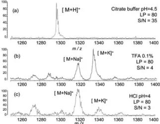
3) on the plate. In some cases, centrifugation was performed to concentrate the nanoparticle dispersion. For peptide samples, the sample aqueous solution was mixed with a citrate aqueous solution (diammonium citrate (200 mmol dm
–3)/citric acid (200 mmol dm
–3) 5:1.1 (v/v); pH = 4.5) to a final concentration of 10 ¥ 10
–6mol dm
–3prior to MALDI-MS. For cationic surfactants with four types of alkyltrimetylammonium bromide (C16TAB, C14TAB, C12TAB, and C10TAB; see Supporting Information), equal concentrations (1 mM) were prepared. AuNPs and PtNPs were dispersed in each surfactant aqueous solution and separated from the solution by centrifugation to remove any excess surfactants. The cationic surfactants trapped by the isolated particles were characterized by SALDI-MS with AuNPs and PtNPs after simple washing with water.
Transmission electron microscopy (TEM) was carried out with a Hitachi HF-2000 field-emission-type TEM (200 kV).
UV-Vis absorption spectra of NP-supported quartz plates in a dried state were obtained at 25˚C using a Jasco Ubest-670 UV/Vis spectrometer.
Results and Discussion
Preparation of nanoparticles
We used in this study a laser ablation process to obtain metal nanoparticles. Among many preparative procedures of metal nanoparticles, the laser ablation process is the most clean wet process, because the particles are obtained from pure metal ingots or flakes without any additives, such as reducing reagents and stabilizing materials.
48,49Even counter ions or ligand molecules of metal sauces do not remain in the nanoparticle dispersion by using this method. The flocculation of NPs was not observed for at least 7 days at the prepared concentration.
48,49Sometimes, the NP dispersion should be concentrated for SALDI-MS application. In such cases, flocculation of NPs was sometimes observed. This preparation process is most suitable for SALDI-MS inorganic matrixes, because naked nanoparticles should be very important for SALDI-MS application in order to avoid any obstacle peaks from organic compounds that are used as stabilizing reagents.
UV-Vis spectra and TEM images
Figure 1(a) shows the absorption spectra of dried AgNPs, AuNPs, CuNPs, and PtNPs deposited on a quartz plate.
Unfortunately, the amounts of particles are not unique in these spectra. The absorption spectra of these NPs show a broad band from the visible region into the ultraviolet region, and characteristic surface plasmon resonance (SPR) bands appear at 420, 520, and 620 nm for AgNPs, AuNPs, and CuNPs, respectively. PtNPs are black, and showed no such specific absorption peak in this region, but do show a relatively high absorbance in the UV region. The absorbance of these metal nanoparticles at a wavelength of 337 nm suggests that these NPs could be used as an assisting material in UV (337 nm nitrogen laser)-SALDI-MS.
The representative TEM images demonstrated that these NPs
have diameters of about 2 – 30 nm. Ag ( ~ 10 – 30 nm) and Cu
(~10 – 20 nm) nanoparticles showed wider size distributions
than Au ( ~ 20 nm) and Pt, as can be seen in the images. In the TEM images, the particles show aggregated structures, except in the case of platinum nanoparticles. These particles were produced in water, and the nanoparticles may form aggregations during the evaporation of water on the carbon-coated copper grids. Pt nanoparticles are smaller (~2 – 5 nm) than the others.
These particles were smaller than Tanaka’s cobalt nanoparticles (~30 nm),
27and the particle size was much smaller than the heat diffusion length during the laser pulse duration, as discussed below (Table 1). Therefore, all of these particles should have enough ability to ionize organic molecules by laser irradiation heating.
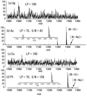
SALDI-MS of angiotensin I
Figure 2 shows the mass spectra of angiotensin I obtained from the AuNPs, PtNPs, CuNPs, and AgNPs as SALDI matrixes. The stainless-steel sample plate, itself, without organic matrixes or metal nanoparticles shows poor ionization/
desorption.
32,51Angiotensin I is a typical peptide molecule for mass spectrometry analyses, and is often used as the material for the first screening of the SALDI ability for peptides. The insets in the figure show expanded views of the molecular ion region.
Using AuNPs and PtNPs, we have successfully ionized angiotensin I in the proton adduct forms of [M+H]
+(m/z = 1298 Da: Au, 130 mV; Pt, 58 mV) in the MS profile. We do not know, unfortunately, the actual value of a laser fluence in the instrument of SALDI-MS (Axima CFR, Shimadzu-Kratos) used in this study, although we know the relative values, denoted by
the laser power, “LP”. The laser power (LP) values used here were LP75 and LP70, respectively, and, furthermore, the signal/
noise ratios of these spectra were high (S/N = 43 and 156, respectively). Also, the isotope peaks of angiotensin I were resolved in the case of AuNPs and PtNPs. A weak signal corresponding to the Na adduct form of [M+Na]
+(m/z = 1320 Da: Au, 20 mV; Pt, 6 mV) appears to be adjacent to the [M+H]
+peak for angiotensin I. In addition, a peak from the fragmentation of angiotensin I appears at m/z = 1250 for the AuNPs, while no such strong peak was observed with the use of the PtNP matrix, even though the difference of LP is relatively small. No ion from angiotensin I was obtained from AgNPs or CuNPs, even at a high laser fluence of LP100 or more.
As can be seen in the spectra in Fig. 2, Ag and Cu showed no ion peak of angiotensin I when using a higher laser power.
Fig. 1 (a) UV-Vis spectra and (b – e) representative transmission electron microscopy images of metal nanoparticles used for SALDI-MS plates: (b) Ag, (c) Au, (d) Cu, and (e) Pt. Ag nanoparticles are relatively large and Pt ones are smaller and unique.
Table 1 Diffusion lengths of the nanoparticle material metals
Metal
Heat capacitya63/
J kg–1 K–1
Heat conductivity63/
W m–1 K–1
Diffusion length/nmb Density63/
103 kg m–3
Ag 10.5 235 429 144 – 277
Au 19.3 129 317 124 – 236
Cu 8.96 384 401 118 – 226
Pt 21.5 133 71.6 55 – 105
a. at 25˚C.
b. Calculated with Eq. (1). t = 3 – 11 ns.