2
0
1
CO
20
alternative oxidase AOX
CO2 capture and utilization CCU
chlorophyll chl
dry cell weight DCW
hydroxyapatite HAP
heatshock protein 70B HSP70B
(infrared gas analyzer) IRGA
light harvest complex LHC
magnesium Ammonium Phosphate MAP
NADPH-dehydrogenase 2 NDA2
non-photochemical quenching NPQ
pulse amplitude aodulated fluorometry PAM fluorometry
polyhydroxybutyrate PHB
photochemical quenching qP
reactive oxygen species ROS
scanning electron microscope SEM
superoxide dismutase SOD
(Solid Surface Continuous Culture System) SSCC
CO2 φ (ICPP, 2015) (CO2)
(Hansen and Sato, 2004)
CO2 CO2
× 37.1 Gton (Harvey, 2018) CO2
CO2 (CO2 capture and storage: CCS) CO2
(CO2 capture and utilization: CCU) (Hasan and Rahman,
2017) CO2 (CCU) CO2 CO2 (Hamed, 2016) (Majid et al., 2014) (Huang et al., 2010)
Botryococcus (Hirose et al., 2013;
Cheng et al., 2019) Pseudochoricystis (Satoh et al., 2010) Δ
Euglena (Krajčovič et al., 2015; Harada et al., 2020) Δ
triacylglycerol (TAG) Parachlorella (Hirai et al., 2016)
CO2 CO2 (open pond) (DCW) 25 g DCW m-2 day-1 10 kg DCW m-2 Δ , , CO2, CO2 3 ton 4
(Shen et al., 2009; Tsuzuki et al., 2012; Blanken et al., 2014; Hans et al., 2014; Schultze et al., 2015; Heimann et al., 2016; Li et al., 2017)
open pond 20–30
CO2 Parachlorella kessleri 11h (Fig. 4-B)( , 2014) P. kessleri Δ (Hu et al., 2008; Hirai et al., 2016)
(Ota et al., 2016) P. kessleri ATP × φ ( ) Magnesium
Ammonium Phosphate(MAP) Hydroxyapatite(HAP)
P. kessleri
CO2
( )
1. Δ
Parachlorella kessleri (NIES-2160, ) 1/5 Gamborg's
B5(1/5 GB5) (Table1-1)(Gamborg et al., 1968) 30°C 80 µmol photons m 2 s 1 •
(fluorescent lamps, FL20S BRF; Toshiba Lighting & Technology Corporation,
Japan) P. kessleri CO2Δ CO2
Δ (Tsuzuki and Miyachi, 1989;
Kaplan and Reinhold, 1999) 2%CO2 48 log phase
(0.04% CO2) 16
Göttingen Chlorella
vulgaris 211-11h Prof. G. H. Schmidt C. vulgaris 11h
( ) ( IAM C-531 NIES NIES-2160) Parachlorella 2. OD730 (DU640; Beckman, USA) 0.1–0.2 5–10 (DCW) ( , 2017) y = 0.35x(R2 = 0.9898) x (OD730) y (mg DCW mL-1) 0.24 P. kessleri DCW (chl) Porra(Porra, 1989) 1 mL
15,000 rpm, 10 min, 4°C (MRX-150, TOMY, Tokyo, Japan)
100% Methanol 1 mL voltex mixer(REAX 2000,
Heidolph, Germany) 15,000rpm, 10min, 4°C
(665nm 650nm) (DU640, Beckman, California, USA) A665
A650 chl (µg mL-1)
Total chl = 4A665 + 25.5A650
chl a = 16.5A665 - 8.3A650
chl b = 33.8A650 - 12.5A665
membrane filter(GF/B; Whatman, Kent, UK) (6.3 cm × 6.3 cm, 590 g m−2,
2 mm 0.5 mm )
0 490 mg chl m 2
LI6400(LI COR, Lincoln, Nebraska, USA) ( 120
65mm)(TREAD LLC, Japan) 30°C (Fig. 1-1A)
CO2
CO2 400 0 2000 µmol CO2 mol 1 air(18 0 89 µmol CO2 L 1 air)
CO2 CO2 3 (
120mg chl m 2 CO
2 400, 2000 µmol CO2 mol 1 air
10.1, 27.9 µmol CO2 mol 1 air ) LED
(Photosynthetic Photon Flux
Density; PPFD) (MQ 200; Apogee Instruments, Logan, Utah, USA)
0 260 µmol photons m 2 s 1 3
4.
P. kessleri (SEM)(JCM−5700; JEOL Ltd.,
Tokyo, Japan) Tissue Tek O.C.T. compound
(Sakura Finetek USA Inc., Torrance, California, USA)
(HM500; Microm International, Walldorf, Germany)
20 µm (BZ X700 Keyence,
1. CO2
IRGA
(CO2 )
P. kessleri glass fiber membrane filter
(0.04% CO2) CO2 (Fig.
1-1A) CO2
CO2 (Fig. 1-1B, ) CO2 glass fiber
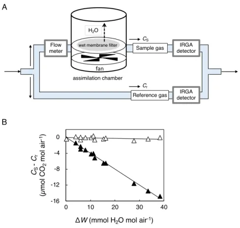
membrane filter ( ) CO2 β CO2 CO2 O2 β CO2 (Eq.(1)) CScl = CSmes(1 + ΔW) (1)
CScl β CO2 (µmol CO2 mol 1 air) CSmes
CO2 (µmol CO2 mol 1 air) W
H2O (mmol H2O mol 1 air)
CO2 Δ Eq.(1) β (Fig.
1-1B, )
A(µmol CO2 m 2 s 1) Eq.(2)
A = F(Cscl Cr) / S (2)
(µmol air sec−1) Cr CO
2
(µmol CO2 mol-1 air) S (m2)
(6.3 cm 6.3 cm) A chl
(Fig. 1-2)
2. CO2
glass fiber membrane filter
120 μmol photons m 2 s 1, CO
2 400 µmol CO2 mol-1 air( ) 2000 µmol CO2
mol-1 air( ) Δ (Fig. 1-2)
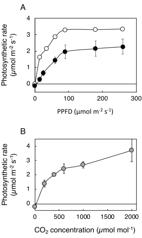
CO2 mg 1 chl h 1 90 µmol CO2 mg 1 chl h 1
(Shiraiwa and Miyachi, 1985) 400 µmol CO2 mol 1 air
(Tsuzuki et al., 2019)
(Fig. 1-2) P. kessleri 400 µmol
CO2 mol 1 air ( )
CO2 glass fiber membrane filter CO2
120 mg chl m 2 400 µmol CO
2 mol 1 air
2000 µmol CO2 mol1 air Δ (Fig. 1-3A)
400 µmol CO2 mol 1 air 2000 µmol CO2 mol 1 air
90 µmol photons
m 2 s 1 0 60 µmol photons m 2 s 1
-400 µmol CO2 mol 1 air 2000 µmol CO2 mol 1 air
0.027 mol CO2 mol 1 photons 0.056 mol CO2 mol 1 photons
400 µmol CO2 mol 1 air CO2
CO2 2000
µmol CO2 mol 1 air 18
120 mg chl m 2, Δ CO
2
(Fig. 3B) CO2 (400 µmol CO2 mol 1 air)
2000 µmol CO2 mol 1 air
CO2 2000 µmol CO2 mol 1 air
CO2
2000 µmol CO2 mol 1 air
CO2 CO2
CO2
120 mg chl m 2 240 mg chl m 2 glass fiber membrane filter chl (55–60 mg chl g 1 DCW), (7 × 10¹⁰ cells g 1 DCW), ( 6 μm) 1 2 SEM 120 mg chl m 2 ( ) (Fig. 1-4Aa) 240 mg chl m 2 2 (Fig. 1-4Ab)
(Fig. 1-4Ba,b) Fig. 1-2
CO2 1-2
400 µmol CO2 mol 1 air (Fig. 1-2, 1-3B)
CO2
CO2 (Hajer. et al., 2020)
CO2 (Two film theory: CO2
) CO2
(Blanken et al., 2014; Gross et al., 2013; Li et al., 2015; Wang et al., 2015; Lai et al., 2020)
4. CO2
CO2 glass fiber membrane filter
CO2
(Fig. 1-5) 2000 µmol CO2 mol 1 air Δ CO2
500 mg chl m 2 glass fiber membrane filter
(2400mg chl m 2 ) glass fiber membrane filter
CO2 (Fig. 1-6) (Fig.1- 6e) 2400 mg chl m 2 (Fig. 1-5) (Fig. 1-6c, d, f) ( 2 mm)(Fig. 1-6c,
d) glass fiber membrane filter 0.2 mm (Fig.
CO2 CO2 CO2 P. kessleri CO2 IRGA P. kessleri chl 190–400 mg chl m 2 leaf area ( 7–14 μmol CO2 m−2 s−1(79.6–97.3 μmol CO2 mg
chl h−1))(Loach, 1967; Oguchi et al., 2003)
CO2 400 μmol CO2 mol air−1 P. kessleri
CO2 P. kessleri CO2 CO2 CO2 (Dillschneider et al., 2013) 5. chl DCW 5.5%
Fig. 1-2 (90 μmol CO2 mg 1 chl h 1) 5.0 mmol CO2 g 1 DCW
h 1 (60 mg C g 1 DCW h 1) DCW 50%
(Tsuzuki et al., 2019) 1 DCW 12%
6.1
(Tsuzuki et al., 2019)
8–9
9.0 µmol CO2 m 2 s 1
19 g DCW m 2 day 1 (Fig. 1-5)
(Kesaano and Sims, 2014; Zhuang et al., 2018)
Fig. 1-1. IRGA CO2 A: B: CO2 ( ) Eq. (1) β ( ) A B IRGA detector CS H2O Cr Flow
meter Sample gas
Reference gas IRGA detector assimilation chamber CS -Cr (µ mo lCO 2 mo lai r -1) -16 -12 -8 -4 0 0 10 20 30 40
ΔW (mmol H2O mol air-1) fan
Fig. 1-2. glass fiber membrane filter P. kessleri
120 µmol photons m-2 s-1 CO
2 400 µmol CO2 mol-1 air ( )
2000 µmol CO2 mol-1 air ( ) (
Fig. 1-3. (A) CO2 (B) glass fiber membrane filter P. kessleri
A: 120 mg chl m-2 400 µmol CO
2 mol-1 air
( ) 2000 µmol CO2 mol-1 air ( ) : 120 mg
chl m-2 120 µmol photons m-2 s-1 (n=3)
-1
0
1
2
3
4
0
100
200
300
PPFD (µmol m
-2s
-1)
P
h
o
to
syn
th
e
ti
c
ra
te
(µ
mo
lm
-2s
-1)
A
-1
0
1
2
3
4
0
500
1000
1500
2000
CO
2concentration (µmol mol
-1)
Fig. 1-4. glass fiber membrane filter P. kessleri A: filter
SEM 120 mg chl m-2 (a) 240 mg chl m-2 (b) filter
Fig. 1-5. glass fiber membrane filter ( ) ( ) P. kessleri
120 µmol photons m-2 s-1, CO
2 2000 µmol CO2 mol-1 air
< 2 >
1 P. kessleri CO2Δ
(chl ) 400 µmol CO2 mol-1 Air
CO2Δ (Fig. 1-2) CO2 CO2 (chl ) 2 1 Ⅱ(PSⅡ) PS chla (P680) ± Pheophytin a (Phe) QA Ⅰ(PSⅠ) P. kessleri Δ chl ( ) chl a/b ( ± ) chl a
Pulse Amplitude Modulated fluorometry (PAM )
1. Δ
P. kessleri 1/5 GB5 30°C 80 µmol photons m 2 s 1 •
(fluorescent lamps, FL20S BRF; Toshiba Lighting & Technology Corporation, Japan)
2%CO2 OD730=0.3
2%CO2 ( CO2Δ ) 48 log phase
(0.04% CO2, CO2Δ ) 16
glass fiber membrane filter(GF/B; Whatman, Kent, UK) 30 mg chl
m-2 1/5 GB5 80 µmol photons m
2 s 1 • 0.04 CO
2
glass fiber membrane filter 0–24 DCW , chl
, PAM RNA
2.
glass fiber membrane filter 105°C, 3
(HR-202i, A&D Inc., Tokyo,
Japan) glass fiber membrane filter
DCW
chl glass fiber membrane filter 30 mL
30 10 650 665 nm 1 chla, chlb Total chl 3. chl a chl a , PAM PAM
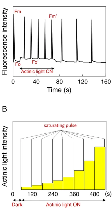
(AquaPen AP 110/C, Photon Systems Instruments, Drásov, Czech) (
)PAM (Junior-PAM, Heinz Walz, Effeltrich, Germany
dark actinic light(80 µmol photon
m-2 s-1) ( : 1200 µmol photons m-2 s-1, 800 ms : 7000 µmol
photons m-2 s-1, 400 ms) 10 160 (Fig. 2-1A)
60 actinic light
(0–500 µmol photon m-2 s-1) ( : 1200 µmol photons m-2 s-1, 800 ms :
Δ Fv/Fm, NPQ, qP, ΦⅡ Perkins (Consalvey et al., 2005)
4. total RNA cDNA
0, 12, 24 h 4°C resuspension buffer
Sepasol®-RNA I Super G kit (Nacalai Tesque, Kyoto, Japan) total RNA
QuantiTect®-Reverse Transcription Kit (Qiagen, Hilden, Germany) cDNA
5. q RT- PCR
cDNA Rotor-Gene®-SYBR® Green PCR Kit (QIAGEN,
Venlo, Nederland) real-time PCR
kit -actin 0 h (ΔΔct ) -actin 5’- ATCAACCTGACAAGGCAACC -3’ 5’- AAACGGCTACCACATCCAAG -3’ Superoxide dismutase (SOD)
RNA RNA-seq GENEWIZ Japan (Saitama, Japan)
RNA-seq ion total RNA-Seq Kit v2
(Life Technologies, Carlsbad, USA) mRNA
Ion PGM sequencer (Life Technologies, Carlsbad, USA)
Fastq Cutadapt v1.9.1
PCR 20
cd-hit Trinity (v2.2.0) (Grabherr et al., 2011)
RNA-seq de novo
unigene sequence file RSEM (v1.2.28) (Limin et al., 2012)
DESeq2 Bioconductor package (Padj > 0.05)
blast sotfware unigene
12 chl
a/b 24 ( ) 12
chl
light harvesting complex (LHC)
Fig. 2-1. PAM Δ A: Δ
Δ Fo Fm
actinic light Fo’ Fm’ B: Δ
60 actinic light
4000
24000
44000
64000
0
40
80
120
160
Actinic light ONA
ct
in
ic
lig
h
t
in
te
n
si
ty
120
240
360
480
0
saturating pulseDark Actinic light ON
< 3 > P. kessleri 4 ( , , , ) ± (Na), (Mg), (P), (S), (K), (Cl), (Fe) P. kessleri (1/5 GB5) N P P ATP P (Ekardt et al., 2015) × P φ Microcystis Dinoflagellates
(Cai et al., 2013)
Microcystis10–30 μmol P L-1 (0.3–0.9 mg P L-1) P
(Saxton, et al., 2012; Tsuzuki et al., 2019)
P 10–30 μmol P L-1 1–8 mg P L-1 P ( ) P P (Magnesium Ammonium Phosphate(MAP) Hydroxyapatite(HAP) ) P (Gonçalves et al., 2017) CO2
(Kesaano et al., 2014; Suparmaniam et al., 2019)
P
(Johnson et al., 2010;
, 2012; Gross et al., 2013;
Kesaano et al., 2014; Tsuzuki et al., 2019;
1
4
)
P
1. Δ
P. kessleri 11h (NIES-2160, ) 1/5 GB5 (220 µM (Pi)
1/5 GB5) (Table1-1) 30°C 2% CO2 80 µmol
photons m 2 s 1 • (fluorescent lamps, FL20S BRF; Toshiba
Lighting & Technology Corporation, Japan)
< 4 >
< >
CO2
( )
(open pond) (Flat
panel)
(Slade and Bauen, 2012; Kesaano and Sims, 2014; Hamed,
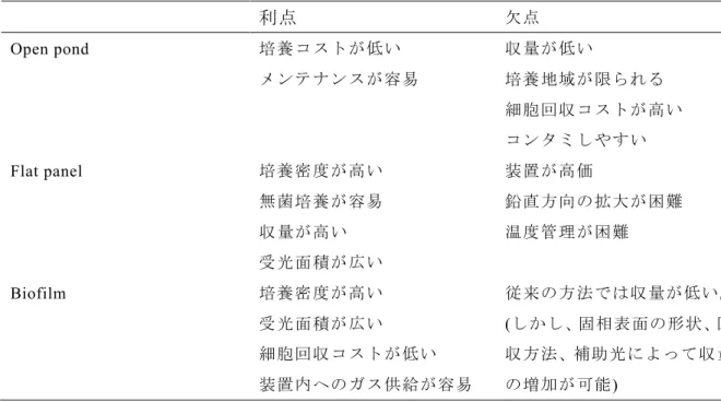
2016)(Table 4-1) CO2
• ( Solid Surface Continuous Culture System ) (SSCC)
1.
Parachlorella kessleri(NIES-2160, ) 1/5 GB5 (Gamborg et al.,
1968) 30°C 80 µmol photons m 2 s 1 • (fluorescent lamps, FL20S BRF;
Toshiba Lighting & Technology Corporation, Japan)
OD730 0.8 1/5 GB5 1/5 GB5 2 OD730 = 1.0 24 2. 1 mL OD730 (DU640; Beckman, USA) 0.1–0.2 DCW y = 0.35x(R2 = 0.9898) x (OD730) y (mg DCW mL-1) 0.24 P. kessleri DCW 3. 5 105–1 107 cells mL-1 1/5 GB5
(cellometer X2; Nexcelom, USA)
4. Δ
4. 1.
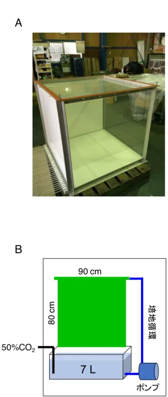
1 1m (Fig. 4-1A) 80 cm 90 cm
(Fig. 4-1B) 50% CO2 7L 1/5 GB5
5.
5 cm 30 cm 1 cm 20 cm LED
8 Haematococcus pluvialis (NIES-144),
Chlamydomonas. reinhardtii cc125, ± Euglena gracilis (NIES-47), β
Nannochloropsis oculate (NIES-2146), Chaetoceros gracilis,
Nostoc commune (NIES-24), Synechocystis sp. PCC 6803,
Arthrospira (Spirulina) platensis
1. 80 cm 90 cm 5 (Fig. 4-2) 15 3.8 g DCW day-1 5.1 g DCW m-2 day-1 12.5 cm2 (Table 4-3) 1.3 g DCW m-1 h-1
(Fig. 4-3A) 30 g DCW m-1 day-1
5 L m-2 min-1
open pond
flat panel (Chisti, 2016; Ting, 2017)
(Table 4-1) open pond 25 g DCW m-2
day-1 flat panel 34 g DCW m-2 day-1 open pond
flat panel open pond
SSCC open pond
Flat panel
SSCC 1 m3 1mm 33 (3cm )
1 kg m-2 day-1
10 m 100 m2
1 ton day-1 1.8 ton day-1 CO
2 500~1000 3.3 ton(2019 3%) 2. P. kessleri SSCC
Haematococcus pluvialis, Chlamydomonas reinhardtii, Eugrena gracilis, Nannochloropsis oculata, Chaetocero gracilis, Synechocystis sp., Arthrospira platensis
(Fig. 4-5) H. pluvialis Δ
Δ (Fig. 4-6) C.
reinhardtii (Fig. 4-7)
SSCC
H. pluvialis Δ
(Shah et al., 2016) SSCC
2 Δ
Table 4-2 Δ
(°C) (µmol m-2 s-1) (day)
H. pluvialis BG-11a(+N, -N) 28 90, 200 9
C. reinhardtii 3/10 HSM b 28 90 5
E. gracilis Hutner’s media c 28 90 9
N. oculata f/2 d 20 60 19
C. gracilis SWM-3 f 28 90 7
N. commune MDM e 28 50 17
Synecocystis sp. BG-11 28 90 7
S. platensis SOT g 28 90 7
a Waterbury and Sranier, 1981; b Harris et al., 2008; c Hutner’s et al., 1950;
Table 4-3 SSCC
a ( , 2014)
(m
2)
(g DCW m
-2day
-1)
7212.5
4.8
aFig. 4-2 SSCC
Hematococcus pluvialis Chlamydomonas reinhardtii
Euglena gracilis Nannochloropsis oculata
Chaetoceros gracilis Synechocystis sp. PCC 6803
Fig. 4-6 SSCC H. pluvialis
: 40 ×
20 µm: 40 ×
20 µm: 40 ×
20 µm: 40 ×
20 µm20 µm 20 µm
Fig. 4-7 SSCC C. reinhardtii
SSCC
< >
Álvarez-Díaz, P.D., Ruiz, J., Arbib, Z., Barragán, J., Garrido-Pérez, M.C., and Perales, J.A. (2015) Wastewater treatment and biodiesel production by Scenedesmus obliquus in a two-stage cultivation process. Bioresour. Technol. 181: 90–96.
Álvarez-Díaz, P.D., Ruiz, J., Arbib, Z., Barragán, J., Garrido-Pérez, M.C., and Perales, J.A. (2017) Freshwater microalgae selection for simultaneous wastewater nutrient removal and lipid production. Algal Res. 24: 477–485.
Bharathiraja, B., Chakravarthy, M., Ranjith Kumar, R., Yogendran, D., Yuvaraj, D., and Jayamuthunagai, J. (2015) Aquatic biomass (algae) as a future feed stock for bio-refineries: A review on cultivation, processing and products. Renew. Sustain. Energy Rev. 47: 634–653. Bhattacharjya, R., Kiran Marella, T., Tiwari, A., Saxena, A., Kumar Singh, P., and Mishra, B. (2020) Bioprospecting of marine diatoms Thalassiosira, Skeletonema and Chaetoceros for lipids and other value-added products. Bioresour. Technol. 318: 124073.
Blanken, W., Janssen, M., Cuaresma, M., Libor, Z., Bhaiji, T., and Wijffels, R.H. (2014) Biofilm growth of Chlorella sorokiniana in a rotating biological contactor based photobioreactor. Biotechnol. Bioeng. 111: 2436–2445.
Bonente, G., Pippa, S., Castellano, S., Bassi, R., and Ballottari, M. (2011) Acclimation of
Chlamydomonas reinhardtii to different growth irradiances. J. Biol. Chem. 287: 5833–5847.
Cai, T., Park, S.Y., and Li, Y. (2013) Nutrient recovery from wastewater streams by microalgae: Status and prospects. Renew. Sustain. Energy Rev. 19: 360–369.
Chan, F.Y. and Torriani, A. (1996) PstB protein of the phosphate-specific transport system of
Escherichia coli is an ATPase. J. Bacteriol. 178: 3974–3977.
Cheng, P., Okada, S., Zhou, C., Chen, P., Huo, S., Li, K., Addy, M., Yan, X., and Ruan, R.R. (2019) High-value chemicals from Botryococcus braunii and their current applications – A review. Bioresour. Technol. 291: 21911.
and biomass in raceway ponds treating raw municipal wastewater. Bioresour. Technol. 191: 481–487.
Christenson, L.B. and Sims, R.C. (2012) Rotating algal biofilm reactor and spool harvester for wastewater treatment with biofuels by-products. Biotechnol. Bioeng. 109: 1674–1684.
Consalvey, M., Perkins, R.G., Paterson, D.M., and Underwood, G.J.C. (2005) PAM fluorescence: A beginners guide for benthic diatomists. Diatom Res. 20: 1–22.
Cuellar-Bermudez, S.P., Garcia-Perez, J.S., Rittmann, B.E., and Parra-Saldivar, R. (2015)
Photosynthetic bioenergy utilizing CO2: an approach on flue gases utilization for third
generation biofuels. J. Clean. Prod. 98: 53–65.
Davis, R., Aden, A., and Pienkos, P.T. (2011) Techno-economic analysis of autotrophic microalgae for fuel production. Appl. Energy. 88: 3524–3531.
Desplats, C., Mus, F., Phan Cuiné, S., Billon, E., Cournac, L., and Peltier, G. (2008) Characterization of Nda2, a plastoquinone-reducing typeII NAD(P)H dehydrogenase in
Chlamydomonas Chloroplasts. J. Biol. Chem. 287: 4148–4157.
Dias, A.S., Lidon, F.C., and Ramalho, J.C. (2009) Heat stress in Triticum: kinetics of Ca and Mg accumulation. Brazilian J. Plant Physiol. 21: 123–134.
Douthe, C., Gago, J., Ribas-Carbó, M., Núñez, R., Pedrol, N., and Flexas, J. (2018) Measuring photosynthesis and respiration with infrared gas analysers. Advances in Plant Ecophysiology
Techniques. Springer International Publishing. pp. 51–75.
Drzymalla, C., Schroda, M., and Beck, C.E. (1996) Light-inducible gene HSP70B encodes a chloroplast-localized heat shock protein in Chlamydomonas reinhardtii. Plant MolecuIar Biol. 31: 1185–1194.
Feilke, K., Streb, P., Cornic, G., Perreau, F., Kruk, J., and Krieger-Liszkay, A. (2015) Effect of Chlamydomonas plastid terminal oxidase 1 expressed in tobacco on photosynthetic electron transfer. Plant J. 85: 219–228.
Flexas, J., Ortuño, M.F., Ribas-Carbo, M., Diaz-Espejo, A., Flórez-Sarasa, I.D., and Medrano,
H. (2007) Mesophyll conductance to CO2 in Arabidopsis thaliana. New Phytol. 175: 501–511.
Gamborg, O.L., Miller, R.A., and Ojima, K. (1968) Nutrient requirements of suspension cultures of soybean root cells. Exp Cell Res. 50: 151–158.
Ganapathy, K., Ramasamy, R., and Dhinakarasamy, I. (2018) Polyhydroxybutyrate production from marine source and its application. Int. J. Biol. Macromol. 111: 102–108.
Ganesan, R., Manigandan, S., Samuel, M.S., Shanmuganathan, R., Brindhadevi, K., Lan Chi, N.T., Duc, P.A., and Pugazhendhi, A. (2020) A review on prospective production of biofuel from microalgae. Biotechnol. Reports. 27: e00509.
de Godos, I., Blanco, S., García-Encina, P.A., Becares, E., and Muñoz, R. (2009) Long-term operation of high rate algal ponds for the bioremediation of piggery wastewaters at high loading rates. Bioresour. Technol. 100: 4332–4339.
Golaman, E., and Jacobs, R. (1961) Determination of Nitrates by Ultraviolet Absorption.
American Water Works Association. 53: 2
Gonçalves, A.L., Pires, J.C.M., and Simões, M. (2017) A review on the use of microalgal consortia for wastewater treatment. Algal Res. 24: 403–415.
Grabherr, M. G., Haas, B. J., Yassour, M., Levin, J. Z., Thompson, D. A., Amit, I., Adiconis, X., Fan, L., Raychowdhury, R., Zeng, Q., Chen, Z., Mauceli, E., Hacohen, N., Gnirke, A., Rhind, N., di Palma, F., Birren, B. W., Nusbaum, C., Lindblad-Toh, K., Friedman, N., Regev, A. (2011) Full-length transcriptome assembly from RNA-seq data without a reference genome. Nat Biotechnol. 29 (7): 644-52.
Gross, M., Henry, W., Michael, C., and Wen, Z. (2013) Development of a rotating algal biofilm growth system for attached microalgae growth with in situ biomass harvest. Bioresour.
Technol. 150: 195–201.
Guillard, R. R. L. and Ryther, J. H. (1962) Studies of marine planktonic diatoms. I. Cyclotella
nana Hustedt, and Detonula confervacea ( Cleve ) Gran. Can. J. Microbiol., 8: 229-239.
Hajer, A.A. and Bayless, D. (2020) Inorganic carbon formation in rotating thin liquid films to support algal growth. Algal Res. 52: 102109.
Hamed, I. (2016) The evolution and versatility of microalgal biotechnology. Compr. Rev. Food
Sci. Food Saf. 15: 1104–1123.
Hansen, J. and Sato, M. (2004) Greenhouse gas growth rates. Proc. Natl. Acad. Sci. USA. 101: 16109–14.
Harada, R., Nomura, T., Yamada, K., Mochida, K., and Suzuki, K. (2020) Genetic engineering strategies for Euglena gracilis and its industrial contribution to sustainable development goals: A review. Front. Bioeng. Biotechnol. 8: 790.
Harvey, C. (2018) CO2 emissions reached an all-time high in 2018. E&E News Environment
Harris, E. H., Stern, D. B., and Witman, G. B. (2008) The Chlamydomonas Sourcebook (Second Edition). Academic Press. Cambridge, USA.
Hasan, M.M. and Rahman, M.M. (2017) Performance and emission characteristics of biodiesel–diesel blend and environmental and economic impacts of biodiesel production: A review. Renew. Sustain. Energy Rev. 74: 938–948.
Heimann, K. (2016) Novel approaches to microalgal and cyanobacterial cultivation for bioenergy and biofuel production. Curr. Opin. Biotechnol. 38: 183–189.
Hirose, M., Mukaida, F., Okada, S., and Noguchi, T. (2013) Active hydrocarbon biosynthesis and accumulation in a green alga, Botryococcus braunii (Race A). Eukaryot. Cell 12: 1132– 1141.
Hollis, L., Ivanov, A.G., and Hüner, N.P.A. (2019) Chlorella vulgaris integrates photoperiod and chloroplast redox signals in response to growth at high light. Planta 249: 1189–1205. Houille-Vernes, L., Rappaport, F., Wollman, F.-A., Alric, J., and Johnson, X. (2011) Plastid terminal oxidase 2 (PTOX2) is the major oxidase involved in chlororespiration in
Chlamydomonas. Proc. Natl. Acad. Sci. USA.
108: 20820-20825.
Hu, Q., Sommerfeld, M., Jarvis, E., Ghirardi, M., Posewitz, M., Seibert, M., and Darzins, A. (2008) Microalgal triacylglycerols as feedstocks for biofuel production: perspectives and advances. Plant J. 54: 621–639.
Huang, G., Chen, F., Wei, D., Zhang, X., and Chen, G. (2010) Biodiesel production by microalgal biotechnology. Appl. Energy. 87: 38–46.
Hyeon, S.-B., Che, F.-S., Cho, C., Takahashi, I., Furushima, M., and Suzuki, A. (1988) Effects of N -substituted glycine analogues on photosynthesis in wheat protoplasts. Agric. Biol. Chem. 52: 1851–1853.
Izui, K., Matsumura, H., Furumoto, T., and Kai, Y. (2004) Phosphoenolpyruvate carboxylase: A new era of structural biology. Annu. Rev. Plant. Biol. l55: 69–84.
Johnson, M.B. and Wen, Z. (2010) Development of an attached microalgal growth system for biofuel production. Appl. Microbiol. Biotechnol. 85: 525–534.
Kang, C.D., Lee, J.S., Park, T.H., and Sim, S.J. (2005) Comparison of heterotrophic and photoautotrophic induction on astaxanthin production by Haematococcus pluvialis. Appl.
Microbiol. Biotechnol. 68: 237–241.
Kaplan, A. and Reinhold, L. (1999) CO2 concentrating mechanisms in photosynthetic
microorganisms. Annu. Rev. Plant Physiol. Plant Mol. Biol. 50: 539–570.
Kesaano, M. and Sims, R.C. (2014) Algal biofilm based technology for wastewater treatment.
Kong, F., Yamaoka, Y., Ohama, T., Lee, Y., and Li-Beisson, Y. (2019). Molecular genetic tools and emerging synthetic biology strategies to increase cellular oil content in
Chlamydomonas reinhardtii. Plant Cell Physiol. 60: 1184–1196.
Krajčovič, J., Matej Vesteg, and Schwartzbach, S.D. (2015) Euglenoid flagellates: A multifaceted biotechnology platform. J. Biotechnol. 202: 135–145.
Lai, Y.J.S., Eustance, E., Shesh, T., and Rittmann, B.E. (2020) Enhanced carbon-transfer and -utilization efficiencies achieved using membrane carbonation with gas sources having a range
of CO2 concentrations. Algal Res. 52: 102098.
Lee, S. H., Oh, H. M., Jo, B. H., and Lee, S. A. (2014). Higher biomass productivity of microalgae in an attached growth system, using wastewater. J. Microbiol. Biotechnol. 24: 11. Li, T., Podola, B., de Beer, D., and Melkonian, M. (2015) A method to determine photosynthetic activity from oxygen microsensor data in biofilms subjected to evaporation. J.
Microbiol. Methods. 117: 100–107.
Li, T., Strous, M., and Melkonian, M. (2017) Biofilm-based photobioreactors: their design and improving productivity through efficient supply of dissolved inorganic carbon. FEMS
Microbiol. Lett. 364: 24–33.
Li, Y., Zhou, W., Hu, B., Min, M., Chen, P., and Ruan, R.R. (2011) Integration of algae cultivation as biodiesel production feedstock with municipal wastewater treatment: Strains screening and significance evaluation of environmental factors. Bioresour. Technol. 102: 10861–10867.
Limin, F., Beifang, N., Zhengwei, Z., Sitao, W., and Weizhong, L. (2012) CD-HIT: accelerated for clustering the next generation sequencing data. Bioinformatics. 28 (23): 3150-3152. Macedo, T.B., Peterson, R.K.D., Dausz, C.L., and Weaver, D.K. (2007) Photosynthetic responses of wheat, Triticum aestivum L., to defoliation patterns on individual leaves. Environ.
Minagawa, J. (2011) State transitions-the molecular remodeling of photosynthetic supercomplexes that controls energy flow in the chloroplast. Biochim. Biophys.
Acta-Bioenerg.1807: 897–905.
Missoum, A. (2019) A mini-review on biofuels from from Chlamydomonas reinhardtii. J.
Microbiol. Biotechnol. Food Sci. 9: 273–279.
Miyauchi, H., Okada, K., Fujiwara, S., and Tsuzuki, M. (2020) Characterization of CO2
fixation on algal biofilms with an infrared gas analyzer and importance of a space-rich structure on the surface. Algal Res. 46: 101814.
Moroney, J., Jungnick, N., DiMario, R., and Longstreth, D. (2013) Photorespiration and
carbon concentrating mechanisms: two adaptations to high O2, low CO2 conditions. Photosyn.
Res. 117: 121–131.
Moseley, J.L., Chang, C.-W., and Grossman, A.R. (2006) Genome-based approaches to understanding phosphorus deprivation responses and PSR1 control in Chlamydomonas
reinhardtii. Eukaryot. Cell 5: 26–44.
Murota, C., Fujiwara, S., Tsujishita, M., Urabe, K., Takayanagi, S., Aoki, M., Umemura, T., Eaton-Rye, J.J., Pitt, F.D., and Tsuzuki, M. (2019) Hyper-resistance to arsenate in the cyanobacterium Synechocystis sp. PCC 6803 is influenced by the differential kinetics of its
pst-ABC transporters and external phosphate concentration exposure. Algal Res. 38: 101410.
Nakajima, Y. and Ueda, R. (2000) The effect of reducing light-harvesting pigment on marine microalgal productivity. J. Appl. Phycol. 12: 285–290.
Nawrocki, W.J., Bailleul, B., Cardol, P., Rappaport, F., Wollman, F.A., and Joliot, P. (2017) Cyclic electron flow in Chlamydomonas reinhardtii. bioRxiv. https://doi.org/10.1101/153288. Nilkens, M., Kress, E., Lambrev, P., Miloslavina, Y., Müller, M., Holzwarth, A.R. and Jahns, P. (2010) Identification of a slowly inducible zeaxanthin-dependent component of non-photochemical quenching of chlorophyll fluorescence generated under steady-state conditions in Arabidopsis. Biochim. Biophys. Acta. 1797: 466–475.
Ohkubo, K., Aburai, N., Miyauchi, H., Tsuzuki, M., and Abe, K. (2017) CO2 fixation and lipid
accumulation in biofilms of the aerial microalga Coccomyxa sp. KGU-D001 (Trebouxiophyceae). J. Appl. Phycol. 29: 1745–1753.
Ota, S., Oshima, K., Yamazaki, T., Kim, S., Yu, Z., Yoshihara, M., Takeda, K., Takeshita, T., Hirata, A., Bisova, K., Zachleder, V., Hattori, M., and Kawano, S. (2016) Highly efficient lipid production in the green alga Parachlorella kessleri: draft genome and transcriptome endorsed by whole-cell 3D ultrastructure. Biotechnol. Biofuels. 9: 13.
Pathak, V.V, Singh, D.P., Kothari, R., and Chopra, A.K. (2014) Phycoremediation of textile wastewater by unicellular microalga Chlorella pyrenoidosa. Cell Mol. Biol. 60: 35–40. Pitt, F.D., Mazard, S., Humphreys, L., and Scanlan, D.J. (2010) Functional characterization of Synechocystis sp. strain PCC 6803 pst1 and pst2 gene clusters reveals a novel strategy for phosphate uptake in a freshwater cyanobacterium. J. Bacteriol. 192: 3512–3523.
Riazunnisa, K., Padmavathi, L., Scheibe, R., and Raghavendra, A.S. (2007) Preparation of
Arabidopsis mesophyll protoplasts with high rates of photosynthesis. Physiol. Plant. 129:
879–886.
Rodolfi, L., Chini Zittelli, G., Bassi, N., Padovani, G., Biondi, N., Bonini, G., and Tredici, M.R. (2009) Microalgae for oil: Strain selection, induction of lipid synthesis and outdoor mass cultivation in a low-cost photobioreactor. Biotechnol. Bioeng. 102: 100–112.
Rathod, J. P., Prakash, G., Vira, C., and Lali, A. M. (2016) Trehalose phosphate synthase overexpression in Parachlorella kessleri improves growth and photosynthetic performance under high light conditions. Prep. Biochem. Biotechnol. 46:803–809
Ruiz, J., Arbib, Z., Álvarez-Díaz, P.D., Garrido-Pérez, C., Barragán, J., and Perales, J.A. (2013) Photobiotreatment model (PhBT): a kinetic model for microalgae biomass growth and nutrient removal in wastewater. Environ. Technol. 34: 979–991.
poly-β-Satoh, A., Kato, M., Yamato, K., Ishibashi, M., Sekiguchi, H., Kurano, N., and Miyachi, S. (2010) Characterization of the Lipid Accumulation in a New Microalgal Species,
Pseudochoricystis ellipsoidea (Trebouxiophyceae). J. Japan Inst. Energy. 89: 909–913.
Saxton, M.A., Arnold, R.J., Bourbonniere, R.A., McKay, R.M.L., and Wilhelm, S.W. (2012) Plasticity of total and intracellular phosphorus quotas in Microcystis aeruginosa cultures and Lake Erie algal assemblages. Front. Microbiol. 3: 3.
Shah, M.M.R., Liang, Y., Cheng, J.J., and Daroch, M. (2016) Astaxanthin-producing green microalga Haematococcus pluvialis: From single cell to high value commercial products.
Front Plant Sci. 7: 531.
Shen, Y., Yuan, W., Pei, Z.J., Wu, Q., and Mao, E. (2009) Microalgae mass production methods. Transactions of the ASABE 52; 1275–1287.
Shi, J., Podola, B., and Melkonian, M. (2014) Application of a prototype-scale twin-layer photobioreactor for effective N and P removal from different process stages of municipal wastewater by immobilized microalgae. Bioresour. Technol. 154: 260–266.
Shimogawara, K. and Usuda, H. (1995) Uptake of inorganic phosphate by suspension-cultured tobacco cells: Kinetics and regulation by Pi starvation. Plant Cell Physiol. 36: 341–351.
Shiraiwa, Y. and Miyachi, S. (1985) Effects of temperature and CO2 concentration on
induction of carbonic anhydrase and changes in efficiency of photosynthesis in Chlorella
vulgaris 11h. Plant Cell Physiol. 26: 543–549.
Slade, R. and Bauen, A. (2013) Micro-algae cultivation for biofuels: Cost, energy balance, environmental impacts and future prospects. Biomass and Bioenergy. 53: 29–38.
Slocombe, S.P., Zúñiga-Burgos, T., Chu, L., Wood, N.J., Camargo-Valero, M.A., and Baker, A. (2020) Fixing the broken phosphorus cycle: Wastewater remediation by microalgal polyphosphates. Front. Plant Sci. 11: 1.
Suparmaniam, U., Lam, M.K., Uemura, Y., Lim, J.W., Lee, K.T., and Shuit, S.H. (2019) Insights into the microalgae cultivation technology and harvesting process for biofuel production: A review. Renew. Sustain. Energy Rev. 115: 109361.
Takeshita, T., Ota, S., Yamazaki, T., Hirata, A., Zachleder, V., and Kawano, S. (2014) Starch and lipid accumulation in eight strains of six Chlorella species under comparatively high light intensity and aeration culture conditions. Bioresour. Technol. 158: 127–134.
Terashima, I., and Saeki, T. (1983) Light environment within a leaf I. Optical properties of paradermal sections of Camellia leaves with special reference to differences in the optical properties of palisade and spongy tissues. Plant Cell Physiol. 24: 1493–1501.
Terashima, I., Sakaguchi, S., and Hara, N. (1986) Intra-leaf and intracellular gradients in chloroplast ultrastructure of dorsiventral leaves illuminated from the adaxial or abaxial side during their development. Plant Cell Physiol. 27: 1023–1031.
Terashima, I., Fujita, T., Inoue, T., Chow, W.S., and Oguchi, R. (2009) Green light drives leaf photosynthesis more efficiently than red light in strong white light: Revisiting the enigmatic question of why leaves are green. Plant Cell Physiol. 50: 684–697.
Ting, H., Haifeng, L., Shanshan, M., Zhang, Y., Zhidan, L., and Na, D. (2017) Progress in microalgae cultivation photobioreactors and applications in wastewater treatment. Int. J. Agric.
Biol. Eng. 10: 1–29.
Toyoshima, M. and Sato, N. (2015) High-level accumulation of triacylglycerol and starch in photoautotrophically grown Chlamydomonas debaryana NIES-2212. Plant Cell Physiol. 56: 2447–2456.
Tsuzuki, M. and Miyachi, S. (1989) The function of carbonic anhydrase in aquatic photosynthesis. Aquat. Bot. 34: 85–104.
Tsuzuki, M., Shiratake, T., Suzuki, M., Saijo, H., Asayama, M., Miyasaka, Y., Okada, K.,
Watanabe, A. (1960) List of algal strains in collection at the Institute of Applied Microbiology.
J. Gen. Appl. Microbiol. 6: 283-292.
Waterbury, J. B. and Sranier, R. Y. (1981) Isolation and growth of cyanobacteria from marine and hypersaline environments. The Prokaryotes. Springer Berlin Heidelberg. pp. 221–223. White, S., Anandraj, A., and Bux, F. (2011) PAM fluorometry as a tool to assess microalgal nutrient stress and monitor cellular neutral lipids. Bioresour. Technol. 102: 1675–1682. Wykoff, D.D., Grossman, A.R., Weeks, D.P., Usuda, H., and Shimogawara, K. (1999) Psr1, a nuclear localized protein that regulates phosphorus metabolism in Chlamydomonas. Proc. Natl.
Acad. Sci. USA. 96: 15336–15341.
Yamori, W., Makino, A., and Shikanai, T. (2016) A physiological role of cyclic electron transport around photosystem I in sustaining photosynthesis under fluctuating light in rice.
Sci. Rep. 6: 1–12.
Yin, Z., Zhu, L., Li, S., Hu, T., Chu, R., Mo, F., Hu, D., Liu, C., and Li, B. (2020) A comprehensive review on cultivation and harvesting of microalgae for biodiesel production: Environmental pollution control and future directions. Bioresour. Technol. 301: 122804. Yuval K., Huang, W., Clowez, S., Saroussi, S., Idoine A., Emanuel, S. L., and Arthur R. Grossman. (2018) The mitochondrial alternative oxidase from Chlamydomonas reinhardtii enables survival in high light. J. Biol. Chem. 294: 1380 –1395.
Zarekarizi, A., Hoffmann, L., and Burritt, D. (2019) Approaches for the sustainable production of fucoxanthin, a xanthophyll with potential health benefits. J. Appl. Phycol. 31: 281–299. Zhang, L., Oh, Y., Li, H., Baldwin, I.T., and Galis, I. (2012) Alternative oxidase in resistance to biotic stresses: Nicotiana attenuata AOX contributes to resistance to a pathogen and a piercing-sucking insect but not Manduca sexta Larvae. Plant Physiol. 160: 1453–1467. Zhou, W., Wang, J., Chen, P., Ji, C., Kang, Q., Lu, B., Li, K., and Liu, J. (2017) Bio-mitigation of carbon dioxide using microalgal systems: Advances and perspectives. Renew Sustain.
Zhuang, L., Wang, J., and Hu, H. (2018) Differences between attached and suspended microalgal cells in ssPBR from the perspective of physiological properties. J. Photochem.
2. ICPP 3
http://www.env.go.jp/earth/ondanka/ipccinfo/IPCCgaiyo/report/IPCChyoukahoukokusho5.ht ml
3. Using the LI-6400 Portable Photosznthesis System. Version 5 (2004) LI-COR Biosciences, Inc. Lincoln, NE, USA
< >