A Unique TBX5 Microdeletion with Microinsertion Detected in Patient with Holt–Oram Syndrome
Mikio Morine,
1Tomohiro Kohmoto,
2,3Kiyoshi Masuda,
2Hidehito Inagaki,
4Miki Watanabe,
2,3Takuya Naruto,
5Hiroki Kurahashi,
4Kazuhisa Maeda,
1and Issei Imoto
2*
1General Perinatal Medical Center, Shikoku Medical Center for Children and Adults, Zentsuji, Japan
2Department of Human Genetics, Institute of Biomedical Sciences, Tokushima University Graduate School, Tokushima, Japan
3Student Lab, Tokushima University Faculty of Medicine, Tokushima, Japan
4Division of Molecular Genetics, Institute for Comprehensive Medical Science, Fujita Health University, Toyoake, Japan
5Department of Stress Science, Institute of Biomedical Sciences, Tokushima University Graduate School, Tokushima, Japan
Manuscript Received: 18 June 2015; Manuscript Accepted: 24 August 2015
Holt–Oram syndrome (HOS) is an autosomal dominant condi- tion characterized by upper limb and congenital heart defects and caused by numerous germline mutations ofTBX5producing preterminal stop codons. Here, we report on a novel and unusual heterozygousTBX5microdeletion with microinsertion (micro- indel) mutation (c.627delinsGTGACTCAGGAAACGCTTTCCT GA), which is predicted to synthesize a truncatedTBX5protein, detected in a sporadic patient with clinical features of HOS prenatally diagnosed by ultrasonography. This uncommon and relatively large inserted sequence contains sequences derived from nearby but not adjacent templates on both sense and antisense strands, suggesting two possible models, which re- quire no repeat sequences, causing this complex microindel through the bypass of large DNA adducts via an error-prone DNA polymerase-mediated translesion synthesis.
Ó2015 Wiley Periodicals, Inc.
Key words:
microindel; TBX5; Holt–Oram syndrome; error- prone DNA polymeraseINTRODUCTION
Holt–Oram syndrome (HOS; MIM# 142900) is a rare autosomal dominant disorder characterized by bilateral upper limb defects involving mainly the pre-axial radial ray and variable congenital heart defects (CHD), most commonly ostium secundum atrial septal defect (ASD) and ventricular septal defect (VSD) [Holt and Oram, 1960; Basson et al., 1994; Huang, 2002]. Although HOS is a highly penetrant disorder, inter- and intra-familial variability is frequently described [Basson et al., 1994; Newbury-Ecob et al., 1996; Brassington et al., 2003]. Mutations in theTBX5gene (MIM#
601620) encoding a member of the T-box family of transcription factors cause HOS [Basson et al., 1997; Li et al., 1997]. More than 70% of individuals who meet strict diagnostic criteria for HOS had TBX5mutations spread throughout coding exons [Heinritz et al., 2005; McDermott et al., 2005]. Most TBX5 mutations cause
premature truncation of the primary transcript, leading to haploinsufficiency.
Among the various types of germline and somatic mutations causing human genetic diseases, deletions with insertions (indels) are a special and uncommon mutation class, defined as a colo- calized insertion and deletion of nucleotides resulting in a net change in the total number of nucleotides, where the two changes are near each other on the DNA [Scaringe et al., 2008]. A micro- indel (microdeletion with microinsertion) is defined as an indel that results in a net change of one to 50 nucleotides [Gonzalez et al., 2007]. Although a deletion followed by an insertion, or vice-versa, has been suggested to occur by simple combinations of the same mechanisms that cause pure microinsertions and pure micro- deletions in microindelogenesis [Chuzhanova et al., 2003],
Mikio Morine and Tomohiro Kohmoto contributed equally to this study.
Conflict of Interest: None.
Grant sponsor: JSPS KAKENHI; Grant number: 26293304.
Correspondence to:
Issei Imoto, M.D., Ph.D., Department of Human Genetics, Institute of Biomedical Sciences, Tokushima University Graduate School, 3-18-15 Kuramoto-cho, Tokushima, Tokushima 770-8503, Japan.
E-mail: [email protected]
Article first published online in Wiley Online Library (wileyonlinelibrary.com): 4 September 2015
DOI 10.1002/ajmg.a.37359
How to Cite this Article:
Morine M, Kohmoto T, Masuda K, Inagaki H, Watanabe M, Naruto T, Kurahashi H, Maeda K, Imoto I. 2015. A uniqueTBX5 microdeletion with microinsertion detected in patient with Holt–Oram syndrome.
Am J Med Genet Part A 167A:3192–3196.
Ó2015 Wiley Periodicals, Inc.
3192
Scaringe et al. [2008] proposed that microindels are not caused predominantly by combinations of the mutational mechanisms of insertion and deletion but occur during error-prone repair of DNA adducts by translesional polymerase in a one-step manner.
Here, we describe a prenatally diagnosed girl with sporadic HOS in whom we found a novel and unusual microindel of theTBX5gene having a 1-nucleotide deletion and a 24-nucleotide insertion with a net gain of 23 nucleotides, resulting in the production of a truncated protein. The inserted sequence with its nearby sequence and a deletion observed in the present case supports the hypothesis that microindels are caused via the activity of a highly error-prone translesional polymerase to bypass a bulky DNA adduct formed by various causes, e.g., genotoxic chemicals and oxidative stress, in the germline, and seems to be consistent with two models, the “Tarzan model” [Scaringe et al., 2008] and the “template switching model” [Sale, 2012].
CLINICAL REPORT
A 29-year-old Japanese woman, gravida 2, para 1, with an unevent- ful spontaneous vaginal delivery two years previously was referred to our tertiary medical centre at 30þ3 weeks of gestation for further evaluation of fetal bilateral upper limb abnormalities and CHD. She had no relevant past medical history and there was no history of consanguinity, structural anomalies, or genetic disorders on either side of the family. Prenatal three-dimensional (3-D) ultrasonography (Voluson E8 Expert; GE Healthcare, Waukesha, WI) showed absence of thumb in the right hand and absence of thumb and index finger in the left hand with bilateral radial deviation of the wrist (Fig. 1A and B). Echocardiography revealed multiple muscular and perimembranous ASD and VSD and a persistent left superior vena cava with sinus bradycardia (Fig. 1C and D). No sign of cardiac failure or hydropic findings were present. Biparietal diameter, abdominal circumference and length of the femur, tibiae, fibulae, and humerus were normal for the gestational age, and all the other organs appeared normal.
Normal fetal growth, amniotic fluid volume and blood flow velocity waveforms in the umbilical and middle cerebral arteries were recorded. Because the parents opted for fetal karyotyping after counselling, an amniocentesis was performed at 33þ2 weeks of gestation and the karyotype analysis revealed a normal female karyotype of 46,XX in all examined metaphases. According to the clinical findings, a prenatal diagnosis of HOS was suspected.
At 41þ1 weeks of gestation, a 2,922 g female with Apgar scores of eight and nine at 1 and 5 min, respectively, was delivered vaginally. On examination, the right upper limb had a radially bowed forearm, radial deviation of the wrist associated with flexion deformity at the elbow and wrist joint caused by absence of the radius and absence of thumb (Fig. 1E). The left upper limb had a shortened middle segment, stiff fingers, and absence of thumb and index finger (Fig. 1F). Radiological examination showed bilateral radial dysplasia with complete absence of the right radius and thumb, the absence of the left radius, thumb and index finger, and hypoplasia of the left humerus (Fig. 1G). Echocardiography con- firmed muscular and perimembranous VSD, large secundum ASD, and persistent left superior vena cava draining into a dilated coronary sinus with sinus bradycardia. In addition, there was a right folded ear and abdominal ultrasonography revealed a large
bladder. Brain ultrasonography and radiography of the spine were normal. On the basis of the established criteria for clinical diagno- sis, the patient was diagnosed with HOS [McDermott et al., 2005].
MATERIALS AND METHODS
This study was approved by the ethical committees of Shikoku Medical Center for Children and Adults and Tokushima University.
Molecular diagnosis was performed using genomic DNA extracted from the patient’s whole blood after informed consent was obtained from her parents. Each coding exon ofTBX5including exon–intron boundaries was amplified by polymerase chain reaction (PCR) using PrimeSTAR1GXL DNA Polymerase (TAKARA Bio, Shiga, Japan) and specific primer sets [Basson et al., 1997; Gruenauer-Kloevekorn and Froster, 2003], and bidirectional sequencing of PCR products was performed using the BigDye1Terminator v. 3.1 Cycle Sequenc- ing kit (Applied Biosystems, Foster City, CA) in an ABI 3130 Genetic Analyzer Sequencer (Applied Biosystems).
RESULTS
Genetic Analysis
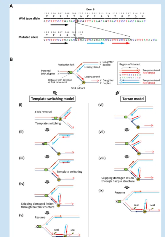
Molecular analysis of the proband identified a novel heterozygous microindel mutation in exon 6 of the TBX5 gene, NM_0001 92.3(TBX5_v001):c.627delinsGTGACTCAGGAAACGCTTTCCTGA (Fig. 2AA), which introduces a stop codon at position 210 (NM_00 0192.3(TBX5_i001):p. (Ala210)) and creates a truncatedTBX5pro- tein of 209 amino acids lacking part of the T-box domain sequences involved in DNA binding [Bruneau et al., 2001]. Given that most previously reportedTBX5mutations cause premature truncation of the primaryTBX5transcript, resulting in haploinsufficiency, this micro- indel is a likely pathogenic mutation for HOS in this patient. Indeed, the mutation is not present in the NHLBI GO Exome Sequencing Project (ESP6500) Variant Server (http://evs.gs.washington.edu/EVS/), the 1,000 Genomes Project database (http://www.1000genomes.org/) or the Human Genetic Variation Database (HGVD, http://www.genome.
med.kyoto-u.ac.jp/SnpDB/), although it has never been reported in the Human Gene Mutation Database professional 2015.2 (HGMD, http://
www.hgmd.org/) or ClinVar (http://www.ncbi.nlm.nih.gov/clinvar/).
Because parental DNA was not available, the mutation was not confirmed as de novo.
DISCUSSION
Both germline and somatic indels are uncommon and tend to shorten the nucleotide sequence, with the majority resulting in a net deletion that also shifts the reading frame [Chuzhanova et al., 2003; Scaringe et al., 2008; Stenson et al., 2014]. In mutation databases including HGMD and ClinVar, only one case of aTBX5microindel, which had a one-nucleotide deletion and a two-nucleotide insertion, was reported [Basson et al., 1999]. In addition, only one case with an insertion of more than three bases, which contained a 6-base duplication, has been reported inTBX5[Debeer et al., 2007]. Thus, the one-base deletion and 24-base insertion with a net 23-base gain detected in our HOS case is the first microindel with an unusually long insertion in theTBX5 gene.
The 24-base inserted sequence contains 8-base sense and anti- sense (reverse complementary) sequences, which are likely to derive from the same nearby but not adjacent template, with three nucleotides of separation and an additional five nucleotides (Fig. 2A). This finding is mostly consistent with the characteristics of microindels reported by Scaringe et al. [2008] from their analysis of somatic microindels. In microindels, the inserted sequences derive from nearby but not adjacent template sequences on the
sense or antisense strand, in contrast to the slippage that character- ises the great majority of pure microinsertions. It was also shown that the mechanisms of microindels, at least those with larger insertions, are highly error-prone overall, with an estimated error rate of 13% per base pair, consistent with the error rates of certain Y-family translesion polymerases [Scaringe et al., 2008]. Thus, the mutation detected in our case seems to be a microindel with a relatively large and complex insertion that arises from the error-
FIG. 1. Clinical photographs of an HOS patient. (A and B) 3-D ultrasonographic images of the fetal upper limbs. The right hand (A) showed absence of thumb, with radial deviation of the wrist, whereas the left hand (B) showed stiff fingers, and absence of thumb and index finger with radial deviation of the wrist. (C and D) Echocardiographic images of the fetus. A four-chamber view (C) showed large perimembranous and muscular VSD, and a three-vessel view (D) showed supernumerary fourth vessels (persistent left superior vena cava, PLSVC) to the left of the pulmonary artery (PA), LV, left ventricle; LA, left atrium; RV, right ventricle; RA, right atrium; AA, ascending aorta; RSVC, right superior vena cava. (E and F) Postnatal appearance of bilateral upper limbs. The right upper limb (E) had a radially bowed forearm, radial deviation of the wrist associated with flexion deformity at the elbow and wrist joint and absence of thumb, whereas the left upper limb (F) had a shortened middle segment, stiff fingers and absence of thumb, and index finger. (G) X-rays of the infant showing the absence of the radius and thumb in the right upper limb and absence of radius, thumb and index finger with hypoplastic humerus in the left upper limb. [Color figure can be seen in the online version of this article, available at http://wileyonlinelibrary.com/journal/ajmga].
FIG. 2. Characterization of theTBX5microindel detected in an HOS patient. A: Schematic representation of wild-type (top) and mutated (bottom) sequences observed inTBX5exon 6 in the HOS patient. Boxes indicate a 1-nucleotide deletion (top) and a 24-nucleotide insertion (bottom). Red and blue arrows indicate a match and reverse complement to the putative template (black arrows), respectively, observed in the inserted sequence. B: Schematic representation of two alternative models causing theTBX5microindel in this case. Replication by normal DNA polymerase is blocked by a DNA adduct (orange circle) located around the deletion on the template DNA (upper). For lagging strand synthesis to progress in the presence of a DNA adduct, a sequential nucleotide synthesis from the template strand (ii and iii) occurs after the association of the two nascent DNA strands (i; “template switching model,” lower left) or the nucleotide synthesis from nascent and template strands (vi and vii) after the dissociation of the nascent strand from the template (vi; “Tarzan model” [Scaringe et al., 2008], lower right) with some errors using an error-prone translesion polymerase. In both models, the translesion polymerase is able to swing across a DNA adduct (iv and viii), resulting in skipping of one nucleotide on the template and the normal polymerase can proceed with replication.
[Color figure can be seen in the online version of this article, available at http://wileyonlinelibrary.com/journal/ajmga].
prone translesion polymerase-mediated post-replication repair to bypass a DNA adduct, blocking the replication of lagging-strand synthesis (Fig. 2B, upper). On the basis of the inserted sequence containing both sense and antisense sequences derived from the nearby template observed in our case, two alternative models for indelogenesis may be presented. To save a cell with a DNA adduct that blocks replication by normal polymerase, an error-prone translesion polymerase complex may be recruited for the progres- sion of lagging-strand synthesis [Scaringe et al., 2008; Sale, 2012].
In the “template switching model” (Fig. 2B; lower left, i–v), the association of the two nascent DNA strands is followed by addi- tional synthesis on the nascent strand while constituting the inserted sequence from two templates sequentially when switching templates [Sale, 2012]. In the “Tarzan model” previously proposed by Scaringe et al. [2008] (Fig. 2B; lower right, iv–ix), the helicase unwinds the nearby nucleotides of the nascent strand from the template so that the translesion polymerase can loop back on itself and back up on the template strand. In both models, the association of sense and antisense sequences synthesized on the nascent strand from the nearby template form a hairpin structure with some additional length acting like a vine that the translesion polymerase uses to swing across the adduct [Scaringe et al., 2008]. In addition, existence of sequences whose template is undetermined in the insertion, other than sense, and antisense sequences derived from the nearby template, supports the involvement of error-prone translesion polymerase. Because of the lack of repeat sequences around the microindel in our case, serial replication of slippage models, a possible alternative mechanism for microindels with large insertion sizes [Chen et al., 2005], may not be involved in the indelogenesis of the microindel in the present case. Although non- homologous end joining after DNA double-strand breaks is unable to be completely excluded as another possible mechanism for microindel generation, the inserted sequence containing both sense and antisense sequences derived from the nearby template is difficult to be explained by this mechanism.
ACKNOWLEDGMENTS
We thank the family members of the patient for their participation in this study. This work was partly performed in the Cooperative Research Project Program of the Medical Institute of Bioregula- tion, Kyushu University, and supported by JSPS KAKENHI Grant Numbers 26293304 (I.I.) from the Ministry of Education, Culture, Sports, Science and Technology, Japan.
REFERENCES
Basson CT, Cowley GS, Solomon SD, Weissman B, Poznanski AK, Traill TA, Seidman JG, Seidman CE. 1994. The clinical and genetic spectrum of the Holt–Oram syndrome (heart hand syndrome). N Engl J Med 330:885–891.
Basson CT, Bachinsky DR, Lin RC, Levi T, Elkins JA, Soults J, Grayzel D, Kroumpouzou E, Traill TA, Leblanc-Straceski J, Renault B, Kucherlapati R, Seidman JG, Seidman CE. 1997. Mutations in human TBX5 cause limb and cardiac malformation in Holt–Oram syndrome. Nat Genet 15:30–35.
Basson CT, Huang T, Lin RC, Bachinsky DR, Weremowicz S, Vaglio A, Bruzzone R, Quadrelli R, Lerone M, Romeo G, Silengo M, Pereira A, Krieger J, Mesquita SF, Kamisago M, Morton CC, Pierpont ME, M€uller CW, Seidman JG, Seidman CE. 1999. Different TBX5 interactions in heart and limb defined by Holt–Oram syndrome mutations. Proc Natl Acad Sci USA 96:2919–2924.
Brassington AME, Sung SS, Toydemir RM, Le T, Roeder AD, Rutherford AE, Whitby FG, Jorde LB, Bamshad MJ. 2003. Expressivity of Holt–
Oram syndrome is not predicted by TBX5 genotype. Am J Hum Genet 73:74–85.
Bruneau BG, Nemer G, Schmitt JP. 2001. A murine model of Holt–Oram syndrome defines roles of the T-box transcription factor Tbx5 in cardio- genesis and disease. Cell 106:709–721.
Chen JM, Chuzhanova N, Stenson PD, Ferec C, Cooper DN. 2005. Complex gene rearrangements caused by serial replication slippage. Hum Mutat 26:125–134.
Chuzhanova NA, Anassis EJ, Ball EV, Krawczak M, Cooper DN. 2003.
Meta-analysis of indels causing human genetic disease: Mechanisms of mutagenesis and the role of local DNA sequence complexity. Hum Mutat 21:28–44.
Debeer P, Race V, Gewillig M, Devriendt K, Frijns JP. 2007. Novel TBX5 mutations in patients with Holt–Oram syndrome. Clin Orthop Relat Res 462:20–26.
Gonzalez KD, Hill KA, Li K, Li W, Scaringe WA, Wang JC, Gu D, Sommer SS. 2007. Somatic microindels: Analysis in mouse soma and comparison with the human germline. Hum Mutat 28:69–80.
Gruenauer-Kloevekorn C, Froster UG. 2003. Holt–Oram syndrome: A new mutation in the TBX5 gene in two unrelated families. Ann Genet 46:19–23.
Heinritz W, Shou L, Moschik A, Froster UG. 2005. The human TBX5 gene mutation database. Hum Mutat 26:397.
Holt M, Oram S. 1960. Familial heart disease with skeletal malformations.
Br Heart J 22:236–242.
Huang T. 2002. Current advances in Holt-Oram syndrome. Curr Opin Pediatr 14:691–695.
Li QY, Newbury-Ecob RA, Terrett JA, Wilson DI, Curtis AR, Yi CH, Gebuhr T, Bullen PJ, Robson SC, Strachan T, Bonnet D, Lyonnet S, Young ID, Raeburn JA, Buckler AJ, Law DJ, Brook JD. 1997. Holt–Oram syndrome is caused by mutations in TBX5, a member of the Brachy-ury (T) gene family. Nat Genet 15:21–29.
McDermott DA, Bressan MC, He J, Lee JS, Aftimos S, Brueckner M, Gilbert F, Graham GE, Hannibal MC, Innis JW, Pierpont ME, Raas-Rothschild A, Shanske AL, Smith WE, Spencer RH, St John-Sutton MG, van Maldergem L, Waggoner DJ, Weber M, Basson CT. 2005. TBX5 genetic testing validates strict clinical criteria for Holt-Oram syndrome. Pediatr Res 58:981–986.
Newbury-Ecob RA, Leanage R, Raeburn JA, Young ID. 1996. Holt–Oram syndrome. Am J Med Genet 65:128–132.
Sale JE. 2012. Competition, collaboration and coordination – determining how cells bypass DNA damage. J Cell Sci 125:1633–1643.
Scaringe WA, Li K, Gu D, Gonzalez KD, Chen Z, Hill KA, Sommer SS.
2008. Somatic microindels in human cancer: The insertions are highly error-prone and derive from nearby but not adjacent sense and antisense templates. Hum Mol Genet 17:2910–2918.
Stenson PD, Mort M, Ball EV, Shaw K, Phillips A, Cooper DN. 2014. The Human Gene Mutation Database: Building a comprehensive mutation repository for clinical and molecular genetics, diagnostic testing and personalized genomic medicine. Hum Genet 133:1–9.