Bot. Mag. Tokyo 84: 429--433, 1971
Short Communication
An Analysis of the Mineral Deposit in the Plant Cell: Analysis of the Stellate Hair of Deu tzia Leaf Using an
X-ray Microanalyzer
NAOKO MIYAMOTO, TAKAO NAMAE and HARUO YOTSUMOTO
Application Department, Electron Optics Division, JEOL LTD., Nakagamicho 1418, Akishimashi, Tokyo 196
The scanning electron microscope used in conjunction with an electron probe X-ray microanalyzer provides information on the elemental composition of specimens by measuring the wave-lengths and intensities of the characteristic X-ray generated from the specimen surface where the fine-focused electron probe irradiates.
This type of equipment has been widely used for the elementary analysis of micro-areas of substances in the fields of metallurgy, mineralogy, ceramics, etc.
Recently it has been possible to use this equipment for the elementary analysis of biological materials. For example, in the field of medical science, hard tissue specimens such as teeth and bones can be analyzed. Now, even soft tissue specimens in animals and plants can be analyzed.
The present paper describes the elementary analysis of stellate hair of Deutzia leaf using the aforementioned equipment.
It has been known for a long time that there are mineral deposits in the plant cytoplasm (Molisch, 1922). As their shape depends upon the species and genus of plants, the mineral deposits are used for morphological identification of plants (Ino, 1949). Mineral deposits exist in the cell wall or on the cell surface. This crystalline substance is considered to be a reserve substance, secretion or excretion of the plant body.
Mineral deposits consist mainly of silicates, calcium and manganese compounds, etc. The main component of the mineral deposit in the leaf, stem and bark cells is calciumoxalate (Ca(Co2)2) (Ogura, 1934). Silicate also exists widely in the cell wall, especially in the epidermal cell and hairs of the stem and leaf.
The hairs have various shapes, which are used as the criteria of classification. They insulate heat and protect the leaves from moisture and damage, thus protecting the plant body. In some kinds of hairs, mineral deposits in their cell wall harden the wall itself. The distribution of the constituent elements of the mineral deposits, however, is not known at all.
Thus, we studied the constituent elements distribution of the stellate hairs on a leaf epidermis of Deutzia scabra Thunb.
430 N. MIYAM0T0 et al. Vol. 84
The equipment used in the present study was the JSM-U3-DDS which comprises a scanning microscope and an X-ray spectrometer. It permits the scanning electron microscopic observation of the specimen surface (resolution : 100 A) and also provides information on constituent elements of the specimen (kinds, distribution and composi-tion ratio of elements) by detecting the characteristic X-ray generated when the specimen is irradiated with electron beams.
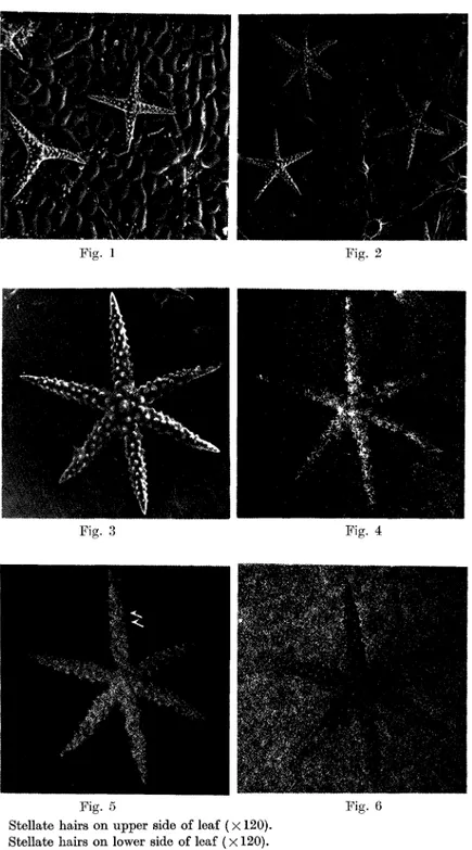
Stellate hairs were observed on the leaf epidermis of the Deutzia scabra Thunb, at a rate of 0.5 hair per square millimeter. Their shapes were varied, ranging from bar-like hairs to six-pronged hairs. Compared with the hairs on the lower side of the leaf, the hairs on the upper side had less prongs; many were three- or four-pronged. Projections were observed on all the stellate hair surfaces (Figs. 1, 2). In this experi-ment, the specimen was coated with carbon in vacuum to give electric conductivity.
The operating conditions were as follows :
accelerating voltage : 15 kV
specimen absorbed current : 10-9A
Figs. 4, 5 and 6 show characteristic X-ray images of each element, obtained without destroying the stellate hair on the lower side of the leaf in Fig. 3. These images show localization of individual elements. The white parts in the image represent a large amount of element, while the dark parts represent a small amount of element. But these images do not represent the quantitative relations between different elements. From another experiment to find the quantitative relations by measuring the intensity of each characteristic X-ray, it is concluded that more calcium is present than potassium or magnesium. The result of this experiment indicating that the stellate hair contains a large amount of calcium agrees with the result of the chemical detection method.
The X-ray images clearly show the difference in localization between calcium and potassium. That is, calcium is concentrated in the stellate hair but is limited to the leaf. Conversely, the potassium content is high in the leaf and low in the stellate hair. Much calcium is seen at the center, tips and projections of the stellate hair, while much potassium is observed between projections.
These facts indicate that at first calcium is formed on the stellate hair and then potassium is deposited inside it. Calcium and potassium are spirally distributed from the center to the tip (Figs. 5, 6).
Figs. 7 and 8 show individual elemental distributions (concentration differences) along the lines A-A' and B-B' on the same prong of the stellate hair transverse section (Fig. 9). The line A-A' was drawn so as to pass between projections, whereas the line B-B' was drawn so as to pass through a projection.
In comparing Fig. 7 with Fig. 8, one readily discerns a distinct difference in the distributions on the right side. In Fig. 7, the right side portion indicates a relatively large amount of potassium, but the amount of calcium is rather small. Fig. 8, however, indicates that the calcium content is very high in the right side projection and gradually decreases towards the left side. The increase of potassium is also slow in the
An Analysis of the Minera 1 Deposit 431
Fig. 1. Stellate hairs on upper side of leaf (X 120). Fig. 2. Stellate hairs on lower side of leaf (X 120). Figs. 3, 4, 5 and 6 (X 360).
Elemental distributions in a Stellate hair. Fig. 3: distribution of magnesium ; Fig. 5 : distribution
potassium. secondary electron of calcium; Fig. 6: image ; Fig. distribution 4: of
432 N. MIYAMOTO et al. Vol. 84
right side portion. This proves that the projections contain a large amount of calcium and a small amount of potassium, and that considerable amounts of potassium are distributed between projections. These findings are consistent with the images shown in Figs. 5 and 6.
Figs. 7, Figs. (Fig.
8 and 9.
7 and 8 show individual elemental distributions along the lines 9) on the stellate hair transverse section (X 2500).
An Analysis of the Mineral Deposit 433
The analytical results obtained with the X-ray microanalyzer should provide a basis for the study of various matters related to the crystalline structure of stellate hair.
As compared with the chemical microquality analysis, this analytical method is significant in that it can reveal the elemental localization corresponding to a specimen micro-area without destroying the specimen. In addition, the method has a further advantage of making clear the localizations of the elements such as magnesium and potassium which cannot be detected with conventional chemical methods. Inclusion in the stellate hairs of these elements which are macronutrients for the plants proceeds
in the following way. The calcium, magnesium and potassium absorbed from the
soil are finally transported to the leaves. Although these elements are normally consumed, their excess absorption results in the deposits of calcium, magnesium and potassium in the leaves. This is a kind of so-called internal excretion.
The present results indicate that the qualitative and quantitative analysis of plant cells and tissues using the X-ray microanalyzer will be one of the effective analytical methods for cell morphology, cell physiology and taxonomy.
We thank Prof. S. Watari, Department of Biology, Chiba University, for his valuable advice. We are also indebted to Prof. T. Ohtsuki, Department of Biology, Medical College of Toyo and Prof. S. Sato, Department of Biology, Japan Women's University for their encouragement and support.
References
FRANKEL, R.S. AND D.W. AITKEN. 1970. Energy-dispersive X-ray emission spectroscopy. Reprinted from applied spectroscopy 24: 557-566.
INO, S. 1949. Plant Histology (Japanese). Uchidarokakuho, Tokyo
MIzumRA, V., S. SHIINA, K. MIAKE, M. ISHIDA, H. NAKAMURA, H. YOTsuMOTo AND T. NAMAE. 1971. Comparative exmaination between the chemical and physical methods to the
demonstration of the ionic localization the tissues. I. In the case of potassium
monate method. Published by the EMSA in the conference proceedings 408-409. MoLIscH, H. 1922. Mikrochemie der Pflanze. Gustav Fischer, Jena.
OGuRA, Y. 1934. Plant Morphology (Japanese). Yokendo, Tokyo.
Russ, J.C. AND E. MCNATT. 1970. Copper localization in cirrhotic rat liver by scanning electron microscopy. Published by the ASTM in the conference proceedings. SCHNEIDER, H. AND P. ZIMMERMANN. 1892. Botanische Mikrotechnik. Gustav Fischer, Jena. WAISEL, Y., AVIA HOFFEN AND A. ESHEL. 1970. Localization of ions in the mesophyll cells of
the succulent halophyte Suaeda monoica Forssk. by X-ray microanalysis. Physiolgia
Plantarum 23: 75-79.
WATARI, S. 1941. Notes on the crystals in the leaves of Saxifragaceous plants. I (Japanese). Shokubutsu oyobi Dobutsu 9: 479-488.
. 1941. Notes on the crystals in the leaves of Saxifragaceous plants. II (Japanese). Shokubutsu oyobi Dobutsu 9: 639-645.