―総説―
AMPK を介した糖・脂質代謝調節に関する研究
横山雄一
a), b), 井口和弘
a), 臼井茂之
a), 平野和行
a)* 要約:グリセロールは肝臓における糖新生や脂質合成の材料であるため、肝臓へのグリセロール流入量の変化は様々な代 謝経路に変調をきたす。アクアポリン 9(AQP9)は、主に肝臓において発現が見られ、水分子のみならず、グリセロー ルや尿素などの低分子溶質をも透過させるチャネル型膜蛋白質である。AMP-activated protein kinase(AMPK)は生体内の エネルギーセンサーであり、糖・脂質代謝の恒常性維持に働くセリン/スレオニンキナーゼである。本研究では、AMPK の活性化剤である、5-aminoimidazole-4-carboxamide-1--D-ribonucleoside(AICAR)を、ヒト肝癌由来 HepG2 細胞に作用さ せたところ、AQP9 mRNA の発現量が顕著に減少することを確認した。レポータージーンアッセイや転写因子 forkhead box a2(Foxa2)遺伝子をノックダウンさせた実験の結果から、Foxa2 は、AICAR による AQP9 遺伝子発現抑制に関わる重要 な転写調節因子であることを見出した。AICAR により活性化された AMPK は、Akt の Thr-308 残基と Ser-473 残基のリン 酸化を促し、それに伴い Foxa2 がリン酸化されて核内から核外へと移行することを明らかにした。したがって、肝臓での グリセロール輸送の観点から AMPK によるコントロールのもとに、AQP9 は肝臓へのグリセロールの流入量を変化させ、 糖・脂質代謝調節に寄与している可能性が示唆された。索引用語:AMP-activated protein kinase(AMPK)、アクアポリン9(AQP9)、forkhead box a2(Foxa2)、Akt/PKB、核外輸 送
Regulation of Glucose and Lipid Metabolism via AMPK
Yuichi YOKOYAMA
a, b), Kazuhiro IGUCHI
a), Shigeyuki USUI
a),
Kazuyuki HIRANO
a)*Abstract: Change in glycerol influx into the liver causes metabolic alternations in many pathways since glycerol is a substrate for
gluconeogenesis and lipogenesis in the liver. Aquaporin 9 (AQP9), a channel membrane protein, is expressed mainly in the liver and permeable to water and small molecular weight solutes such as glycerol and urea. AMP-activated protein kinase, AMPK, functioning as a serine/threonine kinase acts as an energy sensor in the homeostasis of glyco- and lipid-metabolism. In this study, we found that 5-aminoimidazole-4-carboxamide-1--D-ribonucleoside (AICAR), an AMPK activator, significantly down-regulated AQP9 mRNA expression in human hepatoma HepG2 cells. Forkhead box a2 (Foxa2) was demonstrated to be one of the transcriptional regulators of AQP9 gene expression repressed by AICAR from the results of a reporter gene assay and knock-down of the Foxa2 gene by siRNA. Activation of AMPK by AICAR promoted the phosphorylation of Akt at Thr-308 and Ser-473 residues and subsequently phosphorylated and excluded Foxa2 from the nucleus. These results suggest the possibility that AQP9 is under AMPK control in the influx of glycerol into the liver and that AQP9 contributes to the glycol- and lipid-metabolism of glycerol transport in the liver.
Key phrases: AMP-activated protein kinase (AMPK), Aquaporin 9 (AQP9), forkhead box a2 (Foxa2), Akt/PKB, nuclear exclusion
1.緒言 近年、食の欧米化に伴う栄養の過剰摂取が、メタボリッ クシンドロームを誘発する要因になっていると問題視さ れている。メタボリックシンドロームの診断基準となる肥 満や高血糖などの生活習慣病は、悪性新生物、脳血管疾患、 a) 岐阜薬科大学医療薬剤学大講座薬剤学研究室(〒501-1196 岐阜市大学西1丁目25-4)
Laboratory of Pharmaceutics, Gifu Pharmaceutical University (1-25-4 Daigaku-nishi, Gifu 501-1196, JAPAN) b) 名古屋大学医学部附属病院薬剤部(〒466-8560 愛知県名古屋市昭和区鶴舞町65)
心疾患等の重篤な疾病を引き起こすリスクファクターで ある。高齢化が進む昨今においては、健康に長生きをする ことへの関心が高まっており、食や健康や疾病がメディア の題材として取り上げられる機会も多い。このような状況 の中で、近年、エネルギー代謝調節に重要な役割を果たし、 それが持つ運動疑似効果や、食欲調節効果によって、抗メ タボリックシンドローム効果が期待されている分子に、 AMP-activated protein kinase(AMPK)がある1)。
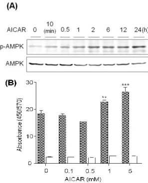
AMPK は、細胞内の AMP/ATP 比の上昇に伴って活性化 されるセリン・スレオニンキナーゼである2)。AMPK が活 性化されると、phosphofruktkinase-2 の活性化、acetyl-CoA carboxylase の阻害、および骨格筋での glucose transporter 4 の細胞膜へのトランスロケーションの促進などによって、 解糖、脂肪酸酸化、さらに糖取り込みの促進といった ATP を産生する経路が亢進する。また同時に、AMPK が活性化 されると糖新生酵素の phosphoenolpyruvate carboxykinase、 glucose-6-phosphatase 、 お よ び 転 写 因 子 sterol regulatory element-binding protein 1 の発現抑制などによって、糖新生 や脂肪合成等の ATP を消費する経路は抑制される。この ような作用によって、AMPK は血糖降下作用や抗肥満効果 を示すことが期待されている。 本研究では、AMPK の活性化によって発現調節される 糖・脂質代謝関連遺伝子として、糖新生や脂質合成の材料 となるグリセロールを透過する aquaporin 9(AQP9)に着 目した。ヒトの AQP には AQP0~12 の 13 種類のサブタイ プが存在し、透過する分子の違いによって 2 つのグループ に分類される。一方は水分子のみを透過する分子種であり、 他方は水分子以外にグリセロールや尿素のような低分子 溶 質 も 透 過 す る 分 子 種 で あ る 。 特 に 後 者 は aquagulyceroporin(AQGP)と総称され、ヒトでは AQP3、 7、9、10 である3)。現在、ヒトにおいてグリセロールチャ ネルとして知られる蛋白質は AQGP のみであり、糖新生 や脂質合成が盛んにおこなわれる肝臓には AQP9 が多く 発現している4, 5)。 AQP9 によって肝臓に取り込まれるグリセロールは、糖 新生や脂肪合成の材料となるため、糖新生や脂肪合成が抑 制される AMPK 活性化時には、AQP9 の発現が抑制される ことが推察される。そこで、ヒト肝癌由来 HepG2 細胞に AMPK 活 性 化 剤 で あ る 5-Aminoimidazole-4-carboxamide ribonucleoside(AICAR)を処理したところ、AQP9 mRNA の発現抑制が認められたので、AICAR による AQP9 転写 調節機序を詳細に検討した。 2.AQP9 遺伝子発現に及ぼす AICAR の影響 AICAR が AQP9 遺伝子発現にどのような影響を及ぼす かを HepG2 細胞において検討した。HepG2 細胞に、各濃 度の AICAR を 24 時間処理した時、または、1 mM の AICAR を各時間処理した時の AQP9 mRNA 発現量を real-time RT-PCR 法にて測定した。AQP9 mRNA 発現は 1 mM の AICAR を 12 時間処理することで有意に抑制された(Fig. 1A、B)。
Fig. 1. Effect of AICAR on the AQP9 mRNA expression in HepG2 cells.
HepG2 cells were incubated with (A) various concentrations of AICAR for 24 h and (B) 1 mM AICAR for the period indicated. HepG2 cells incubated without AICAR were used as a control. The expression of mRNA was quantified by the real-time RT-PCR method after total RNA was extracted. The results were normalized with the 2-microglobulin mRNA levels and the mRNA level of the control was taken as 100%. Data show the mean ± S.D. from five experiments. **p<0.01, ***p<0.001 vs. control. 3.AICAR による AMPK の活性化を介した AQP9 mRNA 遺伝子発現抑制 3.1.AMPK のリン酸化およびその活性に及ぼす AICAR の影響 AICAR は AMPK の活性化剤として用いられるが、 AICAR の作用発現に AMPK の活性化を介さないという報 告も少数存在する。そこで、HepG2 細胞において、AICAR が実際に AMPK を活性化するかどうか、また、AICAR に よる AQP9 mRNA 発現抑制に AMPK の活性化が関連して
いるか否かについて検討した。1 mM AICAR で処理すると AMPK のリン酸化は時間依存的に亢進した(Fig. 2A)。
また、100 M~5 mM の AICAR で 1 時間処理し、AMPK 活性を測定したところ、1 mM と 5 mM の処理濃度にて AMPK 活性の有意な上昇が認められ、その活性は AMPK 阻害剤 Compound C の前処理によって阻害された(Fig. 2B)。
Fig. 2. Effect of AICAR on the AMPK activation.
(A) HepG2 cells were incubated with 1 mM AICAR for the period indicated. Cell lysate was prepared with lysis buffer containing SDS and 2-mercaptoethanol and forty g of protein from the lysate was separated by electrophoresis with a 10% SDS-polyacrylamide gel. After proteins in the gel were electroblotted on a PVDF membrane, AMPK alpha subunit (AMPK) and phosphorylated AMPK were probed with anti-AMPKantibody and anti-phospho-AMPK (Thr-172) antibody, respectively, and visualized using a secondary antibody-peroxidase conjugate and the ECL system. (B) HepG2 cells were incubated with (open column) or without (filled column) 10 M Compound C for 1 h and subsequently treated with various concentration of AICAR for 1 h. Cell lysates were prepared with lysis buffer and relative AMPK activities were measured with AMPK Kinase Assay Kit. The data were indicated by measurement of dual wavelengths of 450/570 nm absorbance. Data show the mean ± S.D. from three experiments. **p<0.01, ***p<0.001 vs. the cells treated without AICAR and Compound C.
3.2.AQP9 mRNA 遺伝子発現に及ぼす AMPK 活性化 の影響
AICAR 処理によって抑制された AQP9 mRNA 発現は Compound C の前処理によって有意に回復した(Fig. 3A)。
また、2 型糖尿病治療薬の metformin(MF)は AMPK の 活性化を介して血糖低下作用を示す。そこで、MF も AICAR と同様に AQP9 mRNA 発現を抑制するかどうかを 調べた結果、MF は AQP9 mRNA 発現を濃度依存的に抑制 した(Fig. 3B)。
Fig. 3. Effect of AMPK on the AQP9 mRNA expression.
(A) HepG2 cells were incubated with various concentrations of Compound C for 1 h and subsequently treated with 1 mM AICAR for 24 h. HepG2 cells incubated without AICAR and Compound C were used as a control. The mRNA expression of AQP9 was analyzed by the real-time RT-PCR method after total RNA was extracted. The results were normalized with the -microglobulin mRNA levels and the mRNA level of the control was taken as 100%. Data show the mean ± S.D. from three experiments. **p<0.01 vs. control, ††p<0.01 vs. the cells treated with AICAR and without Compound C. (B) HepG2 cells were incubated with various concentrations of metformin for 24 h. HepG2 cells incubated without metformin were used as a control. The expression of mRNA was quantified by the real-time RT-PCR method after total RNA was extracted. The results were normalized with the -microglobulin mRNA levels and the mRNA level of the control was taken as 100%. Data show the mean ± S.D. from five experiments. **p<0.01, *** p<0.001 vs. control. 4.AQP9 転写活性に及ぼす AICAR の影響 AQP9 転写開始点から上流-2000 bp、-1480 bp、- 512 bp および-278 bp から+80 bp までのプロモーター領 域をpGL3 ルシフェラーゼベクターにクローニングし、そ れぞれ、プラスミド 2000pGL3、1480pGL3、512pGL3 および278pGL3 とした。これらを HepG2 細胞に導入し、 AICAR の AQP9 プロモーター活性に及ぼす影響について 調べた。AICAR は完全には AQP9 転写活性を抑制しなか ったが、AQP9 プロモーター配列の内、-1480 bp から- 512 bp までの領域、および -512 bp から-278 bp まで の領域に AICAR の関与によって調節を受ける DNA 配列 が存在することが示唆された(Fig. 4A)。従って、上記の 2 種類の配列間に結合領域を持ち、かつ AICAR によって 活性調節される転写因子を検索したところ、Foxa2 を見出 した。そこで、Foxa2 が AQP9 の転写活性を調節するかど うかを確認するため、HepG2 細胞に Foxa2 siRNA をトラ ンスフェクトし、Foxa2 発現を抑制したところ、AQP9 mRNA 発現は有意に抑制された(Fig. 4B)。従って、転写 因子 Foxa2 は AQP9 mRNA の転写亢進に寄与していること が示唆された。
Fig. 4. Transcriptional regulation of AQP9 gene by AICAR.
(A) HepG2 cells were co-transfected with the reporter vector containing the promoter region of AQP9 gene and a reference plasmid, phRL-TK. Transfected cells were maintained in the regular culture medium for 24 h and further cultured with or without 1 mM AICAR for 24 h. Cell lysates were prepared after the incubation and luciferase activities were measured with a Dual-Luciferase Reporter Assay System. Firefly luciferase activity was normalized with Renilla luciferase activity and is expressed relative to the control treated without AICAR. Data represent the mean ± S.D. from five experiments. **p<0.01, ***p<0.001 vs. 2000pGL3 treated with AICAR. (B) Foxa2 siRNA and control siRNA were transfected to HepG2 cells with lipofectamine 2000 and incubated for 24h. The suppression of Foxa2 mRNA levels was measured by RT-PCR and AQP9 mRNA levels were quantified by the real-time RT-PCR method after total RNA was extracted. The results were normalized with the 2-microglobulin mRNA levels and the mRNA level of control siRNA treatment gruoup was taken as 100%. Data show the mean ± S.D. from three experiments. **p<0.01 vs. cells treated with control siRNA. (C) HepG2 cells were incubated with 1 mM AICAR for the period indicated. The expression of Foxa2 mRNA was quantified by the real-time RT-PCR method after total RNA was extracted. The results were normalized with the 2-microglobulin mRNA levels and the mRNA level of 0 h was taken as 100%. Data show the mean ± S.D. from four experiments. **p<0.01 vs. 0 h.
AICAR による Foxa2 を介した AQP9 mRNA 発現抑制機 構を調べるため、先ず AICAR 処理による Foxa2 mRNA 発
現量の変化を real-time RT-PCR 法にて測定した(Fig. 4C)。 Foxa2 mRNA 量は AICAR 処理開始から 16 時間後までは変 化せず、24 時間処理後に、わずかに減少した。AICAR に よる AQP9 mRNA 発現の抑制作用は AICAR 処理 12 時間 後には確認されているので、AICAR による Foxa2 発現調 節が AQP9 の発現抑制には関与しないことが示された。
5.AICAR による AQP9 遺伝子発現への PI3K/Akt 経路 の関与 5.1.AICAR による Akt のリン酸化 Leclerc ら6)は 、 急 性 リ ン パ 性 白 血 病 細 胞 に お い て 、 AICARによってAMPKが活性化されると、AktのThr-308残 基、およびSer-473残基のリン酸化が亢進することを報告し ている。そこで、本実験系における、AICARによるAktの リン酸化亢進について検討した。Fig. 5に示すように、Akt の2つのリン酸化部位(Thr-308、Ser-473)は、1 mM AICAR 処理開始から6時間後をピークとして、リン酸化されるこ とが確認された。
Fig. 5. Phosphorylation of Akt by AICAR in HepG2 cells.
HepG2 cells were treated with 1 mM AICAR for the period indicated. Cell lysate was prepared with lysis buffercontaining SDS and 2-mercaptoethanol, and 40 g protein from the lysate was separated by electrophoresis with a 10% SDS–polyacrylamide gel. After proteins in the gel were electroblotted on a PVDF membrane, phosphorylated Akt (Thr-308), phosphorylated Akt (Ser-473), and Akt were probed with anti-phospho-Akt (Thr-308) antibody, antiphospho-Akt (Ser-473) antibody, and anti-Akt antibody, respectively, and visualized using a secondary antibody-peroxidase conjugate and the ECL system.
5.2. PI3K/Akt経路に及ぼすAICARの影響 血糖降下ホルモンであるinsulinはPI3K/Akt経路を活性化 することが良く知られている。また、insulinはAQP3、AQP7、 AQP9のmRNA発現を抑制することが報告されている。そ こで、HepG2細胞においてinsulinのAQP9 mRNA発現に及 ぼす影響を確認したところ、AQP9 mRNA発現量はinsulin 処理によって濃度依存的に抑制された(Fig. 6A)。
さ ら に 、 AICARによる AQP9 mRNA発現の抑制への Akt1/2インヒビターとPI3K阻害剤であるWortmanninの影 響について検討した(Fig. 6B)。その結果、AICAR処理に よって抑制されたAQP9 mRNA発現は、Akt1/2インヒビタ
ーの前処理により回復が見られたが、Wortmanninでは回復 しなかった。この結果から、AICARはAMPKの活性化を介 してAktのリン酸化を亢進するが、insulinのようにPI3K経 路の活性化を介さないことが示唆された。
Fig. 6. Effects of PI3K and Akt on AQP9 mRNA expression.
(A) HepG2 cellswere incubated with various concentrations of insulin for 12 h or (B) preincubated with inhibitors for 1 h and then treated with 1 mM AICAR for 24 h. HepG2 cells incubated without insulin, AICAR, and inhibitors were used as a control. The expression of AQP9 mRNA was quantified by real-time RT-PCR after total RNA was extracted. The results were normalized with the 2-microglobulin mRNA levels and the mRNA level of the control was taken as 100%. Data show the mean ± S.D. of four experiments. *** p < 0.001 vs. control, ††
p < 0.01 vs. cells treated with AICAR and without Akt inhibitor.
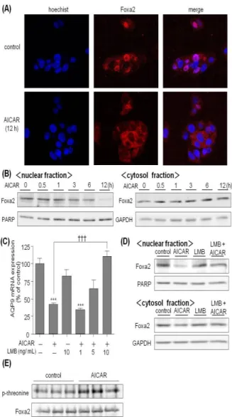
6.AICARによるFoxa2の調節
Fig. 4 では、AICAR による AQP9 遺伝子発現の抑制に、 転写因子 Foxa2 が関与していることを示したが、1 mM AICAR 24 時間処理は Foxa2 mRNA 発現を 35%程度しか抑 制させなかったため、Foxa2 による AQP9 mRNA 発現調節 は転写因子の量的な制御機構では無く、Howell と Stoffel ら7)の報告のように、Foxa2 の核外輸送による制御が行わ れているのではないかと考えられた。そこで、HepG2 細胞 内の Foxa2 の局在性を調べるため、Foxa2 抗体を用いた免 疫染色法および細胞分画を行った各画分について Western blotting を行った(Fig. 7A、B)。HepG2 細胞に 1 mM の AICAR を 12 時間処理することで、Foxa2 は核画分から細 胞質画分へ移行した。Foxa2 タンパクの核外移行メカニズ ムとして、核外輸送体 CRM1 が Foxa2 分子中の核外輸送 シグナル(NES)に結合することで、Foxa2 タンパクを核 外に輸送することが知られている。そこで、AICAR によ る Foxa2 の核外移行に対する CRM1 の関与について検討 した。CRM1 の阻害剤である leptomycin B(LMB)を HepG2 細胞に 1 時間前処理し、その後 AICAR で 12 時間処理した。 その結果、AICAR による Foxa2 の核外移行は LMB によっ て阻害されることが確認された(Fig. 7D)。さらに、Fig. 7C
Fig. 7. CRM1-dependent nuclear export of Foxa2 by AICAR.
(A) HepG2 cells were treated with or without 1 mM AICAR for 12 h. After permeabilizing and blocking the fixed preparation with 0.05% Triton-X and 1% BSA, respectively, cells were probed with anti-Foxa2 antibody and subsequently visualized using a secondary antibody-rhodamine conjugate (red). Nuclear staining was performed with Hoechst (blue). The preparation was examined under a confocal laser scanning microscope. (B) HepG2 cells were treated with 1 mM AICAR for the period indicated, (C) preincubated with or without 10 ng/mL leptomycin B (LMB) for 1 h and then treated with 1 mM AICAR for 12 h. HepG2 cells incubated without AICAR and LMB were used as a control. Cells were subsequently fractionated to cytosol and nuclear fractions. Ten micrograms of protein from cytosol fraction and 5 lg protein from nuclear fraction were separated by electrophoresis with a 10% SDS–polyacrylamide gel. (D, E) HepG2 cells were treated with 1 mM AICAR for 12 h or without AICAR as a control. Cell lysate was prepared with lysis buffer and was subsequent to immunoprecipitation with anti-Foxa2 antibody. The precipitant was collected by protein A/G agarose and separated by electrophoresis with a 10% SDS–polyacrylamide gel. After proteins in the gel were electroblotted on a PVDF membrane, PARP, GAPDH, Foxa2 and phosphorylated threonine residues in Foxa2 were probed with corresponding specific antibodies and visualized using a secondary antibody-peroxidase conjugate and the ECL system.
では、AICAR によって抑制された AQP9 mRNA 発現への CRM1 の関与について検討した。AICAR によって抑制さ れた AQP9 発現は 10 ng/mL の LMB によってコントロール レベルまで完全に回復した。次に、Foxa2 は Akt による Thr-156 残基のリン酸化を受け核外移行するため、AICAR による Foxa2 のリン酸化について検討した。Foxa2 の Thr-156 のリン酸化抗体は市販されていないため、1 mM の AICAR で 12 時間処理した HepG2 細胞から、Foxa2 タンパ クを免疫沈降し、スレオニンのリン酸化状態を Western blotting で測定した。その結果、AICAR 処理によって Foxa2 のリン酸化が亢進することが確認された(Fig. 7E)。 7.結論 本検討では、HepG2細胞にAICARを処理した結果、AQP9 発現が抑制された。AICARによるFoxa2の調節機構は、 AICARによって活性化されたAMPKがAktのThr-308残基 とSer-473残基をリン酸化し、それに引き続いて転写因子 Foxa2をリン酸化し、リン酸化されたFoxa2が核内から核外 へ移行することにより、AQP9遺伝子の転写が阻害される という一連のメカニズムが示唆された。AICARによる肝臓 のAQP9発現の抑制は、糖新生や脂質合成の原料となるグ リセロールがAQP9を介して肝臓に流入するのを阻害する ことで、これらのエネルギー消費経路を抑制するのに寄与 していると考えられる。実際に、レプチンレセプター変異 型肥満モデルマウスにおいてAQP9をノックアウトするこ とで、血中グリセロール濃度の顕著な増加と、血糖値の有 意な低下が認められる8)。従って、AQP9の発現抑制は、肝 臓へのグリセロールの流入を抑制し、その結果、糖新生や 脂質合成といったエネルギー消費経路の抑制作用に繋が ると考えられる。 AICARによって活性化されたAMPKがAktのThr-308残 基とSer-473残基をリン酸化し、それに続いて、Foxa2がリ ン酸化されて核外移行することによって、AQP9発現が抑 制されることが示唆された。InsulinによるFoxa2の核外移 行は、Foxa2構成アミノ酸配列中のロイシンリッチな核外 移行配列(NES)に核外輸送体CRM1が結合することで行 わ れ る が 、 本 検 討 に お い て も CRM1 の 阻 害 剤 で あ る leptomycin BによってAICARによるAQP9の発現抑制に回 復が認められたことから、AICARによるFoxa2の核外移行 もCRM1依存的である事が考えられる。Insulinは、Foxa2 をリン酸化し、核外移行することにより、血糖降下作用の 発現を調節している。近年、Stoffelら7)によって、肝臓細 胞におけるFoxa2の局在性が、糖尿病におけるインスリン 抵抗性の発現に寄与していると報告された。本検討の結果 から、Foxa2によってAQP9発現は亢進されているが、Foxa2 が常時核外に移行しているインスリン抵抗性病態におい ては、AQP9の発現が健常人に比べて低下していることが 予測される。実際、Gema Frühbeckら9)の報告では、2型糖 尿病患者ではAQP9発現が顕著に減少しており、これ以上 の高血糖が亢進するのを防ぐために、患者の生体はAQP9 発現を抑制することで、糖新生の原料となるグリセロール が肝臓内に流入するのを抑制しているのではないかと報 告している。また、AMPKの発現量や酵素活性は、インス リン抵抗性モデルマウスでは、コントロールマウスに比べ て高くなっており、インスリン抵抗性病態に見られる脂肪 代謝の異常を改善するためにAMPK活性が常時高くなっ ているのではないかとの報告もある。これらの報告から、 薬剤などによって AMPKを活性化させ、肝臓細胞内の Foxa2を不活性化し、AQP9発現を抑制することで、肝細胞 外からのグリセロールの流入を減少させ、血糖や脂質の異 常が改善する可能性が考えられる。 AMPKの活性化によりAQP9の発現が抑制されると、肝 臓への糖新生や脂質合成の原料となるグリセロールの流 入が阻害されるため、AQP9はグリセロール輸送の観点か ら、糖代謝、脂質代謝に寄与していると考えられる。本検 討で確認された、AMPK-Akt-Foxa2経路はエネルギー代謝 調節に重要な役割を果たしていると考えられる。 8.謝辞 本研究に際して終始御懇切なる御指導と御鞭撻を受け 賜わりました、岐阜薬科大学 薬剤学研究室 平野和行教 授に深甚なる謝意を表します。 また、本論文に対して貴重な御意見と御指導を賜りまし た岐阜薬科大学 木方正特任教授に厚く御礼申し上げま す。 本研究に際し、常に適切な御助言と御指導を賜わりまし た岐阜薬科大学 薬剤学研究室 臼井茂之准教授、井口和 弘助教、ならびに岐阜薬科大学附属薬局 窪田傑文助教に 深謝いたします。また、御協力を賜りました薬剤学研究室 諸氏に感謝の意を表します。 9.引用文献
1) Zhang B. B., Zhou G., Li C., Cell Metab, 9, 407-416 (2009).
2) Kahn B. B., Alquier T., Carling D., Hardie D. G., Cell Metab, 1, 15–25 (2005).
3) Ishibashi K., Mol. Med, 40, 816-823 (2003).
4) Tsukaguchi H., Shayakul C., Berger U. V., Mackenzie B., Devidas S., Guggino W. B., Van Hoek A. N., Hediger M. A., J. Biol. Chem, 273, 24737-24743 (1988).
5) Tsukaguchi H., Weremowicz S., Morton C. C., Hediger M. A., Am. J. Physiol, 277, F685-F696 (1999).
6) Leclerc G. M., Leclerc G. J., Fu G., Barredo J. C., J. Mol. Signal, 5, 15-27 (2010).
7) Howell J. J., Stoffel M., J. Biol. Chem, 284, 24816-24824 (2009).
8) Rojek A. M., Skowronski M. T., Füchtbauer E. M., Füchtbauer A. C., Fenton R. A., Agre P., Frøkiaer J., Nielsen S., Proc. Natl. Acad. Sci. U.S.A, 104, 3609-3614 (2007).
9) Crute B. E., Seefeld K., Gamble J., Kemp B. E., Witters L. A., J. Biol. Chem, 273, 35347-35354 (1998).
10.特記事項
本総説は、岐阜薬科大学博士論文(甲 128 号)の内容を 中心にまとめたものである。