Differentiation of Genital Organs and
Androgenic Gland in the Kuruma Prawn Penaeus
japonicus
著者
NAKAMURA Kaworu
journal or
publication title
鹿児島大学水産学部紀要=Memoirs of Faculty of
Fisheries Kagoshima University
volume
41
page range
87-94
別言語のタイトル
クルマエビにおける生殖器官と造雄腺の分化
Differentiation of Genital Organs and Androgenic Gland
in the Kuruma Prawn Penaeus japonicus
Kaworu Nakamura*
Keywords : Penaeus japonicus, genital organs, androgenic gland, differentiation
Abstract
For the kuruma prawn Penaeus japonicus, histological studies were conduct ed to reveal the differentiation time of the genital organ and the androgenic gland during postlarval periods from 20th day-stage (P20) to P110. The time schedule of masculinization showed sequent appearances of the vas deferens, external sex character, androgenic gland, and testis. The androgenic gland cells of undevel oped rough surfaced endoplasmic reticula seemed to be unfunctional, indicating their negative role in sex differentiation of the genital organ.
The androgenic gland has been known to be the only hormonal organ in crusta ceans, which participates in masculinization inducing secondary sex characters of male appendages in amphipods and commencing spermatogenesis in amphipods, isopods,
and decapodsx). It inhibits the vitellogenesis of females by its implantation or causes
sex reversal in each sex by its ablation or implantation in species belonging to the
above orders1,2). Especially for Natantia species feminization and masculinization
have been reported in the Malaysian prawn Macrobrachium rosenbergii by andro
genic gland ablation from males and its implantation into females, respectively3,4).
However, regarding its inducing role in sex differentiation of the genital organs some problem seems to be still present especially in decapod species. Namely, con cerning the timing of gametogenesis or development of the genital apparatus, there are few reports in respective species. Especially for the kuruma prawn Penaeus japo
nicus, its description has been only referred to the differentiation sequence of the gen
ital organs including androgenic gland1].
Present study was conducted to reveal the time schedule of differentiations of the genital organ and also of the androgenic gland during larval periods of the kuruma
* Laboratory of Propagation Physiology, Faculty of Fisheries, Kagoshima University, 50-20 Shimoarata 4, Kagoshima, 890 Japan
88 Mem. Fac. Fish. Kagoshima Univ., Vol.41 (1992)
prawn, and to reply to a question, whether this gland participates or not in the sex
determination.
Materials and Methods
Postlarvae of the kuruma prawn Penaeus japonicus were used. First samplings were carried out for larvae at stages from postlarva 20 (20th day-postlarva, P20) to PllO (body weight of PllO, 5.7-6.7 9 ) with intervals of 5 days, during May 28 to August 26, 1991. Each sampling contained 10 individuals. They were fixed in Bouin
solution for over 12 h, and supplied to histological preparations stained with hemato
xylin and eosin. Besides, a second sampling was conducted for two males of P65 (body weight 2.4 9 and 2.5 9 ) on October 29, 1991. All the above material had been reared
in 40,000X1.7 (m3) or 5,500X1.7 (m3) pond of a semi-intensive prawn farm. For the
second sampling, a distal portion of the vas deferens, i.e. ejaculatory bulb, was ex tirpated with neighbouring tissues from each larva and fixed in cold 5 % glutaralde-hyde buffered with 0.1 M 7-collidine (pH7.8). After a 2h fixation, the tissue were rinsed several times in cold 0.2M /-collidine with about lh intervals. Then, they were postfixed in cold 1% osmium tetroxide buffered with 0.1 M /-collidine. They were dehydrated with ethanol series and QY1, then embedded in epon 812. They
were sliced by a Porter-Blum microtome MT-1, and stained with uranylacetate and lead-acetate. The ultrastructural observation was conducted under 100 kV with an electron microscope of Hitachi 300H.
Results
Differentiation Schedules of Sex Characteristics
The gonad was observed at P20 on the dorsal surface of the midgut gland beneath the pericardium and lateral to the descending aorta. It consisted of many masses of primordial germ cells and mesodermal cells, showing simple layered cords. This sim ple layer became the multilayer after P40. The male gonoduct, i.e. vas deferens, was recognized also at P20. Its distal portion, i.e. ejaculatory bulb, was yet slender in shape and linked with the connective tissue under the cuticle of the coxopodite of the fifth pereiopod. For that of P30, a slightly developed lumen and a wall of the connec
tive tissue were observed (Figs. 1A and IB). Externally and opposite to the median
line the wall of the ejaculatory bulb showed a distribution of cellular assembly. It may have been a primordium of the androgenic gland. However, differing from that of the completely formed organ its structure showed constitutions of the fibrillar
connective tissue and dispersed cells (Fig. IB).
The endopodite of the first pleopod in males appeared as a large and triangular lobe, i.e. petasma, at P50. In females, its endopodite was a small and slender lobe.
^pP/?|0,ii.;>.' spp
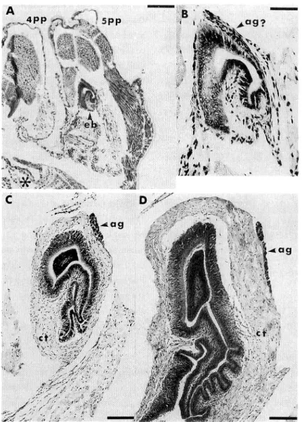
Fig. 1. Cross sections of the right ejaculatory bulb in postlarvae, in dicating the position of the androgenic gland. A, 30th day-stage of postlarva (P30) ; B, partial magnification of A ; C, P60 ; D, P100. Scale bar, A=200/<m ; B =45 ,«m ; C and D = 100 pm. ag and ag?, androgenic gland and its probable pri-mordium ', ct, connective tissue ; eb, ejaculatory bulb ; 4pp, fourth pereiopod i 5pp, fifth pereiopod ; *, part of the lami nar sheath of the fifth thoracic ganglion.
The ejaculatory bulb became to possess thick connective tissue externally and involved lumen surrounded by a simple layered columnar epithelium according to the time lapse
(Figs. 1C and ID). Nuclei of the epithelial cells showed an extensively crowded condi
90 Mem. Fac. Fish. Kagoshima Univ., Vol.41 (1992)
Fig. 2. Postlarval gonads sectioned cross ly. A, undifferentiated gonad of P40 ; B, testis of P60 ; C, ovary of P85 ; D, testis of P85. Scale bar is
50 ,'im.
sufficiently observed. During P50 to P60, the slender androgenic gland was recog nized as a cellular mass independent of the connective tissue of the postero-external wall of the ejaculatory bulb. Nuclei of these crowded cells were dense, small and
flattened. Such characters were maintain
ed at least until PllO, and they differed
from those round and translucent charac
ters of the pubescent androgenic gland (Nakamura, unpublished), indicating such a young androgenic gland to be unfunc-tional. Sexual discrimination of the go nad was possible for the first time at P60
(Fig. 2B). The testis of P60 showed many of small blocks consisting of spermato gonia surrounded by mesodermal cells like
as that of P85 (Fig. 2D ). The ovary of
P60 was not presented in the figure due to its indistinct preparations. That of P85 showed centrally arranged oogonia being
enclosed by mesodermal cells (Fig. 2C).
Ultrastructure of Androgenic Gland Cells
The young androgenic gland consisted of various cells in size ranging from 10 f-m
to 20 fJ-m. Each cell was surrounded by a thick membrane of which thickness was pe culiar and indefinite (Figs. 3B and 3C). This membrane showed a laminar structure
corresponding to that of 'une lame basale' reported in other decapod species5,6'. How
ever, the cell membrane of the latter androgenic gland was thin like the case of com
mon cells.
The nuclear size varied between 4 ftm and 10 fJ-m according to cellular size. The shape was elongated or drop-like, sometimes irregularly contoured. This irregular contour of the nucleus has been already reported in the adult prawn, Penaeus
kera-thurus1'. The nucleus possessed much chromatin along the nuclear membrane and the nucleoli which were situated centrally.
The cytoplasm showed distributions of mitochondria, some of which were large,
and rough surfaced endoplasmic reticula (rer) (Figs. 3B and 3C). Rer were not so
well developed and not arranged lamellarly as in the case of the pubescent kuruma
prawn7' and crab Ocypode quadrata6>. In the latter case, the gland cells were referred as having a very abundant lamellar rer and also a discrete Golgi apparatus as well
Fig. 3. Electron microscopical specimens of the androgenic gland in P65 postlarvae. A, trimming slice stained with toluidine blue, indicating the position of the andro genic gland ; B and C, electron micrograms of the androgenic gland cell. Scale bar, A=100/-<m ; B and C— Ip-m. ag, androgenic gland I cm, cell membrane ; ct, connective tissue of the ejaculatory bulb i ep, epithelium of the ejaculatory bulb I m, mitochondria ', n, nucleus ; rer, rough surfaced endoplasmic reticulum.
as many lysosomes. In the young gland of the present larvae, the Golgi apparatus was not well developed, lacking in lamellar arrangements and dense micro- or large vesicles. Lysosomes were indistinct.
Discussion
The revealed sequence of appearances of the genital organs in this study support
92 Mem. Fac. Fish. Kagoshima Univ., Vol.41 (1992)
seemed to suggest that the vas deferens including the ejaculatory bulb and external sexual character differentiated genetically without participation of the androgenic
gland.
However, nevertheless the presence of controversial results in crabs, crayfish and kuruma prawn, Charniaux-Cotton and Payen proposed in their review1* that sexual dif ferentiation of males in malacostracans needed the androgenic gland. The basis of this proposition seemed to depend eventually on a Katakura's hypothesis8) that andro
genic hormone was first produced by the male determining gene and was responsible
for the development of the androgenic gland primordia. However, Katakura's hy
pothesis8) was deduced by isopod experiments which were lacking in the blank test if
they were examined from a strict standpoint • namely, the incubation experiment of the primordial gonad without androgenic gland hormone and its negation of the tes ticular differentiation were not provided. Such an in vitro experiment seemed neces sary to assert the hypothesis mentioned above. Further, the presence and secretory
site (s) of this hormone have been yet unsubstantiated in such a larval period of sex
ual differentiation in any crustacean species.
In the crayfish Pontastacus leptodactylus leptodactyus, primordia of probable
(according to the author) androgenic gland appeared at the subterminal of the vas
deferens after sexual differentiation of the gonad and before rudimental appearance
of the external sexual character9). This appearance pattern differed from the cases of
crabsl). In the latter, the primordium of the androgenic gland was recognized gener
ally after or just at the occurrence of the gonadal sex differentiation which was ac
companied by complete formation of the vas deferens1}. On the contrary, in the
am-phipod Orchestia gammarella and the prawn M. rosenbergii, the following results
have been reported1]. Differentiation of the male external characteristics began after
differentiation of the androgenic gland. And these results seemed to be the strong ground of Charniaux-Cotton and Payen's proposition.
Therefore, the most important point seemed how to prove the hypothesis of the existence on the androgenic hormone which may have settled the above-mentioned con tradictions among malacostracans and how to build a general concept concerning en docrine control of sex or genetic sex determination.
One of the previous contradictions was found also in the relation between the an drogenic gland and the testis. The time lag between both differentiations obtained in the present study seemed to indicate that the former participated in inducing the lat
ter differentiation.
However, from the light and the electron microscopical observations, the follow ing considerations supported the autodifferentiation of the gonad in terms of inde pendence from the androgenic gland : 1) beginning of the gonadal sex differentiation may have preceded the morphological discrimination, although its time lag was un certain in such a morphological study. The ovary and testis were distinguished in the
present study at P60. The histology of the gonad seemed possible to expect that the
initiation of their differentiation has occurred far before P60 ; 2) during the period from P50 to PllO, the cells of the young androgenic gland did not show so active con
dition of secretory function as that of the pubescent individuals reported1,2,7).
Then, the differentiation of the testis also seemed to have been induced genetical ly without participation of the young androgenic gland in the kuruma prawn. This supposition of gonadal sex differentiation differed from the common concept or pre
vious proposition introduced by Charniaux-Cotton and Payen1}.
In the subadult or adult M. rosenbergii, masculinization of females was induced
by androgenic gland implantation4). And also feminization was achieved by androgen
ic gland ablation3. However, maybe due to technical difficulty, these operations of
the gland were always conducted together with removal or implantation of the ejac ulatory bulb. And the vas deferens, not the ejaculatory bulb, was implanted into the control group of M. rosenbergii. Anatomically, the ejaculatory bulb was certainly a distal part of the vas deferens, though their constructions had some different mor
phology each other10) • the convoluted lumen with its glandular epithelium of the ejac
ulatory bulb developed extremely and expanded, differing from the case of the vas deferens at puberty. This phenomenon and early differentiation of the ejaculatory bulb seemed to indicate its important function related to not only the sperm storage
but also masculinization. The following sentences in the previous reference4) seemed
noticeable : in some masculinized females by implantation of the androgenic gland, living vasa deferentia implants were found but the associated androgenic glands were missing. In these cases, the androgenic glands may have separated from the vasa def
erentia in the host and were not recovered with the vasa deferentia at the termination
of the experiment. Then, it was possible to consider also function of the ejaculatory
bulb on the above-mentioned results.
It has been known that the androgenic gland needed the protocerebral factor of
males for its maintenance in Natantia Leander serratus and Crangon crangonn), al
though in the isopod Porcellio dilatatusu), female protocerebrum showed the same
action as that of the male. Based on these Natantia references, the disappeararance of the implanted androgenic gland in female M. rosenbergii was considered as an in terpretation to have been caused dire to the absence of the brain factor. Then, the an drogenic gland ought to have degenerated. However, the masculinization was referred
to have occurred.
Therefore, the interpretation of the androgenic gland as the only organ inducing sex determination proposed a re-examination especially in decapod malacostracans.
Acknowledgments
94 Mem. Fac. Fish. Kagoshima Univ., Vol.41 (1992)
Aquatic Botany, Faculty of Fisheries, Kagoshima University, for his kind support in the treatment and observations under the electron microscope. This work was indebt ed to Kazunori Matsuzaki and Ken-Ichiroh Yonekura, the staff and the chief of Aira Prawn Farming, Iwasaki Industry Co., for samplings and employment of postlarval prawns.
References
1) H. Charniaux-Cotton and G. Payen (1985) : Sexual differentiation. "The Biology of Crustacea" (ed. by D. E. Bliss), Vol.9, pp. 217-299, Academic Press, New York. 2) H. Charniaux-Cotton and G. Payen (1988) : Crustacean reproduction. "Endocrinology
of Selected Invertebrate Types" (ed. by H. Laufer and R. G. H. Dower), pp. 279-303, Alan R. Liss Inc., New York.
3) C. Nagamine, A. W. Knight, A. Maggenti and G. Paxman (1980a) : Effects of andro genic gland ablation on male primary and secondary sexual characteristics in the Malaysian prawn, Macrobrachium rosenbergii (de Man) (Decapoda, Palaemonidae), with first evidence of induced feminization in a nonhermaphroditic decapod. Gen.
Comp. Endocrinol., 41, 423-441.
4) C. Nagamine, A. W. Knight, A. Maggenti and G. Paxman (1980b) • Masculinization of female Macrobrachium rosenbergii (de Man) (Decapoda, Palaemonidae) by andro genic gland implantation. Gen. Comp. Endocrinol., 41, 442-457.
5) G. Payen, J. D. Costlow, Jr. and H. Charniaux-Cotton (1971) : Etude comparative de l'ultrastructure des glandes androgenes de Crabes normaux et pedonculectomises pen dant la vie larvaire ou apres la puberte chez les especes : Rhithropanopeus harrisii
(Gould) et Callinectes sapidus Rathbun. Gen. Comp. Endocrinol., 17, 526-542. 6) G. Payen (1972) : Etude ultrastructurale de la degenerescence cellulaire dans la glande
androgene du Crabe Ocypode quadrata (Fabricius). Z. Zellforsch., 129, 370-385. 7) G. G. Payen, L. Chim, A. Laubier-Bonichon and H. Charniaux-Cotton (1982) : The an
drogenic gland of the shrimp Penaeus japonicus Bate. Description, role, and control by the eye stalks. Gen. Comp. Endocrinol., 46, 384.
8) Y. Katakura (1984) : Sex differentiation and androgenic gland hormone in the terres trial isopod Armadillidium vulgare. Symp. Zool. Soc. Lond., 53, 127-142.
9) G. Payen (1973) : Etude descriptive des principales etapes de la morphogenese sexuelle chez un Crustace Decapoda a developpement condense, l'Ecrevisse Potastacus
leptodac-tylus leptodacleptodac-tylus (Eschscholtz, 1823). Ann. Embryol. Morphol., 6, 179-206.
10) J. E. King (1948) : A study of the reproductive organs of the common marine shrimp,
Penaeus setiferus (Linnaeus). Biol. Bull., 94, 244-262.
11) A. Touir (1977) : Donnees nouvelles concernant l'endocrinologie sexuelle des Crustaces Decapodes Natantia hermaphrodites et gonochoriques. Mise en evidence d'un controle neurohormonal du maintien de l'appareil genital male et des glandes androgenes ex-erce par le protocerebron median. C. R. Acad. Sci. Paris, Ser. D, 285, 539-542. 12) J. J. Legrand, P. Juchault, J. P. Mocqu and G. Noulin (1968) : Contribution a l'etude
du controle neurohumoral de la physiologie sexuelle male chez les Crustaces Isopodes terrestres. Ann. Embryol. Morphol., 1, 97-105.