1
Research article 1 2 3 4Light-dependent induction of Edn2 expression and attenuation of retinal pathology by
5
endothelin receptor antagonists in Prominin-1- deficient mice
6 7 8 9
Yuka Kobayashi
1, Shizuka Watanabe
2, Manabu Shirai
3, Chiemi Yamashiro
1, Tadahiko Ogata
1,
10
Fumiaki Higashijima
1, Takuya Yoshimoto
1, Takahide Hayano
4, Yoshiyuki Asai
4,
11
Noriaki Sasai
2,5,*and Kazuhiro Kimura
1,5,*12 13 14 15
1
Department of Ophthalmology, Yamaguchi University Graduate School of Medicine, 1-1-1
Minami-16
kogushi, Ube 755-0046, Japan
17
2
Developmental Biomedical Science, Division of Biological Sciences, Nara Institute of Science and
18
Technology, 8916-5 Takayama-cho, Ikoma 630-0192, Japan
19
3
Omics Research Center (ORC), National Cerebral and Cardiovascular Center, 6-1 Kishibe Shinmachi,
20
Suita, Osaka 564-8565, Japan
21
4
Department of Systems Bioinformatics, Yamaguchi University Graduate School of Medicine, 1-1-1
22
Minami-kogushi, Ube 755-0046, Japan
23 5 Co-senior authors 24 25 26 27
*Address for correspondence: 28
Kazuhiro Kimura (e-mail: [email protected]) or
29
Noriaki Sasai (e-mail: [email protected])
30 31
Running Title 32
Blocking endothelin relieves retinopathy
2
Abstract 34
Retinitis pigmentosa (RP) and macular dystrophy (MD) are prevalent retinal degenerative diseases
35
associated with gradual photoreceptor death. These diseases are often caused by genetic mutations that
36
result in degeneration of the retina postnatally after it has fully developed. The Prominin-1 gene (Prom1)
37
is a causative gene for RP and MD, and Prom1- knockout (KO) mice recapitulate key features of these
38
diseases including light-dependent retinal degeneration and stenosis of retinal blood vessels. The
39
mechanisms underlying progression of such degeneration have remained unknown, however. We here
40
analysed early events associated with retinal degeneration in Prom1-KO mice. We found that
41
photoreceptor cell death and glial cell activation occur between 2 and 3 weeks after birth. High-throughput
42
analysis revealed that expression of the endothelin-2 gene (Edn2) was markedly up-regulated in the
43
Prom1-deficient retina during this period. Expression of Edn2 was also induced by light stimulation in
44
Prom1-KO mice that had been reared in the dark. Finally, treatment with endothelin receptor antagonists
45
attenuated photoreceptor cell death, gliosis, and retinal vessel stenosis in Prom1-KO mice. Our findings
46
suggest that inhibitors of endothelin signalling may delay the progression of RP and MD and therefore
47
warrant further study as potential therapeutic agents for these diseases.
48 49
Keywords 50
prominin-1, photoreceptor, glial cell, retinal degeneration, endothelin-2, endothelin receptor antagonists
3
1. Introduction 52
Both retinitis pigmentosa (RP) and macular dystrophy (MD) are inherited retinal disorders
53
associated with progressive photoreceptor cell death [1]. These diseases have a combined prevalence of 1
54
in 3000 to 4000 people worldwide. Initial symptoms include nyctalopia (night blindness) and visual field
55
deficits, which are followed by loss of visual acuity and colour blindness and eventually by complete
56
blindness. Although >60 genes encoding various types of protein - including membrane proteins,
57
transcription factors, splicing regulators, and enzymes related to the visual cycle - have been implicated in
58
RP and MD [1], these conditions remain incurable, with effective therapeutic strategies remaining to be
59
established, and they have profound effects on the quality of life.
60
The Prominin-1 gene (Prom1, also known as AC133, CD133, and RP41) encodes a pentaspan
61
transmembrane glycoprotein that is expressed in photoreceptor cells of the retina as well as in kidney and
62
testis [2]. Several mutations of Prom1 have been identified in individuals with RP or MD [3-5], with all
63
such mutations resulting in amino acid substitutions or carboxyl-terminal truncations of the encoded
64
protein. The mechanisms underlying RP and MD associated with Prom1 mutations have been investigated
65
by studies of several lines of Prom1-knockout (KO) mice [5-7]. Although photoreceptor cells develop
66
normally in these KO mice, they begin to degenerate after birth, resulting in a progressive loss of the outer
67
nuclear layer (ONL) of the retina and recapitulation of the signs of RP and MD. The retinal vasculature
68
also becomes attenuated with disease progression [7].
69
We previously showed that photoreceptor cells of the Prom1-KO mouse retina degenerate in
70
response to light stimulation. Such mice reared in a completely dark setting thus manifested a marked
71
delay in the loss of photoreceptor cells. We therefore suggested that the mutant retinal cells are
72
hypersensitive to light stimulation and experience phototoxicity [6]. The visual cycle was also found to be
73
impaired in the Prom1-KO cells, and treatment based on chemical compounds that modulate the visual
74
cycle was found to mitigate the mutant phenotype [6].
75
The Prom1 protein localises to the connecting cilium and outer segment of both rod and cone
76
photoreceptors [3]. Ultrastructural analysis revealed the structure of the outer segment to be severely
77
disorganised in photoreceptor cells of Prom1-KO mice, whereas other photoreceptor components -
78
including the inner segment, nucleus, and axon - remained largely intact [6, 7]. Biochemical analysis has
79
shown that two tyrosine residues in the carboxyl-terminal region of Prom1 are phosphorylated by the
80
tyrosine kinases Src and Fyn, although the physiological implications of such phosphorylation remain to
81
be elucidated [8]. Prom1 has also been shown to interact with the p85 regulatory subunit of
82
phosphatidylinositol 3-kinase (PI3K) and to be essential for both the self-renewal and tumourigenic
83
capacity of glioma stem cells [9]. In addition, Prom1 has been detected in cilia, which are protrusive
84
structures at the cell membrane and key signalling hubs [10], and to be essential for maximisation of
85
Hedgehog signalling in neural stem cells [11]. We recently showed that Prom1 activates the small GTPase
86
Rho and regulates chloride conductance triggered by intracellular calcium uptake [12].
4
To characterise the mechanisms underlying the role of Prom1 dysfunction in retinal degeneration
88
and thereby to provide insight into potential treatments for Prom1 mutation-associated RP and MD, we
89
here investigated the initial manifestations of such degeneration. We analysed Prom1 expression as well
90
as the ONL transition in Prom1-KO mice. We then performed a high-throughput expression analysis to
91
identify genes responsible for degeneration of the Prom1-deficient retina. Our results implicated an
92
inflammatory pathway dependent on the endothelin 2 gene (Edn2), and we found that a chemical
93
treatment targeted to endothelin signalling mitigated the deterioration of retinal structure and function in
94
Prom1-KO mice.
5
96 2. Methods 97 2.1. Mice 98Prom1-KO mice were established previously (CDB0623K, http://www2.clst.riken.jp/arg/methods.html),
99
and they were reared on a hybrid genetic background of C57BL/6 and CBA/NSlc strains. The targeting
100
vector for Prom1 ablation contained the lacZ (β-galactosidase) gene, with the result that expression of this
101
latter gene reflects that of Prom1. Both the Prom1-KO mice and their wild-type (WT) littermates were
102
kept on a 12-hour-light, 12-hour-dark cycle, with the cage racks being covered with blackout curtains and
103
all procedures including feeding and cage maintenance being performed in the absence of light (<0.5 lux)
104
during the dark phase. For experiments involving light stimulation, mice were exposed for 3 h to a light
105
panel (LED viewer 5000; Shinko, Tokyo, Japan) placed on top of the cage, which resulted in a light
106
intensity of 3800 lux at the bottom of the cage. For chemical treatment, mice received intraperitoneal
107
injections (2 mg/kg) of each of the endothelin receptor antagonists 123 (ab141005, Abcam) and
BQ-108
788 (ab144504, Abcam) on postnatal day (P) 14, P19, and P24. The mice were then subjected to analysis
109
on P28.
110 111
2.2. RNA extraction and RT-qPCR analysis 112
The retina, retinal pigment epithelium (RPE), and testis were dissected from mice killed by cervical
113
dislocation. Total RNA was extracted from the isolated tissue and was subjected to reverse transcription
114
(RT) with the use of a NucleoSpin RNA extraction kit (U955C, Takara) and PrimeScript RT reagent kit
115
(RR037, Takara), respectively. The resulting cDNA was subjected to quantitative polymerase chain
116
reaction (qPCR) analysis with a CFX qPCR machine (Bio-Rad) and with primers listed in supplementary
117
table S1. The amplification data were analysed with the comparative Ct method, and gene expression
118
levels were normalised by that of the glyceraldehyde-3-phosphate dehydrogenase gene (Gapdh).
119 120
2.3. High-throughput expression analysis 121
Total RNA samples were prepared from three (P14) or four (P21) retinas of WT or Prom1-KO mice and
122
were used to synthesise cDNA libraries with a TruSeq stranded-mRNA library preparation kit (Illumina,
123
20020594). The libraries were sequenced with the NextSeq 500 platform (Illumina). In total,
124
approximately twenty million reads/sample were mapped with the CLC genomics workbench software
125
(Qiagen) [13]. The sequencing data were deposited in the DNA Data Bank of Japan (DDBJ) public
126
database, with the accession number of SSUB016168. Gene ontology (GO) term analysis was performed
127
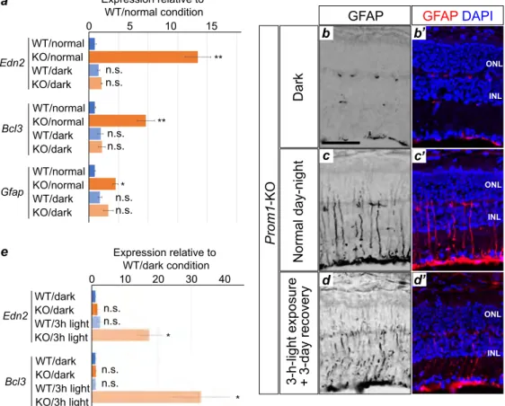
according to the Kyoto Encyclopaedia of Genes and Genomes database (KEGG,
128
https://www.genome.jp/kegg).
129 130
2.4. Immunofluorescence analysis, β-galactosidase and isolectin staining, and TUNEL analysis 131
6
For immunofluorescence analysis, the enucleated retina was fixed for 2 h with a mixture of 1%
132
paraformaldehyde and 0.2% glutaraldehyde in phosphate-buffered saline (PBS), incubated overnight in
133
PBS containing 15% sucrose, embedded in O.C.T. compound (Sakura), and sectioned at a thickness of 12
134
µ m. The sections were exposed to mouse monoclonal antibodies to GFAP (G3893; Sigma) or rabbit
135
polyclonal antibodies to Iba-1 (019-19741; Wako), and immune complexes were detected with
Cy3-136
conjugated secondary antibodies (715-166-151 and 715-166-152 for mouse and rabbit, respectively;
137
Jackson Immunoresearch). Nuclei were counterstained with 4',6-diamidino-2-phenylindole (DAPI) with
138
the use of DAPI Fluoromount-G (0100-20; Southern Biotech). Sections were also stained for
β-139
galactosidase (β-gal) activity with the use of a staining kit (11828673001, Roche). Apoptotic cells were
140
detected by TUNEL analysis with digoxigenin-labelled dUTP (S7105, Merck Millipore), terminal
141
deoxynucleotidyl transferase (3333566001, Merck), and rhodamine-conjugated antibodies to digoxigenin
142
(11207750910, Roche). For preparation of flat-mount samples, the retina was fixed for 150 min with 4%
143
paraformaldehyde and the RPE was peeled off. The samples were subjected to isolectin staining by
144
consecutive exposure to 5% dried skim milk and Alexa Flour 488-conjugated GS-IB4 (I21411, Thermo
145
Fisher Scientific) as described previously [14]. Images were acquired with an LSM 710 confocal
146
microscope (Zeiss) for immunofluorescence, β-gal, and TUNEL staining, or with a BZ-X710 microscope
147
(Keyence) for flat-mount preparations. Imaging data were processed and integrated with Photoshop
148
(Adobe) and Illustrator (Adobe) software, respectively.
149 150
2.5. Statistical analysis 151
Quantitative data are presented as means ± s.e.m. Differences between two or among more than two
152
groups were evaluated with the two-tailed Student’s t test and by one-way analysis of variance (ANOVA)
153
followed by Tukey’s post hoc test, respectively. Statistical analysis was performed with Prism software
154
(Graphpad), and a p value of <0.05 was considered statistically significant.
155 156
7
3. Results 157
3.1. Prom1 is expressed in the retina from perinatal to adult stages 158
We previously showed that retinal cells in Prom1-KO mice appear to develop normally before the onset of
159
degeneration [6]. We here first examined the spatiotemporal expression of Prom1 in the mouse retina.
160
Given that our Prom1-KO mice harbour the lacZ gene at the Prom1 locus, we performed staining for
β-161
gal activity in the heterozygous mutant mice at birth as well as at P2 (figure 1a-a”), P14 (figure 1b-b”),
162
P21 (figure 1c-c”), and P42 (figure 1d-d”). At all the stages analysed, β-gal staining was localised
163
predominantly to the outer layers in the retina, with more sporadic staining also apparent in the inner
164
nuclear layer (INL). Given that retinal phenotypes of Prom1-KO mice are not obvious until 2 weeks after
165
birth, these results suggested that Prom1 expression precedes the onset of function of the encoded protein
166
in postnatal retinal homeostasis.
167 168
3.2. The Prom1-KO mouse retina manifests both apoptosis and an inflammatory response at 3 169
weeks after birth 170
We previously showed that the retina of Prom1-KO mice appears normal at P14 and begins to
171
degenerate soon after the animals first open their eyes at P14 [6]. We therefore investigated whether the
172
Prom1-deficient retina might undergo apoptosis in response to light exposure. Whereas the TUNEL assay
173
revealed few apoptotic cells in the retina of WT or Prom1-KO mice at P14 (figure 2a and b), a significant
174
increase in the number of TUNEL-positive cells, located mainly in the ONL, was detected at P21 in the
175
Prom1-KO retina (figure 2c–e). These results suggested that programmed cell death by apoptosis begins
176
to occur in the ONL of the retina between 2 and 3 weeks after birth in Prom1-KO mice.
177
Glial fibrillary acidic protein (GFAP) is an intermediate filament protein that is expressed by
178
Müller glia in response to retinal injury [15, 16]. Similarly, Iba-1 is a scaffold protein that is expressed in
179
microglia and which is up-regulated during an inflammatory response [17, 18]. We therefore next
180
examined whether the Prom1-KO retina might undergo light-induced inflammation by analysing the
181
expression of these two proteins. Immunofluorescence analysis revealed that, whereas both GFAP and
182
Iba-1 were essentially undetectable in the WT or Prom1-KO retina at P14 (figure 2f–i), a marked increase
183
in the extent of staining for both proteins was observed in the Prom1-KO retina at P21 (figure 2j–m),
184
suggesting that the increased cell death that occurs in the ONL of the mutant mice after birth is
185
accompanied by the activation of glial cells.
186 187
3.3. Inflammation-related gene expression is up-regulated in the Prom1-KO mouse retina 188
We next sought to identify genes whose expression might be affected by Prom1 deficiency by
189
subjecting the retina of WT and Prom1-KO mice at P14 and P21 to high-throughput expression analysis
190
based on RNA sequencing. Gene expression at P14 tended to vary within each genotype, and the only
191
gene whose expression differed significantly between genotypes was Prom1 itself (figure 3a,
192
supplementary table S2), suggesting that Prom1 does not significantly influence the gene expression
8
profile at P14. In contrast, the expression of various genes differed between the two genotypes at P21
194
(figure 3b, supplementary table S3). The expression of 1,081 and 766 genes was thus up- and
down-195
regulated, respectively, in the Prom1-KO retina with a p value of <0.01. In particular, expression of Edn2
196
was the most consistently and markedly up-regulated in the Prom1-KO retina. The expression of genes
197
associated with the inflammatory response - such as Ifi44l, Serpina3n, S100a6, Bcl3, and Gfap - was also
198
increased in the Prom1-KO retina at P21. Conversely, the expression of genes related to RP or of those
199
essential for retinal development and functional homeostasis - including Fscn2 (RP30) [19], Prph2 (RP7)
200
[20], Nr2e3 (RP37) [21], Kcnv2 [22], Elovl2 [23], Pde6b (RD1) [24], and Ttc21b [25] - was
down-201
regulated in the Prom1-KO retina at P21 (supplementary table S3). GO term analysis revealed that several
202
signalling pathways, including apoptotic (TNF) and infectious-related signal (Epstein-Barr virus infection)
203
signals, were affected by the loss of Prom1 (figure 3c).
204
We also investigated whether the observed effects of Prom1 deficiency on gene expression were
205
specific to the retina. Given that Prom1 is expressed in the retina, RPE, and testis [2], we performed
RT-206
qPCR analysis of RNA prepared from these tissues of WT and Prom1-KO mice at P21. Consistent, with
207
the results of our RNA-sequencing analysis, the expression of Edn2, Bcl3, and Gfap was increased in the
208
retina of Prom1-KO mice (figure 3d). However, the expression of these genes in the RPE and testis did
209
not differ between the two genotypes, indicating that the effect of Prom1 on their expression is specific to
210
the retina. Together, these various data suggested that Prom1 deficiency results in up-regulation of
211
inflammation-related genes and down-regulation of genes essential for functional homeostasis of
212
photoreceptor cells at 3 weeks after birth.
213 214
3.4. Inflammation-related gene expression is increased by light stimulation in the Prom1-KO mouse 215
retina 216
To determine the mechanism underlying the up-regulation of specific gene expression apparent in the
217
retina of Prom1-KO mice at P21, we examined whether light stimulation might play a role. We therefore
218
compared such gene expression between P21 retinas obtained from Prom1-KO mice reared under a
219
normal day-night cycle or in the dark. RT-qPCR analysis revealed that, whereas the expression of Edn2,
220
Bcl3, and Gfap did not differ between Prom1-KO and WT mice reared in the dark condition, marked
up-221
regulation of the expression of each of these genes was apparent specifically in Prom1-KO mice raised
222
under the normal day-night condition (figure 4a). Consistent with these results, immunofluorescence
223
analysis showed that the number of GFAP-positive cells in the retina was smaller for Prom1-KO mice
224
reared in the dark compared with those reared under the normal condition (figure 4b and c). To examine
225
further the effect of light on gene expression, we maintained Prom1-KO mice and their WT littermates
226
under the dark condition for 3 weeks, exposed them to a bright light for 3 h, and then allowed them to
227
recover for 3 days in the dark. The retina was then dissected and subjected to RT-qPCR and
228
immunofluorescence analyses. Light stimulation resulted in a marked increase both in the expression of
229
Edn2 and Bcl3 (Figure 4d) and in the number of GFAP-positive cells (figure 4e) in the retina of
9
KO mice but not in that of WT mice. Collectively, these results thus suggested that the up-regulation of
231
Edn2, Bcl3, and Gfap expression apparent in the retina of Prom1-KO mice is an immediate response to
232
light stimulation, and that the inflammatory response mediated by these genes is one of the primary events
233
leading to degeneration of the mutant retina.
234 235
3.5. Endothelin receptor antagonists attenuate Gfap expression and gliosis in the Prom1-KO mouse 236
retina 237
Endothelin acts at specific receptors [26, 27] to increase both the number of GFAP-positive
238
Müller cells [28] and retinal cell death [29]. Given the elevated expression of Edn2 and Gfap apparent in
239
the retina of Prom1-KO mice, we hypothesised that Edn2 might induce aberrant proliferation of glial cells
240
and GFAP expression in association with retinal degeneration in these animals. We therefore examined
241
the possible effects of endothelin receptor antagonists in the mutant mice.
242
The drugs BQ-123 and BQ-788, which target endothelin receptors A and B, respectively [30],
243
were both injected intraperitoneally into Prom1-KO mice at P14, P19, and P24, and the mice were
244
analysed at P28. Whereas GFAP-positive cells were not observed in the retina of WT mice, they were
245
detected in that of Prom1-KO mice treated with dimethyl sulphoxide (DMSO) vehicle (figure 5a and b).
246
However, the number of GFAP-positive cells was markedly reduced in the mutant mice by treatment with
247
BQ-123 and BQ-788 (figure 5c). Staining of retinal flat-mount preparations with fluorescently labelled
248
isolectin to detect vascular endothelial cells also revealed fewer retinal vessels in Prom1-KO mice than in
249
WT mice and that this difference was attenuated by treatment of the mutant animals with 123 and
BQ-250
788 (figure 5d–g).
251
RT-qPCR analysis showed that the expression of Edn2, Bcl3, and Gfap was increased in the retina
252
of Prom1-KO mice at P28 compared with that in WT mice. Whereas the expression of Edn2 and Bcl3 in
253
the mutant retina was not affected by treatment with BQ-123 and BQ-788, that of Gfap was significantly
254
attenuated (figure 5h), suggesting that up-regulation of Gfap expression in the mutant retina is mediated
255
by endothelin receptor signalling but that that of Edn2 and Bcl3 expression is not.
256
Finally, we examined the effect of BQ-123 and BQ-788 treatment on the number of apoptotic
257
cells in the retina of Prom1-KO mice. The TUNEL assay revealed that the marked increase in the number
258
of such cells apparent in the mutant retina at P28 was significantly attenuated by administration of the two
259
drugs (figure 5l), suggesting that endothelin receptor signalling contributes to loss of retinal cell
260
homeostasis.
10
4. Discussion 262
We have here described early manifestations of the retinal degeneration that occurs in Prom1-KO
263
mice and identified related genes. We thus detected the aberrant presence of glial cells and the expression
264
of genes associated with the inflammatory response in the mutant retina. Given that the expression of
265
these genes was not activated in the retina of Prom1-KO mice maintained in the dark condition, this
266
inflammatory response appears to be dependent on light stimulation. Finally, we found that the
267
deterioration and gliosis characteristic of the mutant retina were ameliorated by the administration of
268
endothelin receptor antagonists.
269
Although we found that Prom1 is expressed in the retina from birth, the loss of Prom1 did not
270
substantially affect the expression level of any gene in the retina at P14, suggesting that Prom1 may not
271
play an essential role in the retina prior to light exposure. We previously showed by RT-qPCR analysis
272
that the expression of both Rdh12 and Abca4, two genes that contribute to the visual cycle, was reduced in
273
the retina of Prom1-KO mice compared with that of WT mice at P14 [6], suggesting that impairment of
274
the visual cycle might lead to retinal degeneration. Although this result is reproducible as assayed by
RT-275
qPCR (supplementary figure S1), the difference in the expression level of each gene between the two
276
genotypes was associated with a relatively high p value in the high-throughput expression analysis
277
performed in the present study (figure 3, supplementary table 2), suggesting this decrease is not critical.
278
In contrast to the lack of an effect of Prom1 deficiency on the gene expression profile of the retina
279
at P14, we detected many genes, including those related to the inflammatory response, as well as
280
signalling pathways whose activity was altered in the Prom1-KO retina at P21. The expression of genes
281
related to phototransduction, for example, was significantly down-regulated in the Prom1-KO retina at
282
P21 (figure 3c, supplementary table S3), indicating that Prom1 may be essential for the transcription of
283
such genes or may form a transcriptional network with them. Of note, we found that the expression of
284
causal genes for RP was also down-regulated in the mutant retina at P21.
285
Of the genes whose expression was up-regulated in the Prom1-KO retina at P21, Edn2 showed the
286
largest fold change. Edn2 encodes a secretory peptide that plays a role in a wide range of biological
287
processes, including smooth muscle contraction and ovulation [31] as well as development of the enteric
288
nervous system [32]. Its expression is also induced in association with the inflammatory response and
289
promotes glial cell proliferation in the central nervous system [33]. Furthermore, consistent with the
290
perturbation of the retinal vasculature in Prom1-KO mice apparent in both the present and a previous [7]
291
study, Edn2 has been found to inhibit retinal vascular development [34]. On the other hand, it was also
292
shown to promote photoreceptor cell survival [35]. These various observations suggest that the role of
293
Edn2 in the photoreceptor degeneration associated with RP and MD is complex.
294
The expression of Edn2 has also been shown to be up-regulated in other mouse models of RP [35],
295
including retina-specific Cdhr1-KO mice [36], with Prom1 and Cdhr1 having been found to interact with
296
each other [4]. In addition to Edn2, the other genes whose expression was affected in the Prom1-KO
11
mouse retina overlapped markedly with those affected in the conditional Cdhr1-KO mouse retina,
298
suggesting that Prom1 and Cdhr1 may function in the same intracellular signalling pathways.
299
Although we found that the expression of Edn2 and Bcl3 in the Prom1-KO retina was induced by
300
light stimulation, the mechanism underlying this effect remains unclear. Nevertheless, our study suggests
301
the possibility that an imbalance in intracellular ions caused by the loss of Prom1 (given that Prom1
302
regulates chloride conductance activated by intracellular calcium uptake [12]) may impair the function of
303
cytoplasmic organelles such as mitochondria and the endoplasmic reticulum, and thereby elicit a stress
304
response. Studies to identify the transcriptional regulatory elements of Edn2 and the corresponding
305
transcription factors and upstream signalling pathways underlying its photoactivation are warranted.
306
Gliosis is a response to injury in the central nervous system and is associated with the appearance
307
of GFAP-positive glial cells [26]. It is also a feature of certain neurodegenerative retinal diseases
308
including RP [37], with gliosis in RP having been found to be related to several RP genes. Targeting of
309
gliosis is therefore a potential clinical strategy to delay disease progression and ameliorate associated
310
symptoms. We have now shown that administration of endothelin receptor antagonists attenuated both the
311
appearance of GFAP-positive glial cells and vascular endothelial constriction in the retina of Prom1-KO
312
mice. These findings indicate that blockade of endothelin signalling may be an effective clinical strategy
313
for the treatment of gliosis. However, caution is warranted with such an approach for the treatment of RP,
314
given the various functions of endothelins and the consequent potential for adverse systemic effects.
315
Intravitreal injection of endothelin receptor antagonists may help to avoid such side effects. Gene therapy
316
targeting endothelin receptor function is also a potential therapeutic approach for RP. Finally, replacement
317
of dead tissue with functional cells through a regenerative medicine approach may be required for the
318
successful treatment of RP and MD [38].
319
In conclusion, our results implicating up-regulation of Edn2 expression in the retinal pathology of
320
Prom1-KO mice suggest that localized pharmacological targeting of endothelin receptor signalling
321
warrants further investigation as a clinical intervention for the prevention or treatment of retinal
322
degenerative diseases such as RP and MD.
12
Ethics. All animal experiments were approved by the animal welfare and ethics committees of both 324
Yamaguchi University (approval numbers J16021 and U16005 for K.K.) and Nara Institute of Science and
325
Technology (approval numbers 1810 and 311 for N.S.) and were performed in accordance with the
326
relevant guidelines and regulations.
327 328
Data availability. Data are available in the main text/figures and in the Supplementary Information. 329
330
Competing interests. The authors declare no competing interests. 331
332
Funding. This work was supported in part by grants-in-aid for scientific research from Japan Society for 333
the Promotion of Science (17H03684 and 20H0326310 to N.S.; 20K09805 to K.K.), as well as by
334
Novartis Pharma.
335 336
Acknowledgements. We thank Erika Yoshihara, Yukari Mizuno, and Ayaka Kataoka for technical 337
assistance as well as other laboratory members for their support and discussion.
338 339
Author Contributions. KK and NS conceived the project; YK, NS, SW, MS, CY, TO, FH, TY 340
performed experiments; YK, NS, MS, TH, YA analysed the data; All authors joined the discussion; NS,
341
KK, YK wrote the manuscript.
13
References 343
1 Ferrari, S., Di Iorio, E., Barbaro, V., Ponzin, D., Sorrentino, F. S., Parmeggiani, F. 2011 Retinitis
344
pigmentosa: genes and disease mechanisms. Curr Genomics. 12, 238-249. 345
(10.2174/138920211795860107)
346
2 Fargeas, C. A., Joester, A., Missol-Kolka, E., Hellwig, A., Huttner, W. B., Corbeil, D. 2004
347
Identification of novel Prominin-1/CD133 splice variants with alternative C-termini and their
348
expression in epididymis and testis. Journal of cell science. 117, 4301-4311. (10.1242/jcs.01315)
349
3 Maw, M. A., Corbeil, D., Koch, J., Hellwig, A., Wilson-Wheeler, J. C., Bridges, R. J.,
350
Kumaramanickavel, G., John, S., Nancarrow, D., Roper, K., et al. 2000 A frameshift mutation in
351
prominin (mouse)-like 1 causes human retinal degeneration. Human molecular genetics. 9, 27-34.
352
4 Yang, Z., Chen, Y., Lillo, C., Chien, J., Yu, Z., Michaelides, M., Klein, M., Howes, K. A., Li, Y.,
353
Kaminoh, Y., et al. 2008 Mutant prominin 1 found in patients with macular degeneration disrupts
354
photoreceptor disk morphogenesis in mice. The Journal of clinical investigation. 118, 2908-2916.
355
(10.1172/JCI35891)
356
5 Michaelides, M., Gaillard, M. C., Escher, P., Tiab, L., Bedell, M., Borruat, F. X., Barthelmes, D.,
357
Carmona, R., Zhang, K., White, E., et al. 2010 The PROM1 mutation p.R373C causes an autosomal
358
dominant bull's eye maculopathy associated with rod, rod-cone, and macular dystrophy. Investigative
359
ophthalmology & visual science. 51, 4771-4780. (10.1167/iovs.09-4561)
360
6 Dellett, M., Sasai, N., Nishide, K., Becker, S., Papadaki, V., Limb, G. A., Moore, A. T., Kondo, T.,
361
Ohnuma, S. 2015 Genetic background and light-dependent progression of photoreceptor cell
362
degeneration in Prominin-1 knockout mice. Investigative ophthalmology & visual science. 56, 164-176.
363
(10.1167/iovs.14-15479)
364
7 Zacchigna, S., Oh, H., Wilsch-Brauninger, M., Missol-Kolka, E., Jaszai, J., Jansen, S., Tanimoto, N.,
365
Tonagel, F., Seeliger, M., Huttner, W. B., et al. 2009 Loss of the cholesterol-binding protein
prominin-366
1/CD133 causes disk dysmorphogenesis and photoreceptor degeneration. The Journal of
367
neuroscience : the official journal of the Society for Neuroscience. 29, 2297-2308.
368
(10.1523/JNEUROSCI.2034-08.2009)
369
8 Boivin, D., Labbe, D., Fontaine, N., Lamy, S., Beaulieu, E., Gingras, D., Beliveau, R. 2009 The stem
370
cell marker CD133 (prominin-1) is phosphorylated on cytoplasmic tyrosine-828 and tyrosine-852 by
371
Src and Fyn tyrosine kinases. Biochemistry. 48, 3998-4007. (10.1021/bi900159d)
372
9 Wei, Y., Jiang, Y., Zou, F., Liu, Y., Wang, S., Xu, N., Xu, W., Cui, C., Xing, Y., Liu, Y., et al. 2013
373
Activation of PI3K/Akt pathway by CD133-p85 interaction promotes tumorigenic capacity of glioma
374
stem cells. Proceedings of the National Academy of Sciences of the United States of America. 110,
375
6829-6834. (10.1073/pnas.1217002110)
376
10 Khatri, P., Obernier, K., Simeonova, I. K., Hellwig, A., Holzl-Wenig, G., Mandl, C., Scholl, C., Wolfl,
377
S., Winkler, J., Gaspar, J. A., et al. 2014 Proliferation and cilia dynamics in neural stem cells
378
prospectively isolated from the SEZ. Scientific reports. 4, 3803. (10.1038/srep03803)
379
11 Singer, D., Thamm, K., Zhuang, H., Karbanova, J., Gao, Y., Walker, J. V., Jin, H., Wu, X., Coveney, C.
380
R., Marangoni, P., et al. 2019 Prominin-1 controls stem cell activation by orchestrating ciliary
381
dynamics. The EMBO journal. 38, (10.15252/embj.201899845)
382
12 Hori, A., Nishide, K., Yasukuni, Y., Haga, K., Kakuta, W., Ishikawa, Y., Hayes, M. J., Ohnuma, S. I.,
383
Kiyonari, H., Kimura, K., et al. 2019 Prominin-1 Modulates Rho/ROCK-Mediated Membrane
384
Morphology and Calcium-Dependent Intracellular Chloride Flux. Scientific reports. 9, 15911.
385
(10.1038/s41598-019-52040-9)
386
13 Robinson, M. D., McCarthy, D. J., Smyth, G. K. 2010 edgeR: a Bioconductor package for differential
387
expression analysis of digital gene expression data. Bioinformatics. 26, 139-140. 388
(10.1093/bioinformatics/btp616)
389
14 Yamaguchi, M., Nakao, S., Arita, R., Kaizu, Y., Arima, M., Zhou, Y., Kita, T., Yoshida, S., Kimura,
390
K., Isobe, T., et al. 2016 Vascular Normalization by ROCK Inhibitor: Therapeutic Potential of
391
Ripasudil (K-115) Eye Drop in Retinal Angiogenesis and Hypoxia. Investigative ophthalmology &
392
visual science. 57, 2264-2276. (10.1167/iovs.15-17411)
393
15 Chang, M. L., Wu, C. H., Jiang-Shieh, Y. F., Shieh, J. Y., Wen, C. Y. 2007 Reactive changes of retinal
394
astrocytes and Muller glial cells in kainate-induced neuroexcitotoxicity. J Anat. 210, 54-65.
395
(10.1111/j.1469-7580.2006.00671.x)
14
16 Lewis, G. P., Fisher, S. K. 2003 Up-regulation of glial fibrillary acidic protein in response to retinal
397
injury: its potential role in glial remodeling and a comparison to vimentin expression. Int Rev Cytol.
398
230, 263-290. (10.1016/s0074-7696(03)30005-1) 399
17 Rojas, B., Gallego, B. I., Ramirez, A. I., Salazar, J. J., de Hoz, R., Valiente-Soriano, F. J.,
Aviles-400
Trigueros, M., Villegas-Perez, M. P., Vidal-Sanz, M., Trivino, A., et al. 2014 Microglia in mouse
401
retina contralateral to experimental glaucoma exhibit multiple signs of activation in all retinal layers. J
402
Neuroinflammation. 11, 133. (10.1186/1742-2094-11-133)
403
18 Omri, S., Behar-Cohen, F., de Kozak, Y., Sennlaub, F., Verissimo, L. M., Jonet, L., Savoldelli, M.,
404
Omri, B., Crisanti, P. 2011 Microglia/macrophages migrate through retinal epithelium barrier by a
405
transcellular route in diabetic retinopathy: role of PKCzeta in the Goto Kakizaki rat model. Am J
406
Pathol. 179, 942-953. (10.1016/j.ajpath.2011.04.018)
407
19 Wada, Y., Abe, T., Takeshita, T., Sato, H., Yanashima, K., Tamai, M. 2001 Mutation of human retinal
408
fascin gene (FSCN2) causes autosomal dominant retinitis pigmentosa. Investigative ophthalmology &
409
visual science. 42, 2395-2400.
410
20 Conley, S. M., Naash, M. I. 2014 Gene therapy for PRPH2-associated ocular disease: challenges and
411
prospects. Cold Spring Harbor perspectives in medicine. 4, a017376. (10.1101/cshperspect.a017376)
412
21 Cheng, H., Khanna, H., Oh, E. C., Hicks, D., Mitton, K. P., Swaroop, A. 2004 Photoreceptor-specific
413
nuclear receptor NR2E3 functions as a transcriptional activator in rod photoreceptors. Human
414
molecular genetics. 13, 1563-1575. (10.1093/hmg/ddh173)
415
22 Holter, P., Kunst, S., Wolloscheck, T., Kelleher, D. K., Sticht, C., Wolfrum, U., Spessert, R. 2012 The
416
retinal clock drives the expression of Kcnv2, a channel essential for visual function and cone survival.
417
Investigative ophthalmology & visual science. 53, 6947-6954. (10.1167/iovs.12-10234)
418
23 Chen, D., Chao, D. L., Rocha, L., Kolar, M., Nguyen Huu, V. A., Krawczyk, M., Dasyani, M., Wang,
419
T., Jafari, M., Jabari, M., et al. 2020 The lipid elongation enzyme ELOVL2 is a molecular regulator of
420
aging in the retina. Aging Cell. 19, e13100. (10.1111/acel.13100)
421
24 Yeo, J. H., Jung, B. K., Lee, H., Baek, I. J., Sung, Y. H., Shin, H. S., Kim, H. K., Seo, K. Y., Lee, J. Y.
422
2019 Development of a Pde6b Gene Knockout Rat Model for Studies of Degenerative Retinal Diseases.
423
Investigative ophthalmology & visual science. 60, 1519-1526. (10.1167/iovs.18-25556)
424
25 Liu, Q., Zhang, Q., Pierce, E. A. 2010 Photoreceptor sensory cilia and inherited retinal degeneration.
425
Advances in experimental medicine and biology. 664, 223-232. (10.1007/978-1-4419-1399-9_26)
426
26 Sarthy, V. P., Sawkar, H., Dudley, V. J. 2015 Endothelin2 Induces Expression of Genes Associated
427
with Reactive Gliosis in Retinal Muller Cells. Curr Eye Res. 40, 1181-1184. 428
(10.3109/02713683.2014.982828)
429
27 Patel, C., Narayanan, S. P., Zhang, W., Xu, Z., Sukumari-Ramesh, S., Dhandapani, K. M., Caldwell, R.
430
W., Caldwell, R. B. 2014 Activation of the endothelin system mediates pathological angiogenesis
431
during ischemic retinopathy. Am J Pathol. 184, 3040-3051. (10.1016/j.ajpath.2014.07.012)
432
28 Rattner, A., Toulabi, L., Williams, J., Yu, H., Nathans, J. 2008 The genomic response of the retinal
433
pigment epithelium to light damage and retinal detachment. The Journal of neuroscience : the official
434
journal of the Society for Neuroscience. 28, 9880-9889. (10.1523/JNEUROSCI.2401-08.2008)
435
29 Kobayashi, T., Oku, H., Fukuhara, M., Kojima, S., Komori, A., Ichikawa, M., Katsumura, K.,
436
Kobayashi, M., Sugiyama, T., Ikeda, T. 2005 Endothelin-1 enhances glutamate-induced retinal cell
437
death, possibly through ETA receptors. Investigative ophthalmology & visual science. 46, 4684-4690.
438
(10.1167/iovs.05-0785)
439
30 Fukuroda, T., Ozaki, S., Ihara, M., Ishikawa, K., Yano, M., Nishikibe, M. 1994 Synergistic inhibition
440
by BQ-123 and BQ-788 of endothelin-1-induced contractions of the rabbit pulmonary artery. Br J
441
Pharmacol. 113, 336-338. (10.1111/j.1476-5381.1994.tb16901.x)
442
31 Cacioppo, J. A., Oh, S. W., Kim, H. Y., Cho, J., Lin, P. C., Yanagisawa, M., Ko, C. 2014 Loss of
443
function of endothelin-2 leads to reduced ovulation and CL formation. PloS one. 9, e96115.
444
(10.1371/journal.pone.0096115)
445
32 Gershon, M. D. 1999 Endothelin and the development of the enteric nervous system. Clin Exp
446
Pharmacol Physiol. 26, 985-988. (10.1046/j.1440-1681.1999.03176.x)
447
33 Yuen, T. J., Johnson, K. R., Miron, V. E., Zhao, C., Quandt, J., Harrisingh, M. C., Swire, M., Williams,
448
A., McFarland, H. F., Franklin, R. J., et al. 2013 Identification of endothelin 2 as an inflammatory
449
factor that promotes central nervous system remyelination. Brain. 136, 1035-1047. 450
(10.1093/brain/awt024)
15
34 Rattner, A., Yu, H., Williams, J., Smallwood, P. M., Nathans, J. 2013 Endothelin-2 signaling in the
452
neural retina promotes the endothelial tip cell state and inhibits angiogenesis. Proceedings of the
453
National Academy of Sciences of the United States of America. 110, E3830-3839.
454
(10.1073/pnas.1315509110)
455
35 Bramall, A. N., Szego, M. J., Pacione, L. R., Chang, I., Diez, E., D'Orleans-Juste, P., Stewart, D. J.,
456
Hauswirth, W. W., Yanagisawa, M., McInnes, R. R. 2013 Endothelin-2-mediated protection of mutant
457
photoreceptors in inherited photoreceptor degeneration. PloS one. 8, e58023. 458
(10.1371/journal.pone.0058023)
459
36 Rattner, A., Nathans, J. 2005 The genomic response to retinal disease and injury: evidence for
460
endothelin signaling from photoreceptors to glia. The Journal of neuroscience : the official journal of
461
the Society for Neuroscience. 25, 4540-4549. (10.1523/JNEUROSCI.0492-05.2005)
462
37 Roche, S. L., Ruiz-Lopez, A. M., Moloney, J. N., Byrne, A. M., Cotter, T. G. 2018 Microglial-induced
463
Muller cell gliosis is attenuated by progesterone in a mouse model of retinitis pigmentosa. Glia. 66,
464
295-310. (10.1002/glia.23243)
465
38 Stern, J. H., Tian, Y., Funderburgh, J., Pellegrini, G., Zhang, K., Goldberg, J. L., Ali, R. R., Young, M.,
466
Xie, Y., Temple, S. 2018 Regenerating Eye Tissues to Preserve and Restore Vision. Cell Stem Cell. 22,
467
834-849. (10.1016/j.stem.2018.05.013)
468 469
16
Figure Legends 470
Figure 1. Prom1 is expressed in the ONL of the retina from perinatal to adult stages. The retina of 471
heterozygous Prom1 mutant mice at P2 (a-a”), P14, (b-b”), P21 (c-c”), and P42 (d-d”) was subjected to
472
staining of β-gal activity (a,b,c,d) as well as to staining of nuclei with DAPI (a’,b’,c’,d’). Merged images
473
are also shown (a”,b”,c”,d”). Data are representative of three retinas at each age. Scale bar in (a) is (50
474
μm) and applies to all images. RPE, retinal pigment epithelium; NBL, neuroblast layer; GCL, ganglion
475
cell layer; OS, outer segments; IS, inner segments; ONL, outer nuclear layer; OPL, outer plexiform layer;
476
INL, inner nuclear layer; IPL, inner plexiform layer.
477 478
Figure 2. Programmed cell death and an inflammatory response in the postnatal Prom1-KO mouse retina. 479
(a–d”) TUNEL staining of the WT (a-a”,c-c”) and Prom1-KO (b-b”,d-d”) mouse retina at P14 (a-b”)
480
and P21 (c-d”). Nuclei were stained with DAPI (a’,b’,c’,d’). Merged images of TUNEL (red) and DAPI
481
(blue) staining are also shown (a”,b”,c”,d”). Arrowheads in (d,d”) indicate apoptotic cells. (e)
482
Quantitation of the proportion of TUNEL-positive cells among all DAPI-stained cells for images similar
483
to those in (a),(b),(c) and (d). Data are means ± s.e.m. for four retinas for each condition. ** p < 0.01; n.s.,
484
not significant (two-tailed Student’s t test). (f–m) Immunofluorescence staining for GFAP (f, h,j,l) and
Iba-485
1 (g,i,k,m) in the retina of WT (f,g,j,k) and Prom1-KO (h,i,l,m) mice at P14 (f–i) and P21 (j–m). Merged
486
images with DAPI staining are also shown (f’,g’,h’,i’,j’,k’,l’,m’). Arrowheads in (m) indicate Iba-1–
487
positive cells. Data are representative of three (P14) or five (P21) retinas for each genotype. Scale bar in
488
(a) is 50 µ m and applies to all images.
489 490
Figure 3. Effects of Prom1 deficiency on gene expression in the retina. (a,b) Volcano plots for RNA-491
sequencing analysis of the retina of Prom1-KO mice relative to that of WT mice at P14 (a) and P21 (b).
492
Genes with a p value of 1 × 10⁻¹⁰ are indicated with the blue arrowhead in (b). A cut-off p value of 1 × 10–
493
2
is indicated by the green dashed line. Data are for three (P14) or four (P21) retinas of each genotype. (c)
494
GO term analysis based on KEGG pathways for genes whose expression differed significantly between
495
the retinas of Prom1-KO and WT mice in the RNA-sequencing analysis at P21. (d) RT-qPCR analysis of
496
Edn2, Bcl3, and Gfap expression in the retina, RPE, and testis of WT and Prom1-KO mice at P21. Data
497
are means ± s.e.m. for three retinas of each genotype. *p < 0.05, **p < 0.01, n.s., not significant
(two-498
tailed Student’s t test).
499 500
Figure 4. Genes whose expression is increased by Prom1 deficiency are up-regulated by light stimulation. 501
(a) RT-qPCR analysis of Edn2, Bcl3, and Gfap expression in the P21 retina of WT or Prom1-KO mice
502
that had been reared either under a normal day-night cycle or in the dark. Data are means ± s.e.m. for four
503
retinas for each condition. *p < 0.05, **p < 0.01, n.s., not significant, versus WT/normal (one-way
504
ANOVA followed by Tukey’s post hoc test). (b and c) Immunofluorescence analysis of GFAP expression
505
in the retina of Prom1-KO mice raised as in (a). Merged images with DAPI staining are also shown. Scale
17
bar in (b) is 50 µ m and applies to all images. Data are representative of four (dark) or seven (normal
day-507
night) retinas. (d) RT-qPCR analysis of Edn2 and Bcl3 expression in the retina of Prom1-KO and WT
508
mice that had been reared in the dark condition for 3 weeks, exposed (or not) to a bright light for 3 h, and
509
then allowed to recover in the dark for 3 days. Data are means ± s.e.m. for five retinas for each condition.
510
*p < 0.05, n.s., not significant, versus WT/dark (one-way ANOVA followed by Tukey’s post hoc test). (e)
511
Immunofluorescence analysis of GFAP expression in the retina of Prom1-KO mice raised in the dark and
512
stimulated with light as in (d). Merged images with DAPI staining are also shown. Data are representative
513
of three retinas.
514 515
Figure 5. Endothelin receptor antagonists attenuate the increase in the number of GFAP-positive cells and 516
vascular stenosis in the retina of Prom1-KO mice. (a–c) Immunofluorescence analysis of GFAP
517
expression in the retina of WT (a) or Prom1-KO (b and c) mice treated with the combination of BQ-123
518
and BQ-788 (c) or with DMSO vehicle (a and b) at P14, P19, and P24 and analysed at P28. Merged
519
images with DAPI staining are also shown. Scale bar in (a), 50 µ m. Data are representative of three retinas
520
per condition. (d–f) Isolectin staining of the retina of mice as in (a) to (c). The boxed regions of the left
521
panels are shown at higher magnification in the right panels. Scale bars, 100 μm. (g) Area of blood vessels
522
measured in images similar to those in (d) to (f). Data are means + s.e.m. for X retinas per condition. *p <
523
0.05, ****p < 0.0001 (one-way ANOVA followed by Tukey’s post hoc test). (h) RT-qPCR analysis of
524
Edn2, Bcl3, and Gfap expression in the retina of the treated mice. Data are means ± s.e.m. for three retinas
525
per condition. **p < 0.01, n.s., not significant (one-way ANOVA followed by Tukey’s post hoc test). (i–k)
526
TUNEL staining for apoptotic cells in the retina of the treated mice. Merged images with DAPI staining
527
are also shown. Scale bar in (i), 50 µ m. (l) Number of apoptotic cells determined from images similar to
528
those in (i) to (k). Data are means ± s.e.m. for three retinas per condition. **p < 0.01, ***p < 0.001
(one-529
way ANOVA followed by Tukey’s post hoc test).
530 531
Supplementary Figure 532
Supplementary figure S1. Expression of Rdh12 and Abca4 is down-regulated in the retina of Prom1-KO 533 mice at P14. 534 535 Supplementary Tables 536
Supplementary table S1. Primers used for this study. 537
Supplementary table S2. RNA-sequencing analysis of the retina of Prom1-KO and WT mice at P14. 538
Supplementary table S3. RNA-sequencing analysis of the retina of Prom1-KO and WT mice at P21. 539
Figure 1
β-Gal DAPI Merge
’ ’’ ’ ’ ’ ’ ’ ’ a a’ a’’ b b b c c c d d d P2 P14 P21 P42 RPE ONL INL IPL OPL GCL GCL NBL ONL INL IPL OPL GCL ONL INL IPL OPL GCL IS OS RPE IS OS RPE IS OS RPE
ONL INL
Figure 2
DAPI TUNEL Merge WT Prom1 -KO a ’ ’’ ONL INL P14 ONL INL ’ ’’ DAPI TUNEL Merge WT Prom1 -KO c ’ ’’ ONL INL P21 ONL INL ’ ’’ e GFAPDAPI GFAP WT Prom1 -KO f ’ g ONL INL P14 h ’ Iba-1DAPI Iba-1 GFAPDAPI GFAP WT Prom1 -KO j j’ k P21 ONL INL ’ m Iba-1DAPI Iba-1 ONL INL ’ ’ ONL INL ONL INL Prom1-KO WT Prom1-KO WT P14 P21TUNEL-positive cells/ DAPI(%)
0 1 2 3 4 ** n.s. ’ ’ a a b b b c c d d d f g h i i k l l m
a Prom1 2 weeks c d
Figure 3
Phototransduction TNF signalling Pertussis MAPK signaling Insulin resistance Osteoblast differentiation PI3K-Akt signalling Epstein-Barr virus infectionKEGG pathway 2 4 6 8 10 0 2 4 6 8 -8 -6 -4 -2 TestisRPE Retina TestisRPE Retina TestisRPE Retina Edn2 Bcl3 Gfap 0Expression relative to WT5 10 15 ** ** * n.s. n.s. n.s. n.s. n.s. n.s. Edn2 Ifi44l Bcl3 -log (p-V alue)
log (fold change)
b 3 weeks 2 4 6 8 10 0 2 4 6 8 -8 -6 -4 -2 2 10 -log (p-value)10 0 2 4 6 8 10 -log (p-V alue) 10
Figure 4
GFAPDAPI GFAP Normal day-night b ’ ONL INL c c’ ONL INL Prom1 -KO Dark b 3-h-light exposure d d’ e ONL INL Expression relative to WT/normal condition a Edn2 0 5 10 15 ** ** Bcl3 Gfap n.s. n.s. * WT/normal KO/normal WT/dark KO/dark WT/normal KO/normal WT/dark KO/dark WT/normal KO/normal WT/dark KO/dark n.s. n.s. n.s. n.s. Edn2 0 10 20 30 * * Bcl3 n.s. n.s. WT/dark KO/dark WT/3h light KO/3h light WT/dark KO/dark WT/3h light KO/3h light n.s. n.s. 40 + 3-day recovery Expression relative to WT/dark conditionWT Prom1 -KO a ’ ONL INL ’ ONL INL DMSO BQ-123/BQ-788 c ’ ONL INL
Figure 5
TUNELDAPI TUNEL WT Prom1 -KO i ’ j j’ DMSO BQ-123/BQ-788 k k’ ONL INL ONL INL ONL INL d d’ ’ e WT Prom1 -KO DMSO BQ-123/BQ-788 f f’ h Edn2 Bcl3 Gfap0Expression relative to WT/DMSO2 10 ** ** 6 8 4 WT/DMSO KO/BQ KO/DMSO WT/DMSO KO/BQ KO/DMSO WT/DMSO KO/BQ KO/DMSO n.s. ** ** n.s. ** ** g TUNEL-positive cells/section 0 2 4 6 8 ** l *** 10 12 Area of blood vessels (%)
0 5 10 15 20 25 30 WT/DMSO KO/BQ KO/DMSO **** * a