ORIGINAL ARTICLE
Outcome of concomitant surgical treatment in patients with coronary artery disease and severe aortic stenosis: A single-center study
Zaiqiang Yu,Kazuyuki Daitoku,Wei Yang,Norihiro Kondo,
Masahito Minakawa,and Ikuo Fukuda Abstract
Background
Combined aortic valve replacement (AVR) and coronary artery bypass grafting (CABG) is mostly performed for patients with aortic stenosis (AS) and coronary artery disease (CAD).
Objectives
We aim to clarify that combined operation of AVR and CABG with adequate perioperative cardiac protection does not increase operative and postoperative risk.
Methods
A total of 217 patients who underwent AVR for aortic stenosis alone or combined AVR and CABG from 1/2002 to 12/2015 were recruited. The aortic valve alone group (group A) had 164 patients, with an average age of 71.6±
8.1 years. The combined operation group (group C) consisted of 54 patients, with an average age of 73.5±8.5 years.
Aortic valve area and pressure gradient showed no significant differences between the two groups. In group C, an average of 2±0.8 vessels had CAD. Cold crystalloid cardioplegia according to left ventricular mass ± a terminal hot shot was used for all patients. Distal graft anastomosis was done after cardiac arrest and cardioplegia (1.5-fold normal) was injected additionally from the graft with severe proximal obstruction of the right coronary artery.
Results
Group C included more patients with diabetes mellitus (DM, 43.4% vs. 26.8%) and low left ventricular ejection fraction (LVEF<50%, 33.96% vs. 16.46%) than group A. On the other hand, the incidence of atrial fibrillation (AF, 3.77%
vs. 13.41%) was significantly less in group C than in group A. Although cardiac arrest time was longer in group C, postoperative CPK-MB was not significantly elevated, except in 4 patients. Postoperative data showed no significant differences between the two groups.
Conclusions
In our department, satisfactory clinical outcomes were obtained with combined operation AVR and CABG.
Sufficient myocardial protection had an important effect on clinical outcomes.
Hirosaki Med.J. 70:99―108,2020 Key words: aortic stenosis; aortic valve replacement; coronary artery disease;
coronary artery bypass grafting; combined operation.
Department of Thoracic and Cardiovascular Surgery,
Hirosaki University Graduate School of Medicine Correspondence: K. Daitoku
Received for publication, June 18, 2019 Accepted for publication, August 29, 2019
Introduction
Patients with aortic stenosis (AS) combined with coronary artery disease (CAD) are in- creasing with the increasing aging of society 1). Patients with this condition require both aortic valve replacement (AVR) and coronary artery bypass grafting (CABG).
Previous studies showed that patients with
AS had a CAD prevalence of 37%, and patients above 70 years of age had a CAD prevalence of over 50% 2, 3). To obtain the best clinical outcomes, patients must undergo combined operation (CO)
of AVR and CABG. However, AS patients with CAD always have worse conditions than isolated AS patients. According to recent reports 4, 5), CO increased operative mortality compared with isolated AVR.
Japan) was cooled to 4°C before perfusion, the amount of which was calculated according to patients’ weight (10-20 mL/kg) and corrected body surface area. After the aorta was clamped, CP solution (20 mL/kg) was first administered antegrade when aortic regurgitation was absent or mild. Selective CP solution (10 mL/kg) was administered via a coronary artery ostium ev- ery 30 minutes after the first injection. Interven- tricular septal thickness (IVST) was measured to evaluate the heart weight of AS patients. The data showed that the IVST of AS patients was 1.02-1.65-fold the standard value. According to these data, CP solution was injected at 1.5-fold the normal dose from the second time for AS patients. In group C, in addition to selective an- tegrade cardioplegia, CP solution was adminis- tered additionally via the anastomosed bypass graft if the coronary artery had severe proximal stenosis or total obstruction. If cardiac arrest time was over 180 minutes, terminal warm blood cardioplegia (TWBC, 10 mL/kg) was used from the aortic root cardioplegia cannula before de-clamping of the aorta. Regarding body temperature management, mild hypothermia in- duced by natural cooling was used, while for pa- tients on hemodialysis, moderate hypothermic cardiopulmonary bypass was used.
The pressure gradient through the aortic valve was assessed by transthoracic echocardi- ography, and in group A, AS was severe in 155 patients, moderate in 7 patients, and mild in 2 patients. In group C, AS was severe in 47 patients, moderate in 5 patients, and mild in 1 patient. All patients underwent AVR with a bio- logical or mechanical valve (181:37) according to the patients’ age and preference. A sufficient effective orifice area was obtained with the least pressure gradient.
In group C, the average number of vessels with CAD was 2 ±0.8, and 34 patients had stenosis of the left anterior descending coronary artery (LAD). The average bypass number was This strong association between AS and CAD
is thought to be the common pathophysiology, including the low-density lipoprotein-mediated inflammatory response resulting in an accelerat- ed atherosclerotic process 6). AS and CAD pa- tients have many of the same risk factors, such as hypertension, diabetes mellitus, hyperlipid- emia, smoking, higher serum osteoprotegerin, and so on 7, 8). In these cases, it is a challenge for cardiovascular surgeons to obtain better clinical outcomes.
In this retrospective clinical study, the preop- erative, operative, and postoperative factors of AS patients with and without CAD were com- pared to verify the hypothesis that combined operation of AVR and CABG with adequate perioperative cardiac protection does not in- crease operative and postoperative risk.
Methods and Operations
From January 2002 to December 2015, 289 patients underwent AVR for AS at Hirosaki University Hospital. Of these patients, 164 who underwent isolated AVR (group A) and 53 who underwent combined AVR and CABG (group C) were compared. Patients with previous car- diac surgery, re-do AVR, or other surgical pro- cedures such as mitral valve repair were ex- cluded. In all patients, aortic valve condition was assessed by transthoracic echocardiography.
Aortic valve area less than 1.0 cm2 and pressure gradient more than 50 mmHg were diagnosed as severe AS, which was calculated by doppler ultrasound. CAD was assessed by coronary an- giography, and stenosis greater than 75% was considered significant. Informed consent was performed before operation for all patients. This retrospective study was approved by the insti- tutional review boards of the Hospital of Hirosa- ki University (No. 2017-1037).
St. Thomas’ II cardioplegic (CP) solution
(Myotector, Mochida Pharmaceutical Co., Tokyo,
1.7±0.9. Distal anastomosis of LITA-LAD by- pass was performed under cardiac arrest in 16 patients, whereas it was performed during beat- ing under cardiopulmonary bypass (CPB) using an intracoronary shunt in 10 patients because of good cardiac function. Reversed autologous sa- phenous vein grafts (GSV) were used in 8 pa- tients who were octogenarians or had a small LITA. Distal anastomoses of the circumflex sys- tem were performed under cardiac arrest.
Statistical analysis
All analyses were performed with KyPlot 5.0
(KyensLab, New York, NY). Continuous variables are expressed as means ± standard deviation
(SD) or as medians (interquartile range), as appropriate. Categorical variables are reported as frequencies and percentages. Comparisons between continuous variables were performed with the Kruskal-Wallis H test, as appropriate.
Categorical variables were compared with the χ2 test. Differences in survival were calculated by Kaplan-Meier curve analysis, and survival rates are reported at 1, 3, and 5 years. Preoperative, intra-operative, and postoperative factors were compared between groups A and C. A p value less than 0.05 was considered to indicate signifi- cance. This study was approved by the Institu- tional Review Board at Hirosaki University, Graduate School of Medicine.
Results
The prevalence of diabetes mellitus (DM)
was significantly greater in group C than in group A (Table 1). However, there were no significant differences between the two groups in age and sex distributions. In group A, the mean age was 71.6±8.1 years, and there were 72 male and 92 female patients. In group C, the mean age was 73.5±8.5 years, and there were 29 male and 24 female patients. Other preopera- tive risk factors did not differ between the two
groups, except for cerebral vascular disease
(CVD) and low left ventricular ejection fraction
(LVEF<50%). In group C, 18 patients had low LVEF confirmed by transthoracic echocardiog- raphy, significantly more than in group A
(p<0.01). The other preoperative hemodynamic parameters did not differ between the two groups, including aortic valve area and pressure gradient. Patients’ Japan score and predicted complications score were higher in group C than in group A. The STS score (6.33±3.63 vs.
3.95±2.82) also showed the same results.
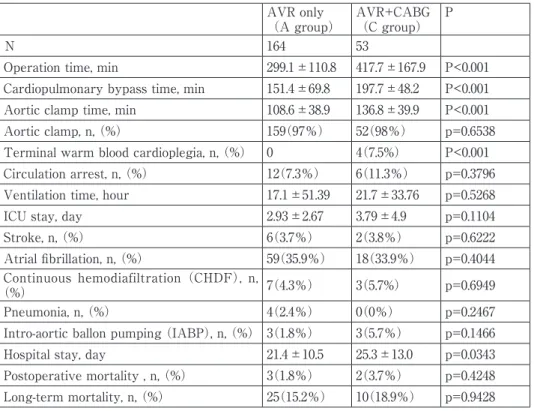
Operative times, CPB times, and aortic clamp times were significantly longer in group C than in group A (Table 2). Postoperative complica- tions, such as low cardiac output syndrome
(LOS), sepsis, renal insufficiency, myocardial ischemia, cerebral ischemia, prolonged ICU stay, and prolonged mechanical ventilation, showed no significant differences between the two groups (Table 2). In group C, only 4 patients showed that the postoperative MB form creatine kinase (CPK-MB) was elevated over 50 IU/L
(Table 3). However, postoperative LVEF was not different between the two groups. Postoper- ative CPK-MB was not elevated in low LVEF patients with our cardiac protection solution
(Table 4). Total cardioplegia dose was showed according to heart disease (Table 5).
Clinical outcomes of groups A and C
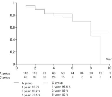
Compared to group A, hospital stay was significantly prolonged in group C. Hospital mortality showed no significant differences between the two groups (Table 2). One-year survival was 95.7% in group A vs. 95.6% in group C, 3-year survival was 90.2% vs. 89.0%, and 5-year survival was 78.5% vs. 82.0%, respectively;
there were no significant differences between the two groups (Figure 1).
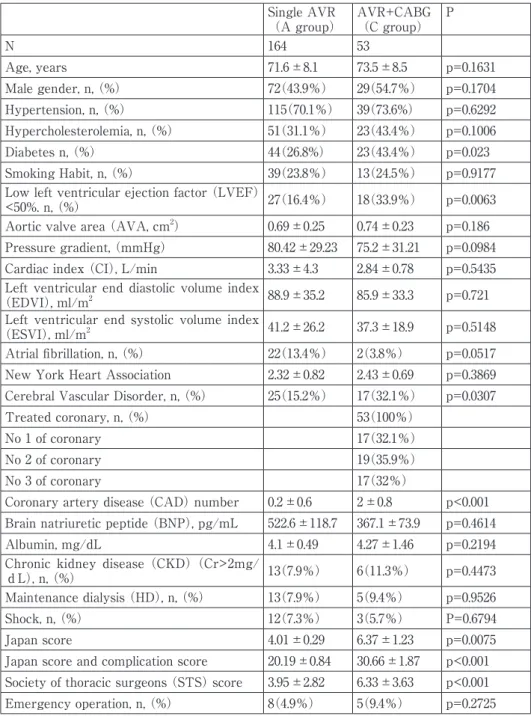
Table 1. Preoperative clinical data of the two groups Single AVR
(A group) AVR+CABG
(C group) P
N 164 53
Age, years 71.6±8.1 73.5±8.5 p=0.1631
Male gender, n, (%) 72(43.9%) 29(54.7%) p=0.1704
Hypertension, n, (%) 115(70.1%) 39(73.6%) p=0.6292 Hypercholesterolemia, n, (%) 51(31.1%) 23(43.4%) p=0.1006
Diabetes n, (%) 44(26.8%) 23(43.4%) p=0.023
Smoking Habit, n, (%) 39(23.8%) 13(24.5%) p=0.9177 Low left ventricular ejection factor (LVEF)
<50%. n, (%) 27(16.4%) 18(33.9%) p=0.0063
Aortic valve area (AVA, cm2) 0.69±0.25 0.74±0.23 p=0.186 Pressure gradient, (mmHg) 80.42±29.23 75.2±31.21 p=0.0984 Cardiac index (CI), L/min 3.33±4.3 2.84±0.78 p=0.5435 Left ventricular end diastolic volume index
(EDVI), ml/m2 88.9±35.2 85.9±33.3 p=0.721
Left ventricular end systolic volume index
(ESVI), ml/m2 41.2±26.2 37.3±18.9 p=0.5148
Atrial fibrillation, n, (%) 22(13.4%) 2(3.8%) p=0.0517 New York Heart Association 2.32±0.82 2.43±0.69 p=0.3869 Cerebral Vascular Disorder, n, (%) 25(15.2%) 17(32.1%) p=0.0307
Treated coronary, n, (%) 53(100%)
No 1 of coronary 17(32.1%)
No 2 of coronary 19(35.9%)
No 3 of coronary 17(32%)
Coronary artery disease (CAD) number 0.2±0.6 2±0.8 p<0.001 Brain natriuretic peptide (BNP), pg/mL 522.6±118.7 367.1±73.9 p=0.4614
Albumin, mg/dL 4.1±0.49 4.27±1.46 p=0.2194
Chronic kidney disease (CKD) (Cr>2mg/
dL), n, (%) 13(7.9%) 6(11.3%) p=0.4473
Maintenance dialysis (HD), n, (%) 13(7.9%) 5(9.4%) p=0.9526
Shock, n, (%) 12(7.3%) 3(5.7%) P=0.6794
Japan score 4.01±0.29 6.37±1.23 p=0.0075
Japan score and complication score 20.19±0.84 30.66±1.87 p<0.001 Society of thoracic surgeons (STS) score 3.95±2.82 6.33±3.63 p<0.001 Emergency operation, n, (%) 8(4.9%) 5(9.4%) p=0.2725
Discussion
Combined operation of AVR and CABG has been reported to provide better clinical outcomes than AVR alone for AS patients with CAD 25). According to current AHA/ACC guidelines, about 49% of these patients need combined AVR and CABG 9). Patients with AS and CAD have been reported to have more postoperative
complications than patients with isolated AS 1). Although a previous study showed that me- chanical ventilation was significantly prolonged in patients who underwent combined operation of AVR and CABG, it was not a predictor of poor surgical outcome 10). But hospital mortality was increased by the combined operation 26). Several studies showed that preoperative mor- tality was not different between combined AVR
Table 2. Intra-operative and postoperative clinical data of the two groups AVR only
(A group) AVR+CABG
(C group) P
N 164 53
Operation time, min 299.1±110.8 417.7±167.9 P<0.001 Cardiopulmonary bypass time, min 151.4±69.8 197.7±48.2 P<0.001 Aortic clamp time, min 108.6±38.9 136.8±39.9 P<0.001
Aortic clamp, n, (%) 159(97%) 52(98%) p=0.6538
Terminal warm blood cardioplegia, n, (%) 0 4(7.5%) P<0.001 Circulation arrest, n, (%) 12(7.3%) 6(11.3%) p=0.3796 Ventilation time, hour 17.1±51.39 21.7±33.76 p=0.5268
ICU stay, day 2.93±2.67 3.79±4.9 p=0.1104
Stroke, n, (%) 6(3.7%) 2(3.8%) p=0.6222
Atrial fibrillation, n, (%) 59(35.9%) 18(33.9%) p=0.4044 Continuous hemodiafiltration (CHDF), n,
(%) 7(4.3%) 3(5.7%) p=0.6949
Pneumonia, n, (%) 4(2.4%) 0(0%) p=0.2467
Intro-aortic ballon pumping (IABP), n, (%) 3(1.8%) 3(5.7%) p=0.1466
Hospital stay, day 21.4±10.5 25.3±13.0 p=0.0343
Postoperative mortality , n, (%) 3(1.8%) 2(3.7%) p=0.4248 Long-term mortality, n, (%) 25(15.2%) 10(18.9%) p=0.9428
Table 3. Myocardial protection of patients with postoperative increasing MB form creatine kinase (CPK-MB)
Case Operation Noradrenaline Postoperative CPK-MB
(IU/L)
Total cardioplegia solution
(ml) Times
1 AVR
CAB3 (LITA-LAD,
GSV-LCX, GSV-RCA)+ 162 3600 5
2 AVR
CAB1(GSV-LAD)
AAR + 104 2250 4
3 AVR
CAB1(GSV-LCX) + 90 3250 4
4 AVR
CAB1(GSV-RCA) + 162 2500 4
AVR: Aortic valve replacement AAR: Ascending aortic replacement LITA: Left internal thoracic artery GSV: Great saphenous vein
LAD: left anterior descending artery LCX: Left circumflex artery RCA: right coronary artery
and CABG and AVR alone, but operative-related morbidity and the incidence of prolonged venti- lation were significantly higher with the com- bined operation 11, 12). However, ventilation time was not prolonged in the combined operation
group in the present study, indicating that pro- longed cardiac arrest time was not a risk factor for postoperative respiratory dysfunction.
The patients with moderate AS and CAD undergoing CABG alone had a higher rate of re-
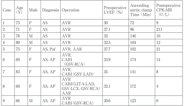
Table 4. Postoperative MB form creatine kinase (CPK-MB) of low LVEF patients in the two groups Case Age(Y) Male Diagnosis Operation Preoperative
LVEF (%)
Ascending aortic clamp Time (Min)
Postoperative CPK-MB
(U/L)
1 75 F AS AVR 30 72 9
2 71 F AS AVR 37.1 96 211
3 78 M AS AVR 32 146 10
4 80 M AS AVR 32.5 104 12
5 75 F AS, Paf AVR, AAR 37.7 102 21
6 69 F AS, AP AVR,
CAB1(GSV-RCA) 33.9 174 14
7 83 F AS, AP AVR,
CAB1(GSV-LAD) 35 141 8
8 60 F AS, AP
AVRCAB3(LITA-LAD, GSV-LCX, GSV-RCA)
AAR
32.1 172 4
9 66 M AS, AP AVR
CAB1(GSV-RCA) 30.6 123 6
Paf: Paroxysmal atrial fibrillation AP: Angina pectoris
AVR: Aortic valve replacement AAR: Ascending aortic replacement F: Female
M: Male
LITA: Left internal thoracic artery GSV: Great saphenous vein
LAD: left anterior descending artery LCX: Left circumflex artery RCA: right coronary artery
Table 5. Total cardioplegia dose.
Case Weight(kg) Total cardioplegia dose (ml) Cardioplegia (ml/kg)
MR(MVP) 50 1000+500+500+500=2500 50
AS(AVR) 55.9 1650+830+830+819=4129 73.8
AS+CAD
(AVR+CAB 3) 76.7 2400+1200+1060+930+1075=6665 86.8 MR: mitral regurgitation
AS: aortic stenosis
CAD: coronary artery disease MVP: mitral valve plasty AVR: aortic valve replacement CAB: coronary artery bypass grafting
operation for AVR within 5 years postoperative- ly. Therefore, patients with moderate AS and CAD need combined AVR and CABG in the early phase 13). Combined AVR and CABG for patients with moderate AS and CAD more than
70 years old should be positively performed if there are no additional preoperative risk fac- tors 14). Concomitant AVR at the time of CABG appears to convey a survival advantage for pa- tients with moderate aortic stenosis 15).
105 Combined aortic stenosis and coronary artery operation
In patients with low LVEF (less than 40%), New York Heart Association (NYHA) functional class of III or IV, chronic obstructive pulmonary disease (COPD), or renal failure preoperatively, long-term survival was significantly decreased 11). Recently, transcatheter aortic valve replacement
(TAVR) has been introduced into clinical use for severe AS patients who cannot tolerate conventional AVR. Although open AVR resulted in good clinical outcomes, TAVR for intermediate risk patients showed lower midterm mortality than surgical AVR 10, 16, 17). The indications for TAVR in AS have been extending beyond high- risk patients recently, so we should consider combined TAVR and CABG or PCI and TAVR as an alternative to concomitant AVR and CABG for patients with AS and CAD in the future.
Previous studies showed that CABG per- formed with AVR could offset the adverse effect of CAD in patients with AS and CAD 18, 19). Pa- tients who underwent combined AVR and CABG showed longer cardiopulmonary bypass time and aortic clamp time than isolated AVR.
Therefore, myocardial protection is the most im-
portant issue. Antegrade coronary artery car- dioplegia perfusion is mostly and safely used, but retrograde perfusion from the coronary si- nus is a safe and effective means of cardioplegia in aortic valve operations 20). Cold blood cardiople- gia reduces the increase in cardiac enzyme levels compared with cold crystalloid cardioplegia in patients undergoing isolated AVR 21). Antegrade crystalloid cardioplegia and retrograde cold blood cardioplegia led to early and late clinical results similar to those achieved with combined ante- grade and retrograde cold blood cardioplegia 22). Continuous retrograde blood cardioplegia is asso- ciated with a good postoperative outcome in heart valve operations 23). Terminal warm blood cardioplegia improved the recovery of myocar- dial electrical activity after coronary artery re-perfusion 27). However, there is no standard solution and enough evidence as a reference. If patients need complicated bypass grafting to coronary arteries, we will consider to perform retrograde cardioplegia to protect from endocar- dial ischemia in our department.
When AS patients have CAD, and it becomes more difficult to obtain enough myocardial pro-
Figure 1 Long-term survival curves
24 Figure 1 Long-term survival curves
tection. A previous study showed that postoper- ative elevation of CPK-MB was found in many patients undergoing combined AVR and CABG 24). In our department, we usually used cooled St. Thomas’ II cardioplegic solution that was antegradely administered without blood first when aortic regurgitation was absent or mild. Advanced AS induces myocardial hyper- trophy and endocardial ischemia, and we inject- ed additional cardioplegic solution from the by- pass graft to the right coronary artery, and we increased the quantity of cardioplegic solution according to the degree of hypertrophy as- sessed by preoperative echocardiography. When cardiac arrest time was over 180 minutes, we used terminal warm blood cardioplegia before staring coronary re-perfusion. Cold blood cardio- plegia was not used, but retrograde coronary si- nus cardioplegia was performed for patients when myocardial protection was insufficient or with complicated bypass grafting. We speculat- ed that hypothermia and hyperkalemia were the most important factors for myocardial pro- tection, so we used cooled crystal cardioplegia not mixed with cold blood, which was not suffi- cient due to decreased potassium and pH chang- es. Although we did not measure the tempera- ture of the myocardium, it was decreased by cold cardioplegia during surgery. In order to sustain cooling and the arrest of the myocardi- um sufficiently, cold cardioplegic solution was injected every 30 minutes with 1.5 fold of nor- mal dose. Bypass grafting was performed in heart arrest including LITA-LAD anastomosis, because beating anastomosis is not good for myocardium protection. Only 4 patients were found to have slight elevation of CPK-MB postoperatively by these efforts in group C, and low LVEF patients did not show increased postoperative CPK-MB, myocardial protection according to our protocol was sufficient.
Conclusions
Combined AVR and CABG could be performed safely with sufficient myocardial protection.
Sufficient myocardial protection during cardiac arrest decreases myocardial ischemia and in- hospital complications, and has an important effect on increasing long-term survival.
Disclosure Statement
There are no disclosures.
Author Contributions
Study conception: ZY, KD Data collection: ZY, KD Analysis: ZY, KD Investigation: ZY, KD Writing: ZY, KD, IF Funding acquisition: None
Critical review and revision: All authors Final approval of the article: All authors Accountability for all aspects of the work: All authors
References
1) Kuwano H, Amano J, Yokomise H. Thoracic and cardiovascular surgery in Japan during 2010:
annual report by The Japanese Association for Thoracic Surgery. Gen Thorac Cardiovasc Surg.
2012;60:680-708.
2) Beach JM, Mihaljevic T, Svensson LG, Rajeswaran J, Marwick T, Griffin B, Johnston DR, et al.
Coronary artery disease and outcomes of aortic valve replacement for severe aortic stenosis. J Am Coll Cardiol. 2013;61:837-48.
3) Roberts WC, Roberts CC, Vowels TJ, Ko JM, Filardo G, Hamman BL, et al. Effect of coronary bypass and valve structure on outcome in isolated valve replacement for aortic stenosis. Am J Cardiol. 2012;109:1334-40.
4) Dè Waard GA, Jansen EK, de Mulder M, Vonk AB, Umans VA. Long-term outcomes of isolated aortic valve replacement and concomitant AVR and coronary artery bypass grafting. Neth Heart J. 2012;20:110-7.
5) Dagenais F, Mathieu P, Doyle D, Dumont È, Voisine P. Moderate aortic stenosis in coronary artery bypass grafting patients more than 70 years of age: to replace or not to replace? Ann Thorac Surg. 2010;90:1495-99; discussion 1499-500.
6) Álvaro D.B. Bordalo, Ângelo l. Nobre, Manuel Dantas, Joāo Cravino. Elevated HDL is the main negative risk factor for coronary artery disease in the elderly patient with calcific aortic valve disease. Rev Port Cardiol. 2012;31:415-24.
7) Kobayashi KJ, Williams JA, Nwakanma LU, Weiss SS, Gott VL, Baumgartner WA, Conte JV.
EuroSCORE Predicts Short- and Mid-Term Mortality in Combined Aortic Valve Replacement and Coronary Artery Bypass Patients. J Card Surg. 2009;24:637-43.
8) Fojt V, PirK J, Kamenicky P. Values of osteoprote- gerin in aortic valve tissue in patients with signifi- cant aortic stenosis depend on the existence of concomitant coronary artery disease. Cardiovasc Pathol. 2016;25:181-4.
9) Nishimura RA, Otto CM, Bonow RO, Carabello BA, Erwin JP, Guyton RA, O’Gara PT, et al. 2014 AHA/
ACC Guideline for the Management of Patients with Valvular heart disease: a report of the American College of Cardiology/American Heart Association Task Force on Practice Guidelines.
Circulation. 2014;129:e521-e643.
10) Carmen Spaccarotella, Annalisa Mongiardo, Salvatore De Rosa, Ciro Indolfi. Transcatheter aortic valve implantation in patients at intermediate surgical risk. Int J Cardiol. 2017;243:161-8.
11) Kai Ming Wang T, Choi D, Ramanathan T, Ruygrok PN. Aortic Valve Replacement With or Without Concurrent Coronary Artery Bypass Grafting in Octogenarians: Eight-Year Cohort Study. Heart Lung Circ. 2017;26:82-7.
12) Sareyyupoglu B, Sundt TM 3rd, Schaff HV, Enriquez-Sarano M, Greason KL, Suri RM, Burkhart HM, et al. Management of Mild Aortic Stenosis at the Time of Coronary Artery Bypass Surgery:
Should the Valve Be Replaced? Ann Thorac Surg.
2009;88:1224-31.
13) Breglio A, Anyanwu A, Itagaki S, Polanco A, Adams DH, Chikwe J. Does Prior Coronary Bypass Surgery Present a Unique Risk for Reoperative Valve Surgery? Ann Thorac Surg. 2013;95:1603-8.
14) Smith WT 4th1, Ferguson TB Jr, Ryan T, Landolfo CK, Peterson ED. Should Coronary Artery Bypass Graft Surgery Patients with Mild or Moderate Aortic Stenosis Undergo Concomitant Aortic Valve Replacement? J Am Coll Cardiol. 2004;44:1241-7.
15) Pereira JJ, Balaban K, Lauer MS, Lytle B, Thomas JD, Garcia MJ. Aortic valve replacement in patients with mild or moderate aortic stenosis and coronary bypass surgery. Am J Med. 2005; 118:
735-42.
16) Conte JV, Gleason TG, Resar JR, Adams DH, Deeb GM, Popma JJ, Hughes GC, et al. Transcath- eter or Surgical Aortic Valve Replacement in Patients with Prior Coronary Artery Bypass Grafting. Ann Thorac Surg. 2016;101:72-9.
17) Petronio AS, Capranzano P, Barbato E, Piazza N, Baumbach A, Haude M, Windecker S. Current status of transcatheter valve therapy in Europe:
results from an EAPCI survey. EuroIntervention.
2016;18:890-5.
18) Di Gioia G, Pellicano M, Gabor GT, Casselman F, Adjedj J, Frank VP, Bernard S, et al. Clinical Outcome of Patients with Aortic Stenosis and Coronary Artery Disease Not Treated According to Current Recommendations. J Cardiovasc Transl Res. 2016;9:145-52.
19) Beach JM, Mihaljevic T, Svensson LG, Rajeswaran J, Marwick T, Griffin B, Johnston DR, et al.
Coronary artery disease and outcomes of aortic valve replacement for severe aortic stenosis. J Am Coll Cardiol. 2013;61:837-48.
20) Menaschè P, Subayi JB, Piwnica A. Retrograde coronary sinus cardioplegia for aortic valve operations: A clinical report on 500 patients. Ann Thorac Surg. 1990;49:556-64.
21) Braathen B, Tonnessen T. Cold blood cardioplegia reduces the increase in cardiac enzyme levels compared with cold crystalloid cardioplegia in patients undergoing aortic valve replacement for
isolated aortic stenosis. J Thorac Cardiovasc Surg.
2010;139:874-80.
22) Lee JH, Jeong DS, Sung K, Kim WS, Lee YT, Park PW. Clinical Results of Different Myocardial Protection Techniques in Aortic Stenosis. Korean J Thorac Cardiovasc Surg. 2015;48:164-73.
23) Flameng WJ, Herijgers P, Dewilde S, Lesaffre E.
Continuous retrograde blood cardioplegia is associ- ated with lower hospital mortality after heart valve surgery. J Thorac Cardiovasc Surg. 2003;125:
121-5.
24) Sherman SK, Fitch JC, Nussmeier NA, Chen JC, Rollins SA, Mojcik CF, Malloy KJ, et al. Impact of Pexelizumab, an ante-C5 complement antibody, on total mortality and adverse cardiovascular out- comes in cardiac surgical patients undergoing car- diopulmonary bypass. Ann Thorac Surg. 2004;77:
942-50.
25) Melby SJ, Zierer A, Kaiser SP, Guthrie TJ, Keune JD, Schuessler RB, Pasque MK, et al. Aortic valve replacement in octogenarians: risk factors for early and late mortality. Ann Thorac Surg. 2007;
83:1651-6.
26) Sakata R, Fujii Y, Kuwano H. Committee for scientific affairs: Thoracic and cardiovascular surgery in Japan during 2009: annual report by the Japanese association for thoracic surgery. Gen Thorac Cardiovasc Surg. 2011;59:636-67.
27) Hattori Y, Yang Z, Sugimura S, Iriyama T, Watanabe K, Negi K, Yamashita M, et al. Terminal warm blood cardioplegia improves the recovery of myocardial electrical activity. A retrospective and comparative study. Jpn J Thorac Cardiovasc Surg.
2000;48:1-8.