Defeating EpCAM+ liver cancer stem cells by targeting chromatin remodeling enzyme CHD4 in human hepatocellular carcinoma
著者 丹尾 幸樹
著者別表示 Nio Kouki journal or
publication title
博士論文本文Full 学位授与番号 13301甲第4446号
学位名 博士(医学)
学位授与年月日 2016‑09‑26
URL http://hdl.handle.net/2297/46456
doi: 10.1016/j.jhep.2015.06.009
Creative Commons : 表示 ‑ 非営利 ‑ 改変禁止
Defeating EpCAM + liver cancer stem cells by targeting chromatin remodeling enzyme CHD4 in human
hepatocellular carcinoma
Kouki Nio, Taro Yamashita ⇑ , Hikari Okada, Mitsumasa Kondo, Takehiro Hayashi, Yasumasa Hara, Yoshimoto Nomura, Sha Sha Zeng, Mariko Yoshida, Tomoyuki Hayashi, Hajime Sunagozaka,
Naoki Oishi, Masao Honda, Shuichi Kaneko
Department of Gastroenterology, Kanazawa University Graduate School of Medical Science, Kanazawa, Ishikawa, Japan
Background & Aims: Hepatocellular carcinoma is composed of a subset of cells with enhanced tumorigenicity and chemoresis- tance that are called cancer stem (or stem-like) cells. We explored the role of chromodomain-helicase-DNA-binding pro- tein 4, which is encoded by the CHD4 gene and is known to epi- genetically control gene regulation and DNA damage responses in EpCAM
+liver cancer stem cells.
Methods: Gene and protein expression profiles were determined by microarray and immunohistochemistry in 245 and 144 hepa- tocellular carcinoma patients, respectively. The relationship between gene/protein expression and prognosis was examined.
The functional role of CHD4 was evaluated in primary hepatocel- lular carcinoma cells and in cell lines in vitro and in vivo.
Results: CHD4 was abundantly expressed in EpCAM
+hepatocel- lular carcinoma with expression of hepatic stem cell markers and poor prognosis in two independent cohorts. In cell lines, CHD4 knockdown increased chemosensitivity and CHD4 overex- pression induced epirubicin chemoresistance. To inhibit the func- tions of CHD4 that are mediated through histone deacetylase and poly (ADP-ribose) polymerase, we evaluated the effect of the his- tone deacetylase inhibitor suberohydroxamic acid and the poly (ADP-ribose) polymerase inhibitor AG-014699. Treatment with either suberohydroxamic acid or AG-014699 reduced the number of EpCAM
+liver cancer stem cells in vitro, and suberohydroxamic acid and AG-014699 in combination successfully inhibited tumor growth in a mouse xenograft model.
Conclusions: CHD4 plays a pivotal role in chemoresistance and the maintenance of stemness in liver cancer stem cells and is therefore a good target for the eradication of hepatocellular carcinoma.
Ó 2015 European Association for the Study of the Liver. Published by Elsevier B.V. All rights reserved.
Introduction
Hepatocellular carcinoma (HCC) is one of the most common causes of cancer death worldwide [1,2]. This is partly due to a lack of effective chemotherapeutic options for patients with advanced-stage disease [3]. Various molecular profiling approaches have been used to identify potential therapeutic tar- gets which are specifically activated in HCC [4–8]. Some studies have indicated the importance of evaluating ‘‘stemness’’ in HCC; it reflects the malignant nature of the tumor and closely correlates with a poor prognosis after surgery [9–12]. Recent evi- dence has also suggested that HCC may conform to the cancer stem cell (CSCs) hypothesis, which proposes that a subset of cells with stem cell features play a fundamental role in tumor mainte- nance and chemo/radiation resistance [13]. CSCs, also called tumor-initiating cells or cancer stem-like cells, possess stem cell features in their self-renewal and differentiation capacity, and contribute to the formation of heterogeneous tumor cell popula- tions. In HCC, several stem cell markers, including CD133, CD90, CD13, epithelial cell adhesion molecule (EpCAM), and CD24, have been reported to enrich side populations of CSCs [13–15]. We recently reported that the stem cell markers EpCAM and alpha-fetoprotein (AFP) can be used to classify HCC subtypes with distinct gene expression profiles and patient prognoses [11]. In particular, the EpCAM
+AFP
+HCC subtype shares the gene expression features of cells from hepatic stem cell-like (HpSC)-HCC, and exhibits resistance to the chemotherapeutic reagent 5-fluorouracil [16,17]. However, the underlying molecu- lar mechanisms which are responsible for the chemoresistance of EpCAM
+CSCs remain to be identified.
Using gene expression profiling approaches, we recently iden- tified the activation of transcription factor Sal-like protein 4 (SALL4) in EpCAM
+CSCs from HpSC-HCC [18]. SALL4 is a
Journal of Hepatology 2015 vol. xxx
jxxx–xxx Keywords: Chromodomain-helicase-DNA-binding protein 4; Liver cancer stem
cells; Histone deacetylase; Poly (ADP-ribose) polymerase; Chemoresistance.
Received 4 December 2014; received in revised form 3 June 2015; accepted 10 June 2015
⇑
Corresponding author. Address: Department of General Medicine/Gastroen- terology, Kanazawa University Graduate School of Medical Science, 13-1 Takara- Machi, Kanazawa, Ishikawa 920-8641, Japan. Tel.: +81 76 265 2042; fax: +81 76 234 4281.
E-mail address: [email protected] (T. Yamashita).
Abbreviations: AFP, alpha-fetoprotein; CHD, chromodomain-helicase-DNA-bind
ing proteins; CSC, cancer stem cell; DSBs, double strand breaks; EpCAM, epithelial
cell adhesion molecule; HCC, hepatocellular carcinoma; HDAC, histone
deacetylase; HpSC, hepatic stem cell-like; NuRD, nucleosome remodeling and
histone deacetylase; PARP, poly (ADP-ribose) polymerase; qRT-PCR, quantitative
reverse transcription-polymerase chain reaction; SALL4, sal-like protein 4; SBHA,
suberohydroxamic acid.
transcription factor which plays a fundamental role in the main- tenance of embryonic stem cells, possibly through interaction with octamer-binding transcription factor 4, sex determining region Y-box 2, and Nanog [19–24]. It has been reported by three independent groups that SALL4 is a biomarker of HCCs with stem-like gene expression signatures and a poor prognosis [18,25,26]. SALL4 was recently found to directly interact with the epigenetic modulator nucleosome remodeling and histone deacetylase (NuRD) complex [27], thereby altering the histone modifications associated with stemness. Indeed, we have demon- strated that SALL4-positive HCCs have high histone deacetylase (HDAC) activity and are chemosensitive to HDAC inhibitors which reduce SALL4 expression [18].
The NuRD complex is a multi-unit chromatin remodeling complex consisting of chromodomain-helicase-DNA-binding pro- teins (CHDs), metastasis-associated proteins, and HDACs [28].
Interestingly, recent studies have indicated that CHD4, a DNA-binding protein which complexes with the NuRD complex, plays a role in the DNA damage/repair network and is recruited to DNA-damaged sites in a poly (ADP-ribose) polymerase (PARP)-dependent manner [29–32]. However, the role of CHD4 in the chemoresistance of EpCAM
+CSCs remains to be elucidated.
In this study, we investigated the role of CHD4, a NuRD complex protein which regulates HDAC activity and the DNA damage response, in the chemoresistance of liver CSCs. We further evalu- ated the efficacy of an HDAC inhibitor in combination with a PARP inhibitor in blocking CHD4 function in EpCAM
+HCCs.
Materials and methods Clinical HCC specimens
For microarray analyses, HCC tissues were obtained from 245 patients who had undergone radical resection from 2002 to 2003 at the Liver Cancer Institute (Fudan University, Shanghai, China). For immunohistochemical analyses, HCC tis- sues and adjacent non-cancerous liver tissues were obtained from 144 patients who had undergone a hepatectomy from 2002 to 2012 at Kanazawa University Hospital, Japan. The pathological diagnoses were performed as previously described [12]. Of these HCC specimens, 38 were obtained fresh and snap-frozen in liquid nitrogen for RNA analysis. An additional fresh HpSC-HCC sample was also obtained from surgical resection and used immediately to pre- pare a single-cell suspension. All tissue acquisition and experimental procedures were approved by the Ethics Committee and Institutional Review Board of each institute and conformed to the 1975 Declaration of Helsinki. All patients provided written informed consent.
Cell culture and reagents
The human liver cancer cell lines HuH7, HuH1, Hep3B, HLE, HLF, and SK-Hep-1 were obtained from the Japanese Collection of Research Bioresources (Osaka, Japan) or the American Type Culture Collection (Manassas, VA) and were rou- tinely cultured in Dulbecco’s modified Eagle’s medium (DMEM) supplemented with 10% fetal bovine serum (FBS). Primary HCC tissue samples were dissected and digested with 1 l g/ml type 4 collagenase solution (Sigma–Aldrich Japan K.K., Tokyo, Japan) at 37 °C for 15 min. Contaminating red blood cells were lysed with ammonium chloride solution (STEMCELL Technologies, Vancouver, Canada) on ice for 10 min. CD45
+leukocytes and annexin V
+apoptotic cells were removed from cell suspensions using an autoMACS-pro cell separator and magnet beads (MiltenyiBiotec K.K., Tokyo, Japan). The HDAC inhibitor suberohydroxamic acid (SBHA) and the PARP inhibitor AG-014699 were obtained from Cayman Chemical (Ann Arbor, MI) and Selleck Chemicals (Houston, TX), respectively. Inhibitor stock solutions were prepared in dimethyl sulfoxide and stored at 20 °C until use. The CHD4 expression plasmid pCMV6-AC-GFP-CHD4 (RG224232) was purchased from Origene Technologies, Inc. (Rockville, MD) and the pcDNA3.1 (V790-20) plasmid, which was used as an empty vector control, was purchased from Invit- rogen (Carlsbad, CA). CHD4-specific and control siRNAs were purchased from
Dharmacon Research, Inc. (Lafayette, CO); the CHD4#1 and CHD4#2 siRNA sequences were ‘‘CCCAGAAGAGGAUUUGUCA’’ and ‘‘GGUUUAAGCUCUUAGAACA’’, respectively. The siRNA constructs were transfected using Lipofectamine 2000 (Invitrogen) in accordance with the manufacturer’s protocol.
Quantitative reverse transcription-polymerase chain reaction (qRT-PCR)
Total RNA was extracted using TRIzol (Invitrogen) in accordance with the manu- facturer’s instructions. The expression of the selected genes was determined in triplicate using a 7900 Sequence Detection System (Applied Biosystems, Foster City, CA). Each sample was normalized relative to 18S ribosomal RNA expression.
The following Applied Biosystems probes used were: CHD4, Hs00172349_m1;
EPCAM, Hs00158980_m1; HDAC1, Hs02621185_sl; AFP, Hs00173490_m1; TERT, Hs00162669_m1; BMI1, Hs00409825_g1; POU5F1, Hs03005111_g1; and 18S, Hs99999901_s1.
Western blotting
Whole cell lysates were prepared using radio-immunoprecipitation assay (RIPA) lysis buffer as previously described [33]. Anti-CHD4 monoclonal (Abcam, Cam- bridge, UK) and anti-b-actin monoclonal (Sigma–Aldrich Japan K.K.) antibodies were used for protein detection. Immune complexes were visualized using enhanced chemiluminescence detection reagents (Amersham Biosciences Corp., Piscataway, NJ) in accordance with the manufacturer’s protocol.
Immunohistochemistry and immunofluorescence
Immunohistochemistry was performed using an anti-CHD4 monoclonal primary antibody (Abcam) and Envision
+kits (DAKO, Carpinteria, CA) in accordance with the manufacturer’s instructions. CHD4 expression was evaluated and categorized as CHD4-high (score, 4–5) or CHD4-low (score, 0–3) based on the CHD4-staining score, which was the sum of the positivity score (0–5%, 0; 5–25%, 1; 25–50%, 2;
and >50%, 3) and staining intensity score (weak, 0; moderate, 1; and strong, 2) for an area. Dual-color immunohistochemistry was performed using Vector red (Vector Laboratories Inc., Burlingame, CA) and the anti-EpCAM antibody VU-1D9 (Oncogene Research Products, San Diego, CA). For immunofluorescence, an Alexa Fluor
Ò488-conjugated anti-mouse immunoglobulin G secondary anti- body (Molecular Probes, Carlsbad, CA) was used. Fluorescence microscopy was essentially performed as previously described [34]. Caspase-3 activation was determined by immunofluorescence using a cleaved caspase-3 (Asp175) antibody (Cell Signaling Technology Inc., Danvers, MA).
Cell proliferation assays and fluorescence-activated cell sorting (FACS)
For cell proliferation assays, 2 10
3cells were seeded in 96-well plates and cell proliferation was evaluated in quadruplicate using the Cell Counting Kit-8 (DOJINDO LABORATORIES, Kumamoto, Japan). For flow cytometry and cell sort- ing, cells were trypsinized, washed, and resuspended in Hank’s balanced salt solu- tion (Lonza, Basel, Switzerland) supplemented with 1% HEPES and 2% FBS. Cells were incubated with the FITC-conjugated anti-EpCAM monoclonal antibody BER-EP4 (DAKO) on ice for 30 min prior to analysis using a FACSCalibur or FAC- SAriaII (BD Biosciences, San Jose, CA).
Measurement of HDAC and PARP activity
HDAC and PARP activities were measured using the Epigenase HDAC Activ- ity/Inhibition Direct Assay Kit (Epigentek Group Inc., Farmingdale, NY) and HT Universal Colorimetric PARP Assay Kit (Trevigen Inc., Gaithersburg, MD), respec- tively. Briefly, nuclear proteins were extracted using NE-PER Nuclear and Cyto- plasmic Extraction Reagents (Thermo Fisher Scientific KK, Tokyo, Japan) and HDAC and PARP activity was measured in triplicate. Calculations were performed in accordance with the manufacturers’ protocols.
Animal studies
Cells (1 10
5HuH7 or primary HCC cells, or 3 10
6HLF cells) were resuspended in 200 l l of a 1:1 DMEM: Matrigel (BD Biosciences) mixture and subcutaneously injected into 6-week-old non-obese diabetic/severe combined immunodeficiency mice (NOD/NCrCRl-Prkdc
scid) which were purchased from Charles River Laboratories, Inc. (Wilmington, MA). For each cell type, 20 mice were inoculated.
Research Article
2 Journal of Hepatology 2015 vol. xxx j xxx–xxx
The experimental protocol was approved by the Kanazawa University Animal Care and Use Committee and conformed to the Guide for the Care and Use of Laboratory Animals prepared by the National Academy of Sciences. The size and prevalence of subcutaneous tumors were recorded as previously described [34]. Once tumors had reached approximately 100 mm
3(HuH7 and primary HCC cells) or 50 mm
3(HLF cells), mice were randomized into treatment groups (n = 5 in each group). Mice were intraperitoneally injected with vehicle, 10 mg/kg AG-014699, 200 mg/kg SBHA, or AG-014699 and SBHA 3 times a week for 14 days.
Microarray studies and statistical analysis
Affymetrix U133A2.0 gene expression data were obtained from the National Center for Biotechnology Information Gene Expression Omnibus database (GEO accession number GSE14520), as previously described [35]. The BRB-ArrayTools software (version 4.3.2) was used for class comparison analyses. Hierarchical clustering analysis was performed using GENESIS software (version 1.7.6). To compare treatment groups in cell proliferation and qRT-PCR assays, Student’s t tests and Chi-square tests were performed in GraphPad Prism software 6.0 (GraphPad Software, San Diego, CA). Kaplan–Meier survival analysis was also per- formed in GraphPad Prism software 6.0 (GraphPad Software). Univariate and multivariate Cox proportional hazards regression analysis was performed using SPSS software (SPSS Inc., Chicago, IL).
Results
High expression of CHD4 in EpCAM
+liver CSCs
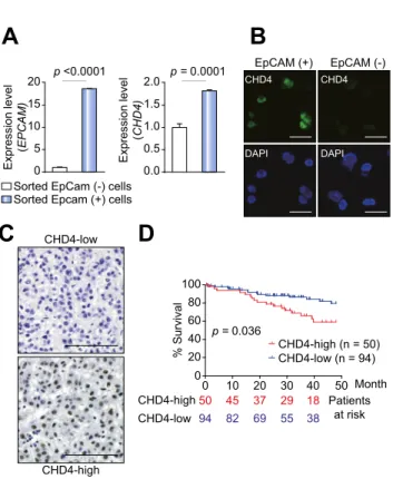
We evaluated the expression of CHD4 in EpCAM
+and EpCAM
cells using a HuH7 single-cell suspension. We found that CHD4 was up-regulated in EpCAM
+cells compared with EpCAM
-cells (Fig. 1A). Using immunofluorescence imaging, a strong nuclear accumulation of CHD4 was identified in the isolated EpCAM
+cells (Fig. 1B and Supplementary Fig. 1A). Similar results were also observed in purified EpCAM
+and EpCAM
cells obtained from a primary HCC specimen (Supplementary Fig. 1B). To explore the relationship between CHD4 and EpCAM expression in HCC and confirm the nuclear accumulation of CHD4 in HCC tissues, we analyzed 144 surgically resected HCC specimens (cohort 1) using immunohistochemistry. Interestingly, the abundance of CHD4
+cells varied dramatically in each HCC specimen (Fig. 1C). We eval- uated the expression of CHD4 in terms of staining intensity and the number of positively stained cells, and categorized HCC tis- sues as CHD4-high or CHD4-low according to the criteria described in the Materials and methods section. Using these cri- teria, 50 of the 144 HCCs (34.7%) were classified as CHD4-high HCCs. An analysis of the clinicopathological characteristics of CHD4-high and CHD4-low HCCs suggested that the CHD4-high HCCs were significantly associated with a poorly differentiated morphology, large tumor size, and EpCAM and CK19 expression (Table 1). The co-expression of CHD4 and EpCAM in HCC tissues was confirmed by dual-color immunohistochemical analysis (Supplementary Fig. 1C). Kaplan–Meier survival analysis indi- cated that, following surgery, CHD4-high HCCs are statistically significantly associated with a poorer survival compared to CHD4-low HCCs (p = 0.036; Fig. 1D). Given that the expression of CHD4 and EpCAM did not completely overlap in the HCC sam- ples (Table 1), we performed a separate evaluation of the effect of CHD4 expression on the survival of EpCAM
+and EpCAM
HCC patients (Supplementary Fig. 1D). As expected, CHD4-low EpCAM
HCCs showed better survival than other subclasses.
CHD4-high EpCAM
+HCCs showed a worse prognosis than CHD4-low EpCAM
+HCCs, but this difference was not statistically significant. Interestingly, CHD4-high EpCAM
HCCs showed a
worse prognosis than CHD4-low EpCAM
HCCs, and this differ- ence was statistically significant (p = 0.03). These analyses sug- gest that CHD4 expression impacts HCC survival, even in EpCAM
HCCs. We therefore evaluated the prognostic value of CHD4 expression on survival, relative to other clinicopathological parameters, in cohort 1 using univariate and multivariate Cox regression analysis (Supplementary Tables 1 and 2). Although high CHD4 expression, tumor size, portal vein invasion, Barcelona Clinic Liver Cancer (BCLC) stage, histology, and serum AFP were all associated with a poor survival in univariate analysis, multi- variate analysis indicated that only the BCLC stage was an inde- pendent risk factor for poor survival in HCC. This suggests that CHD4 expression might be a confounding factor in HCC and is not sufficient, on its own, for evaluating the survival outcome of HCC.
Transcriptomic characteristics of CHD4-high HCCs
To evaluate the molecular profiles of CHD4-high HCCs, we ana- lyzed an Affymetrix gene expression dataset of 245 primary HCC tissues (cohort 2). We defined top-tertile of all HCCs (83 of 245) as CHD4-high when ordered according to the CHD4 signal intensities. The difference between CHD4 gene expression in
A
C CHD4-low D
CHD4-high
B
EpCAM (+) EpCAM (-)
CHD4 CHD4
DAPI DAPI
Sorted EpCam (-) cells Sorted Epcam (+) cells
0.0 0.5 1.0 1.5 2.0
Expression level ( CHD4)
p = 0.0001
0 5 10 15 20
Expression level ( EPCAM)
p <0.0001
.CHD4-low (n = 94) CHD4-high (n = 50)
Patients at risk CHD4-high
CHD4-low 50 94 82
45 37 29 69 55 38
18
% Survival
0 20 40 60 80 100
0 10 20 30 40 50 p = 0.036
Month
Fig. 1. High expression of chromodomain-helicase-DNA-binding protein 4 (CHD4) in epithelial cell adhesion molecule (EpCAM)
+cancer stem cells (CSCs).
(A) Quantitative reverse transcription-polymerase chain reaction (qRT-PCR) analysis of CHD4 and EpCAM in HuH7 cells. EpCAM
+and EpCAM
HuH7 cells were isolated by FACS sorting; (B) Immunofluorescence analysis of CHD4 in EpCAM
+and EpCAM
HuH7 cells (scale bar, 50 l m). (C) Immunohistochemical analysis of CHD4 in surgically resected hepatocellular carcinomas (HCCs).
Representative photomicrographs of CHD4-high (right panel) and CHD4-low (left
panel) HCC tissues are shown (scale bar, 100 l m). (D) Kaplan–Meier survival
curves for 144 patients who underwent surgery. CHD4-high HCCs (n = 50)
showed a poor prognosis compared with CHD4-low HCCs (n = 94).
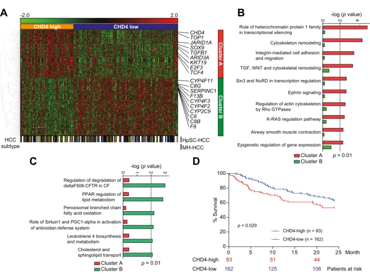
CHD4-high and CHD4-low HCCs was 1.62-fold (Supplementary Fig. 2A). Similarly, the difference between CHD4 gene expression in CHD4-high and CHD4-low HCCs defined by immunohisto- chemistry was 1.86-fold in cohort 1, evaluated by qRT-PCR (Supplementary Fig. 2B). This demonstrates a concordance between the gene and protein expression criteria for CHD4-high and CHD4-low HCCs in the two cohorts. The clinicopathological characteristics of CHD4-high and CHD4-low HCCs in cohort 2 (Supplementary Table 3) indicate that serum AFP was signifi- cantly higher in CHD4-high HCCs than in CHD4-low HCCs (p = 0.015). We performed a class comparison analysis with t tests (p <0.001) and permutation tests (p <0.001, computed based on 10,000 random permutations) of the class labels (CHD4-high and CHD4-low) using BRB-ArrayTools (version 4.3.2) to evaluate the genes differentially expressed between the classes and to control the number and proportion of false discoveries of genes identified. We identified a total of 724 genes that were differen- tially expressed with P1.2-fold difference between the two HCC subclasses (the complete gene list with parametric p values, false discovery rates, and permutation p values of identified genes is available in Supplementary Table 4). Of these genes, 329 were up-regulated and 395 were down-regulated in CHD4-high HCCs (Fig. 2A). Interestingly, stemness-related genes, such as SOX9 and KRT19, and Wnt/TGF-b pathway related genes, including TGFB1 and TCF4, were up-regulated in CHD4-high HCCs. Genes associated with the epigenetic control of transcription, such as JARID1A and ARID3A, were also up-regulated in CHD4-high HCCs.
Furthermore, the genes related to mature hepatocyte function (e.g., those involved in serum production and xenobiotic metabo- lism), such as F8, F13B, C6, C8B, CYP4F2, and CYP2C9, were down-regulated in CHD4-high HCCs. We proceeded to evaluate the expression of CHD4 in EpCAM
+AFP
+HCCs (HpSC-HCCs) and EpCAM
AFP
HCCs (Mature hepatocyte-like HCCs; MH-HCCs).
These are HCC subclasses which we previously proposed as being associated with the stem/maturational status of HCCs [11]. As expected, CHD4 expression was significantly higher in HpSC-HCCs compared with MH-HCCs (p = 0.0026; Supplemen- tary Fig. 3). Consistent with this, pathway analysis indicated that the Wnt/TGF-b pathway, Sin3/NuRD transcriptional regulation, and the epigenetic regulation of gene expression were all acti- vated in CHD4-high HCCs (Fig. 2B). In contrast, in CHD4-low
HCCs, pathways associated with mature hepatocyte function, such as PPAR-regulated lipid metabolism, and cholesterol and sphingolipid transport, were activated (Fig. 2C). The statistically significant poor prognosis of CHD4-high HCCs was independently validated in the cohort 2 microarray data (p = 0.029; Fig. 2D). This suggests that CHD4-high HCCs represent a HCC subtype with a stem cell-like gene expression signature and a poor prognosis.
CHD4 confers chemoresistance in EpCAM
+HCCs
To evaluate the functional role of CHD4 in HCC, we transiently knocked down CHD4 gene expression in EpCAM
+HuH7 cells using two CHD4-specific siRNAs (CHD4#1 and CHD4#2). CHD4 expression decreased in HuH7 cells transfected with CHD4 siR- NAs compared with those transfected with a control siRNA (Fig. 3A). Interestingly, CHD4 knockdown enhanced chemosensi- tivity to epirubicin in HuH7 cells (Fig. 3B). Similar results were also obtained using HuH1 and Hep3B cells (Supplementary Fig. 4A and 4B). We further evaluated the effect of CHD4 overex- pression in HuH1 cells. We transfected HuH1 cells with either an empty vector control (pcDNA 3.1) or a CHD4-encoding plasmid (pCMV6-CHD4) and confirmed the up-regulation of CHD4 tran- script and protein (Fig. 3C). CHD4 overexpression clearly enhanced epirubicin chemoresistance in HuH1 cells (Fig. 3D).
Again, similar results were obtained using HuH7 and Hep3B cells (Supplementary Fig. 4C and 4D). Taken together, these data sug- gest that CHD4 plays a pivotal role in the chemoresistance of EpCAM
+HCC.
Overcoming EpCAM
+HCC by targeting CHD4 through HDAC/PARP inhibition
The above data demonstrate that the activation of CHD4 may play a fundamental role in the chemoresistance of EpCAM
+CSCs to cytotoxic reagents, suggesting that CHD4 suppression may be useful for the eradication of EpCAM
+CSCs. We recently demon- strated that HDACs are activated in EpCAM
+HCCs, and that HDAC inhibitors successfully inhibit the growth of EpCAM
+HCC cell lines [18]. We evaluated the expression of HDAC1 in 38 surgically resected snap-frozen specimens using qRT-PCR. The analysis indicated a concordant up-regulation of HDAC1 in CHD4-high Table 1. Clinicopathological characteristics of CHD4-high and CHD4-low HCCs.
Parameter CHD4-high (n = 50) CHD4-low (n = 94) p value
*Mean age, y (SD) 62.8 ( 9.5) 63.6 ( 9.9) 0.63
Sex: male/female 37/13 67/27 0.85
Mean AFP level, ng/ml (SD) 9931 (55,278) 639.5 (2220) 0.11
Histologic grade
: I − II/II − III/III − IV 5/30/15 16/71/7 0.0014
Mean tumor size, mm (SD) 41.8 (28.2) 31.6 (19.0) 0.048
Macroscopic portal vein invasion: yes/no 14/36 18/76 0.29
BCLC stage: 0/A/B/C 4/23/7/16 20/47/7/20 0.09
Virus status: B/C/B + C/NBNC 17/22/2/9 21/57/1/15 0.19
EpCAM: positive/negative 26/24 30/64 0.02
CK19: positive/negative 15/35 9/85 0.004
CHD4, chromodomain-helicase-DNA-binding protein 4; HCC, hepatocellular carcinoma; SD, standard deviation; AFP, alpha-fetoprotein; BCLC, Barcelona Clinic Liver Cancer;
B, hepatitis B surface (HBs) antigen positive; C, hepatitis C virus (HCV) antibody positive; B + C; HBs antigen positive and HCV antibody positive; NBNC, HBs antigen negative and HCV antibody negative; EpCAM, epithelial cell adhesion molecule.
⁄
Mann–Whitney U test, Chi-square test, or Fisher’s exact test.
⁄⁄