Contents lists available atScienceDirect
Pathology - Research and Practice
journal homepage:www.elsevier.com/locate/prp
Mesenchymal stem cells up-regulate the invasive potential of prostate cancer cells via the eotaxin-3/CCR3 axis
Yukako Ishidaa, Akira Kidoa,⁎, Manabu Akahaneb, Shingo Kishic, Shinji Tsukamotoa,c, Hiromasa Fujiic, Kanya Honokic, Yasuhito Tanakac
aDepartment of Rehabilitation Medicine, Nara Medical University, Nara, Japan
bDepartment of Public Health, Health Management and Policy, Nara Medical University, Nara, Japan
cDepartment of Orthopedic Surgery, Nara Medical University, Nara, Japan
A R T I C L E I N F O
Keywords:
Mesenchymal stem cells Cancer microenvironment Eotaxin-3
CCR3
Homing chemokines
A B S T R A C T
This study aimed to clarify the role of mesenchymal stem cells (MSCs) as a component of the cancer micro- environment. We investigated the homing-related chemokine expression levels of MSCs treated with a prostate cancer cell line (PC-3) -conditioned medium. Among several homing chemokines, an antibody array revealed that expression of eotaxin-3 (but not eotxin-1 and -2) was highly enhanced in MSCs treated with PC-3-condi- tioned medium. A gene expression array showed significantly increased expression of CCR3, a receptor of eo- taxin-3, in PC-3. In a matrigel invasion assay, interferon-gamma, a specific inhibitor of eotaxin-related homing, significantly reduced the transmigration of PC-3 cells, under co-cultured condition with MSCs, in a dose-de- pendent manner (P < 0.05). Consistent with these results, anti-CCR3 antibody successfully reduced PC-3 mi- gration under the co-cultured condition. Thesefindings suggest that MSCs to modulation of the invasive po- tential of prostate cancer cells via the eotaxin-3/CCR3 axis.
1. Introduction
Solid tumors are composed of a heterogeneous cell population that includes a variety of tumor stromal cells. These cells create the tumor microenvironment, which, through cellular interactions, plays an active role in tumor progression [1]. Mesenchymal stem cells (MSCs) are multipotent stromal cells, which are able to differentiate into a variety of tissues, such as fat, cartilage, bone and possibly muscle [2]. Being part of the tumor microenvironment, MSCs are considered to create niches and facilitate tumor growth and metastasis [2]. Previously, we reported that MSCs increased tumor cell proliferation under hypoxia [3], and promoted engraftment and metastatic colonization in a rat osteosarcoma model [4]. Pathway analysis of the gene expression profile showed that genes involved in adhesion, cytokine-cytokine re- ceptors and extracellular matrix pathways were highly expressed in MSCs [4].
The eotaxin family is a group of homing-related CC cytokines, in- itially regarded as an eosinophil chemoattractant [5]. Eotaxin is in- volved in the recruitment of inflammatory cells such as eosinophils and neutrophils [5]. In the oncologicalfield, several groups have reported that eotaxin-1 is a strong candidate as a biomarker of prostate cancer [6,7], and that the eotaxin/CCR3 axis promotes invasive potential in
prostate cancer [8].
In this study, we focused on the role of the eotaxin/CCR3 axis in the interaction between prostate cancer cells and MSCs as a component of the cancer microenvironment in patients with prostate cancer. We in- vestigated the chemokine expression of MSCs treated with a prostate cancer cell line (PC-3) -conditioned medium (CM). Furthermore, func- tional invasion assays were performed under co-cultured conditions with a specific inhibitor, interferon gamma (IFN-γ), or anti−CCR3 antibody, and assessed the role of eotaxin/CCR3 axis on PC-3 invasion.
2. Materials and methods
2.1. Cell lines
The human prostate cancer cell line PC-3 was obtained from ATCC (Rockville, MD) and tested under mycoplasma-free conditions. The cells were grown under standard conditions in Dulbecco’s modified Eagle’s medium (Bio Whittaker, Verviers, Belgium) supplemented with 10%
heat-inactivated fetal calf serum (Hyclone-Perbio, Brebieres, France) and antibiotics/antimycotics (Gibco, Rockville, MD). Human MSCs were purchased from Lonza and maintained as adherent cultures in complete Mesenchymal Stem Cell Growth Medium (MSCGM BulletKit;
https://doi.org/10.1016/j.prp.2018.06.012
Received 12 April 2018; Received in revised form 12 June 2018; Accepted 22 June 2018
⁎Corresponding author at: Department of Rehabilitation Medicine, Nara Medical University, 840 Shijo-cho, Kashihara, Nara, 634-8522, Japan.
E-mail address:[email protected](A. Kido).
0344-0338/ © 2018 Elsevier GmbH. All rights reserved.
T
Lonza Japan, Tokyo,Japan) at 37 °C/5% CO2. Media was changed every 2 days, and the cells were split when they reached 80–90% confluence.
The cells were used at early passage (< 5 passages) for all experiments.
2.2. Conditioned medium preparation
Conditioned medium (CM) was generated as follows: 90% con- fluent, passage 2–4 PC-3 or MSCs in T75 tissue cultureflask, were washed 3 times with phosphate-buffered saline (PBS) and transferred to a serum-free DMEM or MSCGM culture medium during 48 h. CM from differentflasks were harvested and pooled. CM were then sterilized on 0.22μmfilters (Millipore, Billerica, MA), and stored at−80 °C until use.
2.3. Cell proliferation assessment
2.3.1. Cell metabolic viability was assessed using an MTS
[3-(4,5-dimethylthiazol-2-yl)-5-(3-carboxymethoxyphenyl)-2-(4- sulfophenyl)-2H-tetrazolium] assay. A single cell suspension was plated in 96-well plates at 2.0 × 10³cells/well and allowed to attach to the plates at 37 °C for 4 h. The cells were then left untreated or treated with the CM for 48 h. After the treatment, cell viability was measured using the Cell Titer 96 Aqueous One Solution Cell Proliferation Assay Reagent (Promega, Madison, WI). Then, 20μl of MTS solution was added to each well, and incubated for 1 h at 37 °C under a CO2 atmosphere. Cell proliferation was measured at an emission wavelength of 492 nm. All experiments were carried out in quadruplicate, and viability was ex- pressed as the ratio of the number of viable cells.
2.4. Measurement of cytokine/chemokine protein expression by antibody array method
Upon confirming the differential mRNA expression of the re- presentative pro-inflammatory cytokines and chemokines in MSCs cultured with PC-3 CM versus controls, we studied the chemokine ex- pression at the protein level using the RayBio Human Cytokine Antibody Array C5 (RayBiotech, Inc. GA), which detected 82 human cytokines/chemokines (Table 1). The protein of the samples was ex- tracted using 2X Ray-Bio Cell Lysis Buffer. After centrifugation, the supernatant was collected and the protein concentration was measured using Protein Assay Reagent (BioRad, Hercules, CA). The protein sample (200μg) was transferred to an antibody array membrane by electroplotting. The antibody-array membrane was blocked using 1×
blocking buffer for 30 min at room temperature. After 3 washes, the membrane was incubated with 2 ml biotin-conjugated anticytokine antibodies for 2 h at room temperature. Then, 2 ml of 1000-fold diluted horseradish peroxidase (HRP)-conjugated streptavidin was added and incubated for 2 h to allow the streptavidin to attach to the biotin. After washing, the membrane was treated with the detection buffer for HRP for 2 min. After the excess detection buffer had been drained off, the membrane was covered with 2 plastic sheets and chemiluminescence detection was performed using Fusion Solo 2 (Vilber-Lourmat, Colle- gien, France). The intensities of the dot signals were calculated using Image J software.
2.5. Total RNA isolation
Total RNA was extracted from MSCs or PC-3 cells, using TRIzol reagent (Invitrogen, Carlsbad, California, USA) following the manu- facturer’s protocol, and RNA purity (A260/280 nm) was assessed using a spectrophotometer (NanoDrop ND-1000, NanoDrop Technologies, Wilmington, Delaware, USA). RNA integrity was evaluated using a bioanalyzer (Agilent 2100, Agilent Technologies, Foster City, California, USA) and only samples with preserved 18S and 28S peaks and RNA integrity number values equal or greater than 5 were selected for gene expression analysis.
Table 1
Target proteins for the human cytokine antibody array.
Angiogenin BDNF BLC (CXCL13) CK beta 8-1 (CCL23) EGF
ENA-78 (CXCL5) Eotaxin-1 (CCL11) Eotaxin-2 (MPIF-2/CCL24) Eotaxin-3 (CCL26) FGF-4
FGF-6 FGF-7 (KGF) FGF-9 Flt-3 Ligand Fractalkine (CX3CL1) GCP-2 (CXCL6) GCSF GDNF GM-CSF
GRO alpha (CXCL1) GRO alpha/beta/gamma HGF
I-309 (TCA-3/CCL1) IFN-gamma IGF-1 IGFBP-1 IGFBP-2 IGFBP-3 IGFBP-4 IL-1 alpha (IL-1 F1) IL-1 beta (IL-1 F2) IL-2
IL-3 IL-4 IL-5 IL-6 IL-7 IL-8 (CXCL8) IL-10 IL-12 p40/p70 IL-13 IL-15 IL-16 IP-10 (CXCL10) Leptin LIF LIGHT (TNFSF14) MCP-1 (CCL2) MCP-2 (CCL8) MCP-3 (MARC/CCL7) MCP-4 (CCL13) M-CSF MDC (CCL22) MIF MIG (CXCL9) MIP-1 beta (CCL4) MIP-1 delta (CCL15) MIP-3 alpha (CCL20) NAP-2 (PPBP/CXCL7) NT-3
NT-4 Oncostatin M Osteopontin (SPP1) Osteoprotegerin (TNFRSF11B) PARC (CCL18)
PDGF-BB PLGF RANTES (CCL5) SCF
SDF-1 alpha (CXCL12 alpha) TARC (CCL17)
TGF beta 1 TGF beta 2 TGF beta 3
(continued on next page)
2.6. Quantitative real-time PCR
First, cDNA samples were prepared from total RNA samples using the High Capacity cDNA Reverse Transcription kit (Applied Biosystems, Carlsbad, California, USA). For each sample, qPCR reactions were car- ried out in duplicate using 10 ng of cDNA and a 96-plex gene card, TaqMan Array Human Chemokines, Fast 96-Well Plate (Applied Biosystems, Carlsbad, California, USA), representing 92 chemokine-re- lated genes and 4 reference genes, in accordance with the manu- facturer’s instructions. These reactions were performed using a real- time PCR system (StepOne, Applied Biosystems, Carlsbad, California, USA)
2.7. Invasion assays in boyden chambers
Migration and invasion assays were performed using HTS Fluoroblok 24-well chambers containingfilters with an 8μm pore size (BD Biosciences, Franklin Lakes, NJ). For the invasion assay, filters were precoated on the upper side with matrigel (20μg per well; BD Biosciences). Serum-free conditioned medium from PC-3 cells was prestained with calcein AM (5μg/ml; Molecular Probes,Eugene,Oregon) for 30 min at 37 °C, and 1 × 10⁵ cells were seeded in serum-free DMEM on the upper compartment of the chambers for 12 h at 37 °C. In the lower compartment of the chamber, 1 × 10⁵ cells of MSCs were seeded 24 h earlier than the seeding of PC-3. Images of fluorescent cells in the lower side of the filter were captured and analyzed by fluorescent microscopy in accordance with the manu- facturer’s protocol. The number of cells was counted using Image J software. All analyses were performed 3 times in triplicate under
identical conditions. Values shown are means ± SEM. For neutraliza- tion of CCR-3, cells were cultured with a concentration of 10μg/ml of anti-CCR3 antibody (MAB155, R&D Systems, MN, USA) for 30 min. In addition, inhibitory assay was performed using IFN-γ(Sigma Aldrich, St. Louis, USA) at concentrations of 1, 10, and 100 ng/mL for 24 h.
2.8. Statistical analysis
Student’st-test was used for comparing the 2 groups. The statistical analyses were performed using the Statcel Ver. 4 program, and p-values less than 0.05 were considered statistically significant. Regarding the gene expression, normalization and fold change were calculated with the ΔΔCt (cycle threshold) method standardized by the geometric averaging of the expression level of the 4 reference genes, using StepOne software ver.2.3 (Applied Biosystems, Foster City, CA). The relative gene expression levels were calculated based on the 2−ΔΔCq method.
3. Results
3.1. Effect of conditioned medium on the cellular proliferation of MSCs and PC-3
First, we studied the effects of PC-3 CM on cellular proliferation.
After a 24 h treatment with PC-3 CM, MSCs showed no significant effect on proliferation. Next, we treated PC-3 cells with MSCs CM for 24 h, and performed an MTS assay and, similarly, found no significant effect on the proliferation of PC-3.
3.2. Chemokine expression of MSCs treated with PC-3 CM
Fig. 1shows the chemokine expression of MSCs treated with PC-3 CM. Among several chemokines, the antibody array revealed that ex- pression of eotaxin-3 (but not eotxin-1 and -2) is significantly higher in MSCs treated with PC-3 CM than in controls.
Table 1(continued)
Thrombopoietin (TPO) TIMP-1
TIMP-2 TNF alpha TNF beta (TNFSF1B) VEGF-A
Fig. 1.Cytokine secretion profiling of MSCs treated with PC-3 CM. (A) Human cytokine array image. MSCs were treated with/without the CM for 48 h, and the supernatant was collected and analyzed by RayBio human cyto- kine antibody array C5. (B) Average net optical signal intensity for chemokines including eotaxin -1, -2, and -3. For each spot, the net optical density level was determined by subtracting the background and normalization to the positive control. The spots of treated MSCs were compared with the corre- sponding spots of the untreated MSCs.
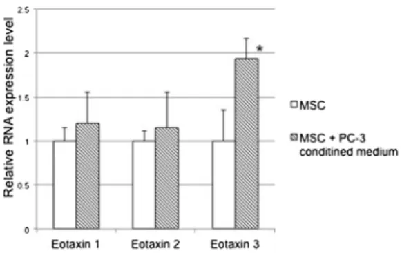
3.3. Relative RNA expression level of eotaxin- 3 in MSCs treated with PC-3 CM
Next, we carried out quantitative real-time PCR to determine the expression level of eotaxin families at mRNA levels. Consistent with the chemokine expression, significantly increased expression of eotaxin-3 was found in MSCs treated with PC-3 CM (P < 0.05), but not eotaxin-1 and 2 (Fig. 2).
3.4. Relative RNA expression level of CCR3 in PC-3 treated with MSCs CM To confirm the expression of CCR3, the functional receptor of eo- taxin-3, in PC-3, we further carried out real-time PCR. CCR3 was ex- pressed in PC-3; however, there was no difference in expression levels between the control and treated PC-3 (Fig. 3).
3.5. Matrigel invasion assay of PC-3 cells s under co-cultured condition with MSCs
To examine whether MSCs affect the in vitro invasion activity of PC- 3 cells, we performed a matrigel invasion assay under the co-cultured condition. In the presence of co-cultured MSCs, PC-3 cells showed a significant increase in cellular invasion activity (131 ± 8.5%, P < 0.05. Fig. 4A–D). Interestingly, interferon significantly reduced the transmigration of PC-3 cells in a dose-dependent manner (P < 0.05). Consistent with these, the neutralization of CCR-3 sig- nificantly inhibited the effect of co-cultured MSCs (P < 0.05,Fig. 4D).
4. Discussion
Eotaxin/CCR3 axis-related chemotaxis may promote malignant in- vasion. Agarwal et al. reported eotaxin-1 as a possible diagnostic marker for prostate cancer [6]. Zhu et al. reported that eotaxin-1 pro- motes prostate cancer cell invasion via the CCR3-ERK pathway with up- regulation of MMP3 expression [8]. More recently, Laurent reported that periprostatic adipocytes act as a driving force for CCR3 expressing prostate cancer [9], suggesting the importance of the biological en- vironment on tumor invasiveness.
In the present study, wefirst reported the importance of eotaxin-3, which is also known to bind to CCR3, as well as other eotaxin families, on prostate cancer invasion. We examined the influence of MSC-CM or the co-cultured condition on the invasive potential of PC-3. Antibody array and quantitative real-time PCR showed that eotaxin-3 (but not eotaxin-1 and -2) significantly increased in PC-3. To assess a functional role, we then investigated whether the co-cultured condition leads to increased cellular transmigration. The co-cultured condition sig- nificantly enhanced cellular invasion, which was reduced by a CCR inhibitor, IFN-γor anti-CCR3 antibody. Although CCR3 antibody are not specific inhibitors for eotaxin-3/ CCR3 axis, we think that the in- hibitory effect on invasion assay is due to eotaxin-3, because other CCR3 binding chemokines did not show marked differences (Fig. 1B).
MSCs secrete a variety of chemoattractant molecules capable of re- cruiting diverse types of immune or progenitor cells. The search for chemotactic cytokines on MSC secretomes results in a large list of CXnC and CC chemokines such as CX3CL1/fractalkine, CXCL1/GROa, CXCL2/GRPb, CXCL5/ENA-78, CXCL8/IL8, CXCL10/IP-10, CXCL-11/i- TAC, CXCL12/SDF-1a, CCL2/MCP-1, CCL3/MIP-1a, CCL4/MIP-1beta, CCL5/RANTES, CCL7/MCP-3, CCL-20/MIP-3 alpha, and CCL-26/eo- taxin-3 [10–15]. These complex networks of chemokines and receptors may provide a benefit in terms of properties of tissue repair [16,17], immuno-regulation [18,19], or hematopoietic effects [20,21]. How- ever, they also possess a mechanism to increase prostate cancer cell transmigration.
Eosinophils are noted in tumor infiltrates in several types of tumors and are usually associated with good prognosis [22], however this has recently emerged as controversial [23]. Poor prognosis was also re- ported in several solid tumors [24]. In prostate cancer, an increase of serum eosinophil counts during Sipuleucel-T, an autologous cellular immunotherapy, is associated with prolonged survival [25]. Co-culture experiments of prostate cancer cells and eosinophils from allergic or asthmatic individuals were reported to result in the growth inhibition [26]. These suggest a multiple aspects of chemokine biology, and it may be different in therapeutic or experimental conditions.
In this study, we focused on the biological relationship between PC- 3 and MSCs cell lines, and successfully demonstrated tumor stromal crosstalk in an in vitro model with essential cellular components. Lin et al. reported that cancer-associated fibroblast up-regulate several chemokines including eotaxin-3, which related to promotion of cancer progression in hepatocellular carcinoma cells [27]. Stich et al. reported that CXCL12 stimulation dose dependently up-regulated migratory in- ducers in MSCs including eotaxin-3 [28]. Together with our data, these results suggest that eotaxin-3 up-regulation can be seen in wide variety of stromal cellular interactions, and MSCs support the cancer micro- environment, which is consistent with previous reports [2–4].
Furthermore, eotaxin-3 secreted from MSCs may be a key player that modulates the aggressiveness of prostate cancer. Inhibition of the eotaxin-3/CCR3 axis in MSCs recruited to prostate cancer may be a possible therapeutic strategy.
Acknowledgement
This work was supported by JSPS KAKENHI Grant Numbers JP24592241, JP15K01381.
Fig. 2.Relative RNA expression level of eotaxin families in MSCs was measured with/without PC-3 CM. Expression of all eotaxin families detected in MSCs.
After PC-3 CM treatment, the expression level of eotaxin-3 was increased sig- nificantly (P < 0.05), but not eotaxin 1, -2.
Fig. 3.Relative RNA expression of CCR3 in PC-3 was measured with/without MSC-CM. CCR3 mRNA expression was detected in quantitative real-time PCR.
There was no significant difference between PC-3 cells with and those without MSC CM.
References
[1] J. Marx, Cancer biology. All in the stroma: cancer’s Cosa Nostra, Science 320 (2008) 38–41.
[2] S.A. Bergfeld, Y.A. DeClerck, Bone marrow-derived mesenchymal stem cells and the tumor microenvironment, Cancer Metastasis Rev. 29 (2010) 249–261.
[3] A. Kido, K. Yoshitani, T. Shimizu, M. Akahane, H. Fujii, S. Tsukamoto, Y. Kondo, K. Honoki, M. Imano, Y. Tanaka, Effect of mesenchymal stem cells on hypoxia- induced desensitization ofβ2-adrenergic receptors in rat osteosarcoma cells, Oncol.
Lett. 4 (2012) 745–750.
[4] S. Tsukamoto, K. Honoki, H. Fujii, Y. Tohma, A. Kido, T. Mori, T. Tsujiuchi, Y. Tanaka, Mesenchymal stem cells promote tumor engraftment and metastatic colonization in rat osteosarcoma model, Int. J. Oncol. 40 (2012) 163–169.
[5] E.M. Campbell, S.L. Kunkel, R.M. Strieter, N.W. Lukacs, Temporal role of chemo- kines in a murine model of cockroach allergen-induced airway hyperreactivity and eosinophilia, J. Immunol. 161 (1998) 7047–7053.
[6] M. Agarwal, C. He, J. Siddiqui, J.T. Wei, J.A. Macoska, CCL11 (eotaxin-1): a new diagnostic serum marker for prostate cancer, Prostate 73 (2013) 573–581.
[7] I. Heidegger, J. Höfer, M. Luger, R. Pichler, H. Klocker, W. Horninger, E. Steiner, S. Jochberger, Z. Culig, Is Eotaxin-1 a serum and urinary biomarker for prostate cancer detection and recurrence? Prostate 75 (2015) 1904–1909.
[8] F. Zhu, P. Liu, J. Li, Y. Zhang, Eotaxin-1 promotes prostate cancer cell invasion via activation of the CCR3-ERK pathway and upregulation of MMP-3 expression, Oncol.
Rep. 31 (2014) 2049–2054.
[9] V. Laurent, A. Guérard, C. Mazerolles, Le Gonidec S, A. Toulet, L. Nieto, F. Zaidi, B. Majed, D. Garandeau, Y. Socrier, M. Golzio, T. Cadoudal, K. Chaoui, C. Dray, B. Monsarrat, O. Schiltz, Y.Y. Wang, B. Couderc, P. Valet, B. Malavaud, C. Muller, Periprostatic adipocytes act as a driving force for prostate cancer progression in obesity, Nat. Commun. 7 (10230) (2016),https://doi.org/10.1038/ncomms10230.
[10] J. Croitoru-Lamoury, F.M. Lamoury, J.J. Zaunders, L.A. Veas, B.J. Brew, Human mesenchymal stem cells constitutively express chemokines and chemokine re- ceptors that can be upregulated by cytokines, IFN-beta, and Copaxone, J. Interferon Cytokine Res. 27 (2007) 53–64.
[11] M. Honczarenko, Y. Le, M. Swierkowski, I. Ghiran, A.M. Glodek, L.E. Silberstein, Human bone marrow stromal cells express a distinct set of biologically functional chemokine receptors, Stem Cells 24 (2005) 1030–1041.
[12] J.F. Ji, B.P. He, S.T. Dheen, S.S. Tay, Interactions of chemokines and chemokine receptors mediate the migration of mesenchymal stem cells to the impaired site in the brain after hypoglossal nerve injury, Stem Cells 22 (2004) 415–427.
[13] A.L. Ponte, E. Marais, N. Gallay, A. Langonné, B. Delorme, O. Hérault, P. Charbord, J. Domenech, The in vitro migration capacity of human bone marrow mesenchymal stem cells: comparison of chemo- kine and growth factor chemotactic activities, Stem Cells 25 (2007) 1737–1745.
[14] V. Sordi, M.L. Malosio, F. Marchesi, A. Mercalli, R. Melzi, Giordano T, N. Belmonte, G. Ferrari, B.E. Leone, F. Bertuzzi, G. Zerbini, P. Allavena, E. Bonifacio, L. Piemonti, Bone marrow mesenchymal stem cells express a restricted set of functionally active chemokine receptors capable of promoting migration to pancreatic islets, Blood 106 (2005) 419–427.
[15] IUIS/WHO Subcommittee on Chemokine Nomenclature, Chemokine/chemokine receptor nomenclature, Cytokine 21 (2003) 48–49.
[16] B. Rüster, S. Gottig, R.J. Ludwig, R. Bistrian, S. Müller, E. Seifried, J. Gille, R. Henschler, Mesenchymal stem cells display coordinated rolling and adhesion behavior on endothelial cells, Blood 108 (2006) 3938–3944.
[17] V.F. Segers, I. Van Riet, L.J. Andries, K. Lemmens, M.J. Demolder, A.J. De Becker, M.M. Kockx, G.W. De Keulenaer, Mesenchymal stem cell adhesion to cardiac mi- crovascular endothelium: activators and mechanisms, Am. J. Physiol. Heart Circ.
Physiol. 290 (2006) H1370–H1377.
[18] L. Sun, D. Wang, J. Liang, H. Zhang, X. Feng, H. Wang, B. Hua, B. Liu, S. Ye, X. Hu, W. Xu, X. Zeng, Y. Hou, G.S. Gilkeson, R.M. Silver, L. Lu, S. Shi, Umbilical cord mesenchymal stem cell transplantation in severe and refractory systemic lupus er- ythematosus, Arthritis Rheum. 62 (2010) 2467–2475.
[19] F. Carrion, E. Nova, C. Ruiz, F. Diaz, C. Inostroza, G. Mönckeberg, F.E. Figueroa, Autologous mesenchymal stem cell treatment increased T regulatory cells with no effect on disease activity in two systemic lupus erythe- matosus patients, Lupus 19 (2010) 317–322.
[20] K. Le Blanc, H. Samuelsson, B. Gustafsson, M. Remberger, B. Sundberg, J. Arvidson, P. Ljungman, H. Lönnies, S. Nava, O. Ringdén, Transplantation of mesenchymal stem cells to enhance engraftment of hematopoietic stem cells, Leukemia 21 (2007) 1733–1738.
[21] L. Fouillard, M. Bensidhoum, D. Bories, H. Bonte, M. Lopez, A.M. Moseley, A. Smith, A. Lesage, F. Beaujean, D. Thierry, P. Gourmelon, A. Najman, N.C. Gorin, Engraftment of allogeneic mesenchymal stem cells in the bone marrow of a patient with severe idiopathic aplastic anemia improves stroma, Leukemia 17 (2003) 474–476.
[22] S. Gatault, F. Legrand, M. Delbeke, S. Loiseau, M. Capron, Involvement of eosino- phils in the anti-tumor response, Cancer Immunol. Immunother. 61 (2012) 1527–1534.
[23] J. Ellyard, L. Simson, C. Parish, Th2-mediated antitumour immunity: friend or foe?
Fig. 4.Invasion of PC-3 cells (1 × 10⁵cells/well) in matrigel-coated Transwell chambers (8-μm pores). The images offluorescent cells migrating through recon- stituted membrane to the lower side of thefilter were captured and counted using Image J software. Representative photographs of the control (A), PC-3 with anti- CCR3 antibody (B), and the counting view of Image J (C) are shown. Compared with PC-3 alone, the invasive potential of the co-cultured control was increased (D, P < 0.05), and was significantly reduced by IFN-γor anti-CCR3 antibody (D, P < 0.05, respectively).
Tissue Antigens 70 (2007) 1–11.
[24] S. Sakkal, S. Miller, V. Apostolopoulos, K. Nurgali, Eosinophils in cancer: favourable or unfavourable? Curr. Med. Chem. 23 (2016) 650–666.
[25] D.G. McNeel, T.A. Gardner, C.S. Higano, P.W. Kantoff, E.J. Small, M.H. Wener, R.B. Sims, T. DeVries, N.A. Sheikh, R. Dreicer, A transient increase in eosinophils is associated with prolonged survival in men with metastatic castration-resistant prostate cancer who receive sipuleucel-T, Cancer Immunol. Res. 2 (2014) 988–999.
[26] P. Furbert-Harris, D. Parish‐Gause, I. Laniyan, K.A. Hunter, J. Okomo-Awich,
T.R. Vaughn, K.C. Forrest, C. Howland, A. Abdelnaby, O.A. Oredipe, Inhibition of prostate cancer cell growth by activated eosinophils, Prostate 57 (2003) 165–175.
[27] Z.Y. Lin, Y.H. Chuang, W.L. Chuang, Cancer-associatedfibroblasts up-regulate CCL2, CCL26, IL6 and LOXL2 genes related to promotion of cancer progression in hepatocellular carcinoma cells, Biomed. Pharmacother. 66 (2012) 525–529.
[28] S. Stich, M. Haag, T. Häupl, O. Sezer, M. Notter, C. Kaps, M. Sittinger, J. Ringe, Gene expression profiling of human mesenchymal stem cells chemotactically in- duced with CXCL12, Cell Tissue Res. 336 (2009) 225–236.