Clinical Significance of Reverse Redistribution Phenomenon for
201Tl Scintigraphy in Nonischemic Disease
Jumpei S
UYAMA*1), Akira S
HINOZUKA1), Hiroto S
ASAMORI1), Norifumi H
OSAKA1), Osamu M
IYAGAMI1), Masako K
ATO1),
Akifumi N
IIYA1), Kyouichi K
ANEKO2)Yasushi A
KUTSU3)and Takehiko G
OKAN1)Abstract : The reverse redistribution phenomenon (RR) on
201Tl SPECT has been focused mainly on ischemic improvement regions after reperfusion therapy or vaso- spastic angina pectoris. However, RR analysis has not been used in the context of non-ischemic disease. The aim of this study was to evaluate the clinical role of RR on
201Tl SPECT in patients without a history of myocardial ischemia. We ret- rospectively enrolled 86 patients showing RR by myocardial perfusion SPECT and studied 75 other patients as a control group. For quantitative analysis, each
201Tl SPECT polar map was divided into 13 segments. Differences between the RR and control group were assessed with respect to patient characteristics and cardiac event-free survival using the Kaplan-Meier method. RR was detected frequently in the inferoposterior wall, septal portion of the anterior wall, and septum. The two groups showed significant differences in rates of heart failure (P < 0.01) , hypertro- phic cardiomyopathy (P < 0.05) , and wall motion abnormality (P < 0.05) , but not in the rate of event occurrence. The study demonstrated that RR on
201Tl SPECT could indicate the existence of myocardial damage ; however, it would not be a factor that determines the prognosis.
Key words : nuclear cardiology, reverse redistribution, Thallium-201, SPECT, myocar- dial perfusion
Introduction
201
Tl has been widely used for myocardial perfusion scintigraphy in the clinical setting since the 1970s.
201Tl has several advantages over the 99mTc-labeled agent including ease of use by a single administration and its usefulness for myocardial viability evaluation.
201Tl reverse redistribution (RR) was defined as either normal exercise perfusion and defective redistribution or an exercise defect worsened at redistribution. Although initially many studies were conducted on ischemic improvement regions after acute coronary syndrome (ACS)
1-3), RR is sometimes noted even in patients not undergoing reperfusion therapy as part of their daily treatment.
Original
1)
Department of Radiology, Showa University School of Medicine, 1—5-8 Hatanodai, Shinagawa-ku, Tokyo 142-8666, Japan.
2)
Division of Cardiology, Department of Medicine, Showa University School of Medicine.
3)
Department of Internal Medicine, Showa University Karasuyama Hospital.
*
To whom corresponding should be addressed.
Recently, RR was also associated with vasospastic angina pectoris (VAP) and non-ischemic diseases
4-6). However, to the best of our knowledge, there has been no unified study on the value of RR analysis. This study thus evaluated the clinical significance of the RR phenomenon on
201Tl scintigraphy in patients without a history of myocardial ischemia.
Material and Methods
201
Tl myocardial perfusion SPECT was performed in patients without a history of ischemic heart disease and with no obvious signs of ischemia as diagnosed by medical examination performed between January 2000 and August 2008. For this study, we included 86 patients based on the following criteria : (1) to exclude the influence of ischemia due to stenosis of a coronary artery, we studied patients not showing reduced blood flow in stress images ; (2) to exclude artefactual influence, we studied patients showing decreased uptake across at least 2 of 13 segments in the rest images. We also studied 75 patients as a control group. These patients had no history of ischemic heart disease and showed normal perfusion in stress myocardial scintigraphy performed between March 2005 and May 2005. The data presented herein corresponds to consecutive data from both groups.
Results are expressed as mean SD for continuous variables and as percentages of the total number of patients for categorical variables. Significance between the groups was determined by unpaired Studentʼs t-test for continuous variables and by the chi-square test for categorical variables. If data were not distributed normally, the Wilcoxon signed-rank test was used.
Kaplan-Meier analysis and long-rank tests were used to compare the event-free survival rates.
Probability levels less than 0.05 were considered statistically significant.
Patients were followed up after the stress myocardial scintigraphy, using the following three items as endpoints : (1) reduced blood flow on stress scintigraphy ; (2) cardiac events requiring hospitalization or PCI ; and, (3) cardiac death. The longest follow-up period for cardiac death is 96 months.
Cardiac scintigraphy was performed in a large rotating field-view gamma camera (ECAM、
Siemens, USA) with low-energy, high-resolution, parallel-hole collimators. Each dataset was acquired over a 180 semicircular arc extending from the right anterior oblique to left posterior oblique position. After fasting, the patients underwent stress by exercise loading or pharmacological loading, and then 110 MBq of
201Tl (Nihon Medi-Physics, Nishinomiya, Japan or Fuji Film RI Pharma, Tokyo, Japan) was intravenously injected. MPS was performed within 10 minutes of the tracer injection (Stress image) . Rest images for assessment of myocardial viability were obtained four hours after the stress images.
The exercise stress studies used the symptom-limited supine ergometer exercise test, performed
by starting the exercise at 25 W and increasing it by 25 W every 3 min. In exercise-stressed
patients, the endpoint of the test was determined by five items as follows : ① shortness
of breath or fatigue ; ② decreased blood pressure ; ③ target heart rate (85% of predicted
maximum heart rate ; (220age) 0.85) ; ④ ST change on ECG ; and, ⑤ high maximum blood
pressure over 230 mmHg.
The pharmacological stress study used three kinds of vasodilators : adenosine ; ATP : 0.15 mg / kg / min, dypilidamole : 0.56 mg / kg, or dobutamine : 340 µg / kg.
The polar map shown in Fig. 1 was adopted to evaluate the images, and two clinical specialists in nuclear medicine conducted the visual evaluations.
Results
The different stress methods used in this study are detailed in Table 1. There were no significant differences in the number of patients undergoing the exercise stress and pharmacological stress test between groups. All the exercise-stressed patients satisfied at least one of the five endpoints (Table 2) . In both groups, item ① (shortness of breath or fatigue)
was most frequent, followed by item ③ (target heart rate) .
On ECG, no case revealed ST elevation at aVR, and the ST depression affected a wide region over six leads in all cases including controls. No patient in the RR group showed
Fig. 1. Schematic polar map of the left ventricular myocardium divided into 13 segments.
Table 1. Number of patients with given type of stress for each patient group Control group RR group
exercise 56 67
adenosine 0 6
ATP 11 10
dipyridamole 2 2
dobutamine 5 1
Exercise : pharmacological 56:18 67:19 P=0.608
marked ST changes.
In 20 (76.9%) of the 26 cases showing both RR and abnormal wall motion, an overlap of the occurrence region was found.
Distributions of the regions in which the RR phenomenon was observed are shown in Table 3. Frequent manifestation of RR was found in Seg. 4 (41 patients, 47.7%) , Seg.10 (35 patients, 40.7%) , Seg. 7 (28 patients, 32.6%) , and Seg. 13 (27 patients, 31.4%) . Conversely, there were sites that showed a low incidence, namely Seg. 2 and Seg. 12 (0 patient) , Seg. 3 and Seg. 8 (1 patient 1.2%) , and Seg. 9 (2 patients 2.4%) .
The baseline characteristics for both groups are shown in Table 4. Wilcoxon tests were conducted for BMI, blood pressure, RBC, uric acid, and creatinine, all of which showed a non-
Table 2. Number of patients with various exercise stress tes end- points for each patient group
control group RR group
① shortness of breath or fatigue 40 (71.4%) 48 (73.8%)
② blood pressure decrease 0 3 (4.6%)
③ target heart rate 15 (26.8%) 11(16.9%)
④ ST change on ECG 1 (1.8%) 0
⑤ high maximum blood pressure 0 3 (4.6%)
Total 56 65
Table 3. Regional perfusion abnormality identified by visual inspection in 86 patients Segment number of cases percentage
1 16 18.6
2 0 0
3 1 1.2
4 41 47.7
5 13 15.1
6 9.3 8
7 28 32.6
8 1 1.2
9 2 2.4
10 35 40.7
11 6 7
12 0 0
13 27 31.4
normal distribution. No significant differences were found between the groups in sex, weight, blood pressure, blood findings (red cell count, uric acid, creatinine level) , history of metabolic disorders (diabetes, hyperlipidemia) , arrhythmia (af, others) , VAP, and the presence of abnormal electrocardiogram.
Heart failure occurred more frequently in the RR group than in the control group (17.4% vs.
4.0%) , and the incidence of hypertrophic cardiomyopathy (HCM) was also significantly higher in the RR group than in controls (36.0% vs. 4.0%) . As for dilated cardiomyopathy (DCM) , there were no significant differences between the RR and control group in the low incidence rates. Functional analysis of the thallium scintigraphy indicated a significantly higher incidence of wall motion abnormality in the RR group than in controls, and no significant difference in
Table 4. Patient characteristics
Control group number RR group number P value
Age 67.7 10.1 75 64.1 14.3 86 0.076
Male 34 (45.3%) 75 50(58.1%) 86 0.105
BMI 23.2 3.2 75 24.0 4.1 81 0.349
RBC 416.9 58.2 65 411.9 54.7 80 0.716
Uric Acid (mg / dL) 5.80 1.66 65 5.85 1.53 80 0.935
Creatinine 0.903 0.828 66 1.417 2.348 80 0.208
Systolic BP 135.7 18.0 71 137.9 24.2 84 0.74
Diastolic BP 77.3 13.3 71 78.8 14.2 86 0.874
Smoker 24(36.4%) 66 27(36.5%) 74 0.987
DM 13(17.6%) 74 22(27.5%) 80 0.142
Dyslipidemia 50(70.4%) 71 44(59.5%) 74 0.167
VAP 6(8.0%) 75 3(3.5%) 86 0.214
DCM 0(0%) 75 3(3.5%) 86 0.103
HCM 3(4.0%) 75 12(14.0%) 86 0.048
Heart failure 3(4.0%) 75 15(17.4%) 86 0.007
Bundle branch
block 9(11.7%) 75 8(9.3%) 86 0.578
Arrhythmia 19(25.3%) 75 10(36.0%) 86 0.143
Af 8(10.7%) 75 13(15.1%) 86 0.403
abnormal wall
motion 12(16.0%) 75 26(30.2%) 86 0.034
EF 65.1 12.1% 75 61.5 15.0% 86 0.092
(BMI=body mass index, DM=diabetes mellitus, DCM=dilated cardiomyopathy, HCM=
hypertrophic cardiomyopathy, Af=atrial fibrillation, EF=ejection fraction)
EF between the groups. There were no significant differences in the presence of abnormal electrocardiogram.
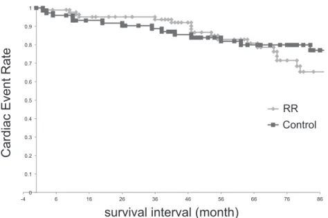
Average observation period during the follow-up was 55.6 30.3 months for the RR group and 59.8 29.9 months for the control group. Cardiac events occurred in 13 patients in the control group and in 17 patients in the RR group, as detailed in Table 5. One patient in the RR group died of chronic heart failure after 11 months of follow-up. Cumulative event-free curves were calculated by the Kaplan-Meier method, and compared by the log-rank test. There were no significant differences in the event occurrence rate between the groups (P=0.38)(Fig. 2) .
Table 5. The number of cases, separated by cause, for a heart event that was an endpoint for follow up in the two groups
Cardiac Events
Control group RR group
Ischemic change at exercise 3 7
Admission due to heart failure 6 4
ACS (death) 3 (0) 5 (1)
Admission due to VAP 1 0
Admission due to Af 0 1
Mean observation period (month) 59.8 29.9 55.6 30.3
(ACS=acute coronary syndrome, VAP=vasospastic angina pectoris, Af=atrial fibrillation)
Fig. 2. Kaplan-Meier event-free survival curves for RR group and control group.
Discussion
The inflow of
201Tl is associated with blood flow from the terminal branch of the coronary artery and cellular oxygen metabolism, whereas the outflow of
201Tl is associated with cellular metabolism and membrane integrity. Causes of reduced uptake in the
201Tl RR phenomenon at rest include myocardial stunning, reduced electrolytic concentration gradient through the cell membrane, and interstitial edema. Reports of RR since the 1990s frequently involved RR after reperfusion therapy for acute coronary syndrome, thus it seems that ischemia-induced dysfunction in metabolism or the cell membrane persists after improved blood flow
7). Also, improvements in wall motion are known to lag behind the blood flow improvement, which is necessary for functional recovery, and it has been documented that lesions with delayed or incomplete functional improvement occur in sites away from those where infarction is complete
1,8). On the other hand, RR phenomenon has also been attributed to an artifact, such as possible signal attenuation due to the breast, diaphragm, or obesity
4,7).
Subsequently, a high occurrence rate of RR for vasospastic angina was also reported.
According to Xiang et al
4), the sensitivity of RR to VAP was 100% and the specificity was 63%. Although there was no significant correlation between the history of VAP and the incidence of RR in this patient group, it does not refute the association between active VAP and RR. Other similar reports cite manifestations of RR in myocardial bridging
5)and collagen disease
6), both of which are considered as stunning involved in ischemia, and similar to VAP as a phenomenon.
With regard to HCM, a significant acceleration in the regional myocardial washout rate in the thickened part as well as the non-thickened part was reported in the 1990s
9). That study showed normal uptake in the inferior wall that had no thickening in two HCM patients with
123