STUDY ON DRUG DELIVERY TARGETING OVARIAN CANCER CELLS
March 2019
APRILIANA CAHYA KHAYRANI
Graduate School of Natural Science Technology (Doctoral Course)
OKAYAMA UNIVERSITY
STUDY ON DRUG DELIVERY TARGETING OVARIAN CANCER CELLS March 2019APRILIANA CAHYA KHAYRANI
STUDY ON DRUG DELIVERY
TARGETING OVARIAN CANCER CELLS
March 2019
APRILIANA CAHYA KHAYRANI
Graduate School of Natural Science Technology (Doctoral Course)
OKAYAMA UNIVERSITY
I
Study on Drug Delivery Targeting Ovarian Cancer Cells
A dissertation submitted by Apriliana Cahya Khayrani in partial fulfilment of the requirements for the Doctor of Philosophy in Engineering in the Graduate School of
Natural Science and Technology, Okayama University, Japan.
March 2019
CONTENTS
Summary ... 1
CHAPTER 1 ... 3
General Introduction ... 3
Ovarian Cancers ... 4
Ovarian Cancer Treatment ... 5
Paclitaxel (PTX) ... 6
Glycosylated Paclitaxel (gPTX) ... 10
Liposomes Based Drug Delivery System ... 12
• Definition and structure of liposomes ... 14
• Liposome Composition ... 15
• Methods of liposomes preparation ... 17
• Characterization of liposomes ... 18
References ... 20
CHAPTER 2 ... 27
CD44 As Target Receptor ... 27
CD44 ... 28
• The structure of CD44 ... 28
• CD44 as target receptor ... 31
Result and Discussion ... 32
Materials and Method ... 36
• Materials ... 36
• Cells Culture and Experimental Animal ... 36
• Preparation of anti-hCD44 MAb ... 36
• Expression of CD44 in Ovarian Cancer Cells line. ... 37
References ... 38
CHAPTER 3 ... 42
Targeting Ovarian Cancer Cells Overexpressing CD44 with Immunoliposomes Encapsulating Glycosylated Paclitaxel ... 42
LIST OF ABBREVIATIONS AND ACRONYMS ... 43
ABSTRACT ... 44
Introduction ... 45
Results ... 47
• Expression of CD44 in human ovarian cancer derived cells ... 47
• Sensitivity of human ovarian cancer derived cells to gPTX. ... 49
• Potential uptake of liposome conjugated with anti-hCD44 MAb ... 50
• Preparation and characterization of gPTX-L and gPTX-IL ... 52
• Cytotoxicity of gPTX, gPTX-L, gPTX-IL In Vitro ... 55
• Suppression of tumor growth In Vivo ... 56
Discussion ... 61
Conclusions ... 64
Materials and Methods ... 65
• Materials ... 65
• Cells Culture and Experimental Animal ... 65
• Preparation of anti-hCD44 MAb ... 66
• Expression of CD44 in Ovarian Cancer Cells line ... 66
• Preparation of Liposome Encapsulating gPTX ... 68
• Evaluation of Cellular Uptake ... 69
• Characterization of Liposome ... 70
• Cytotoxicity Assay ... 71
• Evaluation of cytotoxic effects of liposome formulation by 24h and 72h treatment. ... 73
• Evaluation of Antitumor Effects of Drugs In Vivo ... 73
• Statistical Analysis ... 73
References ... 75
ACKNOWLEDGEMENT ... 81
LIST OF PUBLICATION ... 83
ORAL AND POSTER PRESENTATION ... 86
Say, "I am only a man like you, to whom has been revealed that your god is one God.
So whoever would hope for the meeting with his Lord let him do righteous work and not associate in the worship of his Lord anyone." (Qur'an 18:110)
“Katakanlah: ‘Sesungguhnya aku ini hanya seorang manusia sepertimu, yang diwahyukan kepadaku: ‘Bahwa sesungguhnya Ilahmu itu adalah Ilah Yang Esa.’
Barangsiapa yang mengharapkan perjumpaan dengan Rabbnya, maka hendaklah ia mengerjakan amal yang shalih dan janganlah ia mempersekutukan seorang pun
dalam beribadah kepada Rabbnya.” (QS. Al-Kahfi: 110)
Summary
Ovarian cancer is the most lethal gynecologic malignancy, it ranks the eighth cause of cancer death in women of worldwide. The standard treatment of progressive ovarian cancer is surgical resections followed by systemic chemotherapy. The National Comprehensive Cancer Network (NCCN) guideline shows as first line chemotherapeutic agents for ovarian cancer the carboplatin, paclitaxel (PTX) or the administration in combination of these two
PTX acts as an anti-cancer agent preventing cells division by promoting and stabilizing the assembly of microtubule structures. Because PTX is highly hydrophobic the mixture of Cremophor EL® and ethanol has been adopted as solvent for the commercial formulation known as Taxol. Nonetheless, administration of Taxol may cause side effects such as hypersensitivity reactions, nephrotoxicity and neurotoxicity. Therefore, the disadvantages of Taxol treatment should urgently be ameliorated by the development of proper drug delivery system of PTX. The highly hydrophobic nature of PTX hinders the loading efficiency into the liposomes resulting in a poor encapsulation yield. Recently, we successfully developed liposomes encapsulating glycosylated paclitaxel (gPTX) in the hydrophilic core. gPTX is a PTX derivative with a glucose moiety coupled at the 7-OH radical. This modification enhanced the hydrophilicity of PTX allowing its solubility in different solvents CEP (Cremophor EL®, ethanol, and PBS; in 12:12:76 ratio) and EG (40% Ethylene Glycol).
Exploiting the difference of the solubility we could prepare stable gPTX liposome (gPTX-L) with sufficient amount of encapsulated drug.
The outer layer of liposomes can be modified with coupled ligands targeting molecules localized on the cell membrane surface. Targeted-drug delivery nanosystems with coupled ligands have been actively utilized to optimize therapeutic
efficacy and minimize systemic toxicity. Ligands for cell surface receptors highly expressed in tumor cell populations have provided a great specificity. The cell membrane receptor CD44 could be one of the most promising candidates to be targeted.
CD44 is a receptor for hyaluronic acid type-1 transmembrane glycoprotein that is implicated in cell–cell and cell–matrix interactions and is associated with malignancy, particularly with metastasis promotion. CD44 has also been considered as a cancer stem cell (CSC) marker in several malignancies of hematopoietic and epithelial origin, and is closely related with tumor progression and drug resistance in several tumors including ovarian cancer. Collectively, CD44 could be a suitable candidate molecule to be targeted by the drug delivery nanosystem as ovarian cancer therapeutic.
In this study, we have optimized the loading efficiency of gPTX achieving higher encapsulation yields of gPTX-L, and have also designed the immunoliposome CD44-targeted gPTX-IL. To generate gPTX-IL, the anti-human CD44 monoclonal antibody (anti-hCD44 MAb) was conjugated to the gPTX-L and its efficacy was evaluated under in vitro and in vivo conditions.
CHAPTER 1
General Introduction
Ovarian Cancers
Ovarian cancer causes more deaths in the United States than any other type of female reproductive tract cancer, with an estimated 22.240 new cases and 14.070 deaths in 2018 [1]. Ovarian cancer accounts for just 2.5% of all female cancer cases, but 5% of cancer deaths because of the disease’s low survival. This is largely because 4 out of 5 ovarian cancer patients are diagnosed with advanced disease that has spread throughout the abdominal cavity. Overall ovarian cancer incidence rates have been decreasing since the mid-1980s, with the pace of the decline accelerating in the early 2000s[1,2].
Figure 1. Normal female reproductive anatomy. (originally published by the National Cancer Institute)
The ovaries are a pair of reproductive glands, each about the size of a grape, located
fallopian tubes into the uterus, where they are fertilized for reproduction. In premenopausal women, the ovaries are the primary source of the hormones estrogen and progesterone, which maintain the health of the female reproductive system. The three major types of ovarian cancer are epithelial, accounting for 90% of cases, germ cell (3%), and sex cord-stromal (2%). Epithelial cancers are further subdivided into serous (52%), endometrioid (10%), mucinous (6%), and clear cell (6%) tumors[3,4].
Ovarian cancer is not easily diagnosed because the most common presenting symptoms of persistent abdominal distension — pain and pressure in the pelvis — can be attributed to a number of causes[5]. Patients may be asymptomatic until an abdominal mass is discovered during routine pelvic examination or until the tumour has metastasised [6], consequently, progression to late stage before diagnosis is seen in the majority of presenting women. Approximately 75% of patients are at International Federation of Gynecology and Obstetrics (FIGO) stages II–IV at the time of diagnosis [5].
Ovarian Cancer Treatment
Surgery is currently the intervention of first choice in ovarian cancer [5]. In advanced cases, tumour debulking is recommended to improve the efficacy of adjunctive therapies. Optimal debulking can be achieved in the majority of patients, and prognosis is directly related to the success of such cytoreductive surgery [6].
Chemotherapy for ovarian cancer has progressed considerably over the past two decades, with treatment for advanced disease moving from the use of alkylating agents to current recommended regimens based on taxanes and platinum compounds [5,6].
Clinical trials performed in the late 1970s demonstrated that cisplatin was an active chemotherapy in advanced or recurrent ovarian cancer with a single agent response rate in the range of 13–30% [7,8]. The next generation of research studies evaluating
combination chemotherapy with cisplatin plus cyclophosphamide revealed that the time to progression and the duration of survival were markedly improved compared with single agents [9]. As the result, the standard combination chemotherapy in the late 1980s and early 1990s was cisplatin plus cyclophosphamide. Carboplatin was introduced in the 1990s as an analog of cisplatin with similar single-agent activity in terms of response and survival rates, but with a significantly improved toxicity profile.
Paclitaxel, an active chemotherapeutic agent introduced in the 1990s, changed the standard of care in ovarian cancer yet again. Two randomized clinical trials comparing cyclophosphamide and cisplatin with paclitaxel and cisplatin demonstrated that the investigational arm had an improved outcome compared with the previous standard combination of cyclophosphamide and cisplatin [10,11].
From the past 20 years, the standard of care in advanced ovarian cancer for ovarian cancer encompasses administration of platinum, paclitaxel, or the combination of platinum plus paclitaxel.
Paclitaxel (PTX)
Between 1960 and 1981, the National Cancer Institute (NCI) and the U.S.
Department of Agriculture (USDA) collaborated on a plant screening program that collected and tested 115,000 extracts from 15,000 species of plants to identify naturally occurring compounds with anticancer activity. In 1963, a crude extract from the bark of the Pacific yew Taxus brevifolia, a scarce and slow-growing evergreen found in the old-growth forests of the Pacific Northwest, was found in preclinical studies to have cytotoxic activity against many tumors [12]. PTX was identified as the active constituent of this extract in 1971 by Mansukh Wani and Monroe Wall. They had isolated and identified the active ingredient and named it taxol (generic name of
PTX), based on its species of origin and the presence of hydroxyl groups [13] and it entered the NCI drug development program.
Figure 2. Structure of PTX (Rowinsky EK, Donehower RC. N Engl J Med 1995;332:1004-1014)
In 1979, Susan Horwitz at Albert Einstein College of Medicine (New York, NY), reported that PTX promotes the assembly of microtubules—polymers composed of repeating subunits of α- and β-tubulin heterodimers. PTX reduces the critical concentration of purified tubulin subunits necessary for polymerization into microtubules in vitro and increases the percentage of tubulin subunits that assemble.
Furthermore, microtubules polymerized in the presence of PTX are protected from the disassembly normally induced by cold or calcium treatment [14]. These effects were in stark contrast to previously identified microtubule poisons, including colchicine and vinca alkaloids, which prevent microtubule polymerization [15]. Similar to its effects on purified tubulin, PTX promotes microtubule polymerization and stabilization in living cells, where it is capable of antagonizing the effects of colchicine and vinca alkaloids [14].
PTX-induced mitotic arrest occurs due to activation of the mitotic checkpoint (also known as the spindle assembly checkpoint), the major cell cycle control mechanism acting during mitosis to prevent chromosome missegregation. The mitotic checkpoint delays separation of the chromosomes, which enter mitosis as replicated pairs of sister chromatids, until each pair has made stable attachments to both poles of the mitotic spindle. This arrangement ensures that each daughter cell will receive one copy of every chromatid. Chromatids connect to spindle microtubules through their kinetochores, protein complexes that assemble on centromeric regions of DNA.
Unattached kinetochores, which have not made stable attachments to microtubules, activate a signal transduction cascade that delays mitotic progression by inhibiting the anaphase-promoting complex/cyclosome [16]. PTX treatment arrests cells in mitosis due to the presence of a small number of unattached kinetochores [17].
In spite of its promising anticancer activity, the development of intravenous PTXs formulation has showed several difficulties due to its poor solubility in water.
According to this, the first commercially available formulation containing PTX (Taxol®) is formulated in a vehicle composed of polyoxyethylated castor oil (Cremophor® EL) and dehydrated alcohol in equal parts. Thus, the current clinical dosage form contains in each millilitre 6 mg of PTX, 527 mg of Cremophor® EL (CrEL) and 49.7% (v/v) of absolute ethanol. This vehicle is associated with a variety of side effects such as hypersensitivity, nephrotoxicity and neurotoxicity, attributable mainly to Cremophor® EL. Importantly, these effects are shown in 25–30% of treated patients [18]. Thus, the amount of CrEL administered (for an average patient for a single dose administration) with these drugs averages 5 mL, whereas the amount of CrEL in Taxol® per administration is the relatively higher, approximately 26 mL [19].
Consequently, all patients receiving Taxol® must be premedicated with corticoids, H2
antagonists and antihistamines to prevent, sometimes fatal, hypersensitivity reactions.
Moreover, CrEL has a direct influence over the cells of the pulmonary and vascular endothelium, causing respiratory difficulties and vasodilatation. Severe reactions as bronchospasms and hypotension have been reported [20]. Furthermore, since both ethanol and CrEL solubilize the plasticizers, Taxol® requires the use of non- plasticized solution containers such as di(2-ethyl-hexyl) pthalate (DEHP) in the polyvinylchloride (PVC) infusion bags/sets (Rowe et al., 2009).
Since CrEL can cause hypotension and hypersensitive reactions, Taxol® should be slowly infused over a period of 3 to 24 h for doses of 135–175 mg/m2 every 3 weeks, respectively [21]. It should be previously diluted at a final concentration of 0.3 to 1.2 mg/mL, thus depending on the dose volumes ranging from 250 to 1000 mL of physiological solution or dextrose 5% may be required. Also, due the risk of drug precipitation upon dilution, Taxol® should be administered using an in-line filter (≤0.22 μm). A considerable number of clinic studies with Taxol® have been performed to date and have revealed highly variable pharmacokinetics [22]. The half- life was found to be in the range of 1.3 and 8.6 h and a large volume of distribution of about 55 L/m2 was also reported [23]. PTX is more than 90% bound to plasma proteins.
The main pathways of elimination are hepatic metabolism followed by biliary excretion. In the liver, metabolism is mediated by the cytochrome P450 (CYP3A4 and CYP2C8) and less than 10% of the dose is excreted intact by urine [24]. This drug has shown a variable pharmacokinetic pattern depending on the infusion time. Early studies for prolonged infusion times (6 or 24 h) were generally suggestive of linear pharmacokinetics, but become nonlinear for shorter durations infusion (3 h) due to saturable elimination [25]. The clinical relevance of nonlinear deposition of the drug is based on the fact that small changes either in dosage or infusion duration might result in systemic exposure levels of PTX too large, thereby increasing the risk of toxicity. For example, 3-h infusions of PTX at 135 mg/m2 resulted in a mean Cmax
of 3.3 mM and a mean AUC of 10.4 μMh, whereas at 175 mg/m2, the mean Cmax and AUC values were 5.9 and 18.0 μMh, respectively [26]. Moreover, various studies have shown that CrEL alters the pharmacokinetics profile of the drug and contribute to the reduction in plasma clearance observed at higher PTX doses. Indeed, PTX may be entrapped within hydrophobic interior of CrEL micelles in plasma, which tend to diminish the free fraction of PTX and, thus making it less available for distribution to tumor [27].
Generally, the main reason for discontinuation of PTX is not the lack of efficacy, but toxicity [28]. Peripheral sensory neuropathy is the most commonly reported neurotoxic effect of PTX which is dose- and infusion-duration related [29]. The symptoms may begin as early as 24–72 h after administration and include numbness, paresthesias and burning pain in a glove and-stocking distribution. Because CrEL can also cause neurotoxicity, PTX-induced neuropathy may be at least, in part, contributed by the vehicle formulation [19]. The other major adverse effect is myelosuppression, which mainly consists of neutropenia and usually becomes the dose-limiting toxicity [30].
Glycosylated Paclitaxel (gPTX)
Side effects of PTX in the cancer treatment due to its highly hydrophobicity resulted in massif research on the development or modification of PTX, including our laboratory. We developed glycosylated PTX (gPTX), a PTX derivative with a glucose moiety coupled at the 7-OH, this modification enhanced the hydrophilicity of PTX allowing practically different solubility in the solvents CEP (Cremophor EL®, ethanol, and PBS; in 12:12:76 ratio) and EG (40% Ethylene Glycol)[31].
The preparation of gPTX is occupied commercially available PTX. It was treated with TESCl and N, N- diisopropylethylamine as a base in methylene chloride,
providing 20- triethylsilyloxyPTX (20-TES-PTX) at 97% yield, which was reacted with protected glucosyloxyacetic acid [32,33] using EDCI/DMAP/CH2Cl2 to furnish an inseparable mixture of 20- TES-7-a-gPTX and 20-TES-7-b-gPTX (20-TES-7- gPTX) at 78% combined yield. Triethylsilyl, trityl and benzyl groups were cleanly removed in a single step through a catalytic hydrogenation as described earlier, providing gPTX at 83% yield (Figure 3A). The 20-TES-7-gPTX was condensed with protected a-glucosyloxyacetic acid or protected b-glucosyloxyacetic acid [33] using EDCI/DMAP/CH2Cl2 to give 20-TES-7-a-gPTX at 78% yield or 20-TES-7-b-gPTX at 76% yield. Deprotection of triethylsilyl, trityl, and benzyl groups was accomplished in the same manner as described earlier providing 7-a-gPTX at 83% yield and 7-b- gPTX at 80% yield, respectively (Figure 3B and C)
B A
O O
O
OCPh3 OBn BnO
OBn
O
O HO AcO
O O
O O O
O
OCPh3 OBn BnO
OBn Ph
OSiEt3 NH Ph
O
O
O HO AcO
O O
O O O
O
OH OH HO
OH Ph
OH NH Ph
O
H OBz
OBzH OAc
OAc O
O HOAc OBz HO O O Ph
OSiEt3 NH Ph
O AcO OH HO
EDCI/DMAP CH2Cl2
H2, 10%Pd/C EtOH/dioxane/0.5%HCl
r.t., 48 h 83%
2'-TES-PTX 2'-TES-7-α-gPTX
7-α-gPTX 78%
α
α α
O O
O
OCPh3 OBn BnO
OBn
O
O HO AcO
O O
O O O
O
OCPh3 OBn BnO
OBn Ph
OSiEt3 NH Ph
O
O
O HO AcO
O O
O O O
O
OH OH HO
OH Ph
OH NH Ph
O
H OBz
OBzH OAc
OAc O
O HOAc HO OBz O O Ph
OSiEt3 NH Ph
O AcO OH HO
EDCI/DMAP CH2Cl2
H2, 10%Pd/C EtOH/dioxane/0.5%HCl
r.t., 48 h 80%
2'-TES-PTX 2'-TES-7-β-gPTX
7-β-gPTX 76%
β β
β
O O O
OCPh3 OBn BnO
OBn
O
O HO AcO
O O
O O O
O
OCPh3 OBn BnO
OBn Ph
OSiEt3 NH Ph
O O
O HO AcO
O O
O O O
O
OH OH HO
OH Ph
OH NH Ph
O
H
OBz OBzH
OAc OAc
O
O HOAc OBz HO O O Ph
OSiEt3 NH Ph
O AcO OH HO
EDCI/DMAP CH2Cl2
H2, 10%Pd/C EtOH/dioxane/0.5%HCl
r.t., 48 h 83%
2'-TES-PTX Et3SiCl
iPr2NEt 97 % CH2Cl2 O
O HOAc OBz HO O O Ph
OH NH Ph
O AcO OH
Paclitaxel(PTX)
2'-TES-7-gPTX 7-gPTX
78%
C
gPTX 2’-TES-7-gPTX
2’-TES-7-PTX PTX
2’-TES-7-PTX 2’-TES-7-α-gPTX
7-α-gPTX
2’-TES-7-PTX 2’-TES-7-β-gPTX
7-β-gPTX
Figure 3. Synthesis of 7-glucosyloxyacetylPTXs (gPTX). (A) scheme of gPTX as a mixture of 7-a-gPTX and 7-b-gPTX. (B) scheme of 7-a-gPTX. (C) scheme of 7-b- gPTX (Shigehiro,T, et al, Journal of Microencapsulation, 2016, 33:2, 172-182).
Liposomes Based Drug Delivery System
Drug delivery is the method or process of administering a pharmaceutical compound to achieve a therapeutic effect in humans or animals. Recently, nanotechnology and nanoscience based drug delivery present a highly positive prospective of bringing benefits to many research areas and applications. Nanosized vehicles have received considerable attention over the past 30 years as pharmaceutical carriers with a wide range of applications, including drug delivery vehicles, adjuvants in vaccinations, signal enhancers/carriers in medical diagnostics and analytical biochemistry, solubilizers for various materials, as well as their role as a support matrix for chemical ingredients and as penetration enhancers in cosmetic products.
More recent developments have reported on the field of liposomal drugs, from the viewpoint of clinically approved products, with cancer therapy representing the main area of interest [34,35]. In this context, liposomes can be used to improve current cancer treatment regimens due to their capacity to increase the solubility of poorly water-soluble antitumor drugs. Moreover, these also act to decrease the mononuclear phagocyte system’s (MPS) uptake by using long-circulating liposomes which promote a passive directing toward the tumor region and can lead to an active directing toward the tumor site by connecting specific ligands to the liposome surface [36]. These strategies minimize drug degradation and inactivation upon administration, as well as increase the drug’s bioavailability and the fraction of drug delivered within the pathological area, thus improving efficacy and/or minimizing drug toxicity.
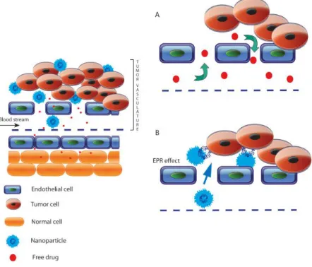
Alongside, in cancer chemotherapy, liposome as nanomedicine has a special interest, it enables the preferential delivery of drugs to the tumoral site, introducing the concept of Enhanced Permeability and Retention (EPR) effect, a particular phenomenon of solid tumors as a result of their anatomical and physiopathological characteristics that makes them different from normal tissues (Fig. 4). The endothelial cells from malignant blood vessels present larger gaps than normal blood vessel junctions (5–10 nm), that range from 100 nm to several hundred nanometres between them. In consequence, solid tumors exhibit selective extravasation and retention of drug-loaded liposomes. Moreover, these liposomes are cleared by the lymphatics in healthy tissues. However, in solid tumors most of these lymphatic vessels are collapsed and compressed, therefore the liposomes are selectively retained [37]. Ideally, nanocarriers, such as micelles or liposomes, by virtue of their size, can escape from the vasculature through the leaky endothelium overlying the tumor and then accumulating preferentially in solid tumors [38].
Figure 4. Schematic representation of enhanced permeability and retention (EPR) effect. Typical blood vessels from solid tumor contain pores of various sizes,
which allow nanoparticles and molecules of drug to enter into the interstitium of tumor tissue. (A) However, due to their small size, anticancer drugs can diffuse freely in and out of the tumor site, hence, only low levels of the drug accumulate in tumor. At the same time, significant concentrations of the agent are found in normal tissues. (B) The size of nanoparticles allows them to extravasate through gaps into the extravascular spaces and accumulate inside the tumor where the carrier releases the drug. (Bernabeu, Ezequiel, et al. International journal of pharmaceutics 526.1-2 (2017): 474-495).[39]
• Definition and structure of liposomes
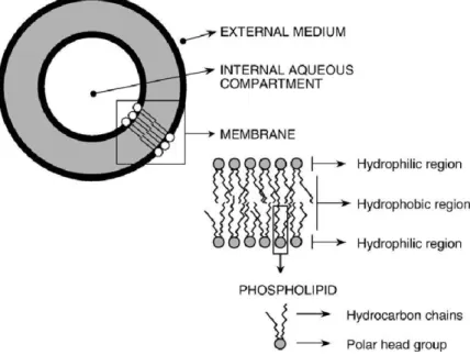
Liposomes are spherical vesicles composed of one or more lipid bilayers, involving an aqueous compartment (Figure 5). These are formed spontaneously when the lipids are dispersed in an aqueous medium by stirring, in turn giving rise to a population of vesicles which may reach a size range from dozens of nanometres to dozens of microns in diameter [40]. The lipid molecules possess head groups which are attracted to water molecules and organize them‐ selves in such a way as to point toward the aqueous cavity, whereas the hydrocarbon tails are repelled by the water molecules and point in the opposite direction.
The head groups of the inner layer point in the direction of the intravesicular fluid, with the tails pointing away from it. As such, the hydrocarbon tails of one layer point toward the hydrocarbon tails of the outer layer, in turn forming the normal bilipid membrane [35]. Once the liposomes have reached both the aqueous and lipid phases, they can encapsulate drugs with widely varying lipophilicities in the lipid bilayer, in the entrapped aqueous volume, or at the bilayer interface [41].
Figure 5. Basic structure and composition of liposomes. (Frézard, Frédéric, Neila M. Silva-Barcellos, and Robson AS dos Santos. Regulatory peptides 138.2-3 (2007): 59-65.) [42]
• Liposome Composition
Liposomes are composed mainly of natural and/or synthetic phospho- and sphingo- lipids with other membrane bilayer constituents, such as cholesterol and hydrophilic polymer conjugated lipids positioned randomly around each liposomal vesicle [43].
Phosphatidylcholine (PC; also known as lecithin) and phosphatidylethanolamine (PE) are the most common phospholipid found in both plants and animals and constitute the major structural parts of biologic membranes [44]. In contrast, the membranes of liposomes and other lipid- based drug delivery systems consist mostly of PC with little PE present [44]. This is because PE has the ability to form non-bilayer structures under physiologic conditions, destabilize membranes, and induce membrane fusion [45]. Other phospholipids, such as phosphatidylserine (PS), phosphatidylglycerol (PG), and phosphatidylinositol (PI), can also be used in the preparation of liposomes, depending on the desired liposomal characteristics [44].
Cholesterol is also an important component in the preparation of liposomes. Once it is incorporated into the liposomal membrane bilayer, cholesterol arranges itself among the phospholipid molecules with its hydroxyl group facing toward the water phase, whereas its tetracyclic ring inserts itself between the first few carbons of the fatty acyl chains into the hydrocarbon core of the membrane bilayer [44]. The incorporation of cholesterol into liposomes helps to decrease the fluidity of the liposomal membrane bilayer, reduce the permeability of water soluble molecules through the liposomal membrane, and improve the stability of the liposomal membrane in biologic fluids, such as blood and plasma [44]. In the absence of cholesterol, liposomes often interact with blood proteins, such as albumin,transferrin, macroglobulin, and high density lipoprotein [43,44,46]. These proteins tend to destabilize liposomes, and thus, decrease their capacity as a drug delivery system [46]. Although cholesterol has the ability to protect liposomes from being destabilized by blood proteins, the loss of liposomal phospholipids cannot be prevented completely [46].
Apart from cholesterol, a small fraction of polymers containing hydrophilic groups, especially polyethylene glycol (PEG), are at times conjugated to the surface of liposomes. PEG is often used for its stealth functions in nanoparticle formulations because it is a hydrophilic and flexible polymer [47]. The conjugation of PEG to the surface of the liposomal phospholipid bilayer reduces the interaction of liposomes with plasma proteins through steric hindrance [48,49]. As a result, this prevents plasma proteins, such as opsonin, from adsorbing to the surface of liposomes, which reduces opsonization and uptake of liposomes by the reticuloendothelial system (RES) [48]. The conjugation of PEG or PEGylation allows liposomes to circulate within the body for a longer period of time, extending their circulation half-life and, consequently, increasing the accumulation of liposomes within tumors [49].
• Methods of liposomes preparation
As mentioned before, liposomes are spontaneously formed when phospholipids are hydrated. Additional steps are often necessary to modify the size distribution and lamellarity of liposomes. Liposome preparation involves three major steps: vesicle formation, vesicle size reduction, and purification. Several preparation methods have been established based on the scale of the production and other considerations, such as drug encapsulation efficiency, the drug’s physicochemical characteristics, and the administration route.
The most commonly used methods for liposome preparation are lipid hydration and the replacement of organic solvents by an aqueous media (reverse-phase evaporation and organic-solvent injection). The lipid hydration followed by vortex or manual stirring, also known as Bangham’s method, consists of dissolving the lipids in a suitable organic solvent, such as chloroform or methanol [50]. This process is then followed by removing the solvent under reduced pressure, by rotary evaporation, until a thin film has been formed. After, the thin film is hydrated in an aqueous medium, above the phase-transition temperature, resulting in the formation of MLV liposomes (Figure 6). This is the simplest method of vesicle formation;
however, it is limited in use due to its low encapsulation ability [51].
All methods based on the replacement of an organic solvent by an aqueous media show that the solvents, whether miscible or immiscible with water, are replaced by an aqueous solution. First, the water-immiscible organic solution containing lipids is injected into the aqueous phase (reverse-phase method), or the stepwise addition of the organic phase (specifically, ethanol) is injected into the aqueous phase (organic solvent injection method), followed by the removal of the solvent. These methods are able to form liposomes with a high encapsulation percentage of both hydrophilic and lipophilic substances. Generally, the incorporation of lipophilic drugs is
performed through their co-dissolution with the lipids [51]. Hydrophilic drugs are dissolved in the aqueous medium, whereas amphiphilic drugs can be dissolved in both mediums. The processes of liposome preparation can result in the formation of large vesicles (MLV) with heterogeneous size distribution; therefore, it is important to calibrate the formulation using a vesicle size reduction method.
Figure 6. Representation of liposome production by lipid hydration followed by vortex or manual stirring. (Lopes, S.; Giuberti, C.; Rocha, T.; Ferreira, D.; Leite, E.; Oliveira, M. Conv. Innov. Approaches -b. 2013, 85–124) [52]
• Characterization of liposomes
The behaviour of liposomes in storage conditions and biological mediums is determined by specific factors, such as the size and surface charge of vesicles, chemical composition, membrane permeability, quantity of entrapped solutes, as well as the quality and purity of raw materials. Thus, it is of utmost importance to have as much information as possible regarding these parameters [40].
Bilayer constituents are responsible for the shelf-life; interactions with biological components, such as specific tissues, cells, and proteins; as well as the kinetics of the release of the entrapped drug in liposomes. The size of the liposomes influences it’s in vivo distribution, as this factor can determine the amount of time that the liposomes will remain in the bloodstream before being removed. By contrast, the surface charge of vesicles influences their physical stability due to the possible occurrence of fusion and/or aggregation phenomena [40]. Therefore, detailed chemical, physical, and physicochemical characterizations are important in an attempt to ensure the efficacy and stabilization of the liposome formulation.
Physical characterization consists of determining the size, surface charge, and lamellarity of the liposomes. As the performance of liposomes in vivo and physical stability strongly depend on the vesicle size, liposome size distribution should be determined during the preparation process and storage. On the other hand, the nature and density of the charge on the liposome surface are important parameters that influence the mechanism and extent of liposome-cell interaction. Furthermore, the retention of the superficial charge for long periods during storage contributes to the high physical stability of the formulation
References
1. Siegel, R.L.; Miller, K.D.; Jemal, A. Cancer statistics, 2018. CA. Cancer J. Clin.
2018, 68, 7–30, doi:10.3322/caac.21442.
2. Howlader N, Noone AM, Krapcho M, Miller D, Bishop K, Kosary CL, Yu M, Ruhl J, Tatalovich Z, Mariotto A, Lewis DR, Chen HS, Feuer EJ, C.K. (eds).
Cancer Statistics Review, 1975-2014 - SEER Statistics Available online:
https://seer.cancer.gov/archive/csr/1975_2014/ (accessed on Jan 23, 2019).
3. Bell, D.A. Origins and molecular pathology of ovarian cancer. Mod. Pathol.
2005, 18, S19–S32, doi:10.1038/modpathol.3800306.
4. American Cancer Society Cancer Facts & Figures 2018; Atlanta, 2018;
5. Lister-Sharp, D.; McDonagh, M.S.; Khan, K.S.; Kleijnen, J. A rapid and systematic review of the effectiveness and cost-effectiveness of the taxanes used in the treatment of advanced breast and ovarian cancer. Health Technol.
Assess. 2000, 4, 1–113.
6. Memarzadeh, S.; Berek, J.S. Advances in the management of epithelial ovarian cancer. J. Reprod. Med. 2001, 46, 621-9; discussion 629-30.
7. Rossof, A.H.; Talley, R.W.; Stephens, R.; Thigpen, T.; Samson, M.K.; Groppe, C.; Eyre, H.J.; Fisher, R. Phase II evaluation of cis- dichlorodiammineplatinum(II) in advanced malignancies of the genitourinary and gynecologic organs: a Southwest Oncology Group Study. Cancer Treat.
Rep. 63, 1557–64.
8. Thigpen, T.; Shingleton, H.; Homesley, H.; LaGasse, L.; Blessing, J. cis- Dichlorodiammineplatinum(II) in the treatment of gynecologic malignancies:
phase II trials by the Gynecologic Oncology Group. Cancer Treat. Rep. 63,
1549–55.
9. Decker, D.G.; Fleming, T.R.; Malkasian, G.D.; Webb, M.J.; Jeffries, J.A.;
Edmonson, J.H. Cyclophosphamide plus cis-platinum in combination:
treatment program for stage III or IV ovarian carcinoma. Obstet. Gynecol. 1982, 60, 481–7.
10. Piccart, M.J.; Bertelsen, K.; James, K.; Cassidy, J.; Mangioni, C.; Simonsen, E.; Stuart, G.; Kaye, S.; Vergote, I.; Blom, R.; et al. Randomized intergroup trial of cisplatin-paclitaxel versus cisplatin-cyclophosphamide in women with advanced epithelial ovarian cancer: three-year results. J. Natl. Cancer Inst. 2000, 92, 699–708.
11. McGuire, W.P.; Hoskins, W.J.; Brady, M.F.; Kucera, P.R.; Partridge, E.E.;
Look, K.Y.; Clarke-Pearson, D.L.; Davidson, M. Cyclophosphamide and Cisplatin Compared with Paclitaxel and Cisplatin in Patients with Stage III and Stage IV Ovarian Cancer. N. Engl. J. Med. 1996, 334, 1–6, doi:10.1056/NEJM199601043340101.
12. Wani, M.C.; Taylor, H.L.; Wall, M.E.; Coggon, P.; McPhail, A.T. Plant antitumor agents. VI. The isolation and structure of taxol, a novel antileukemic and antitumor agent from Taxus brevifolia. J. Am. Chem. Soc. 1971, 93, 2325–
7.
13. Wall, M.E.; Wani, M.C. Camptothecin and taxol: discovery to clinic--thirteenth Bruce F. Cain Memorial Award Lecture. Cancer Res. 1995, 55, 753–60.
14. SCHIFF, P.B.; FANT, J.; HORWITZ, S.B. Promotion of microtubule assembly in vitro by taxol. Nature 1979, 277, 665–667, doi:10.1038/277665a0.
15. Malawista, S.E.; Bensch, K.G. Human polymorphonuclear leukocytes:
demonstration of microtubules and effect of colchicine. Science 1967, 156, 521–2, doi:10.1126/SCIENCE.156.3774.521.
16. Kops, G.J.P.L.; Weaver, B.A.A.; Cleveland, D.W. On the road to cancer:
aneuploidy and the mitotic checkpoint. Nat. Rev. Cancer 2005, 5, 773–785, doi:10.1038/nrc1714.
17. Waters, J.C.; Chen, R.H.; Murray, A.W.; Salmon, E.D. Localization of Mad2 to kinetochores depends on microtubule attachment, not tension. J. Cell Biol.
1998, 141, 1181–91, doi:10.1083/JCB.141.5.1181.
18. Weiss, R.B.; Donehower, R.C.; Wiernik, P.H.; Ohnuma, T.; Gralla, R.J.; Trump, D.L.; Baker, J.R.; Van Echo, D.A.; Von Hoff, D.D.; Leyland-Jones, B.
Hypersensitivity reactions from taxol. J. Clin. Oncol. 1990, 8, 1263–8, doi:10.1200/JCO.1990.8.7.1263.
19. Gelderblom, H.; Verweij, J.; Nooter, K.; Sparreboom, A. Cremophor EL: the drawbacks and advantages of vehicle selection for drug formulation. Eur. J.
Cancer 2001, 37, 1590–1598, doi:10.1016/S0959-8049(01)00171-X.
20. Singla, A.K.; Garg, A.; Aggarwal, D. Paclitaxel and its formulations. Int. J.
Pharm. 2002, 235, 179–192, doi:10.1016/S0378-5173(01)00986-3.
21. Panchagnula, R. Pharmaceutical aspects of paclitaxel. Int. J. Pharm. 1998, 172, 1–15, doi:10.1016/S0378-5173(98)00188-4.
22. Gianni, L.; Kearns, C.M.; Giani, A.; Capri, G.; Viganó, L.; Lacatelli, A.;
Bonadonna, G.; Egorin, M.J. Nonlinear pharmacokinetics and metabolism of paclitaxel and its pharmacokinetic/pharmacodynamic relationships in humans.
J. Clin. Oncol. 1995, 13, 180–90, doi:10.1200/JCO.1995.13.1.180.
23. Wiernik, P.H.; Schwartz, E.L.; Strauman, J.J.; Dutcher, J.P.; Lipton, R.B.;
Paietta, E. Phase I clinical and pharmacokinetic study of taxol. Cancer Res.
1987, 47, 2486–93.
24. Rowinsky, E.K.; Donehower, R.C. Paclitaxel (Taxol). N. Engl. J. Med. 1995, 332, 1004–1014, doi:10.1056/NEJM199504133321507.
25. Sonnichsen, D.S.; Relling, M. V. Clinical Pharmacokinetics of Paclitaxel. Clin.
Pharmacokinet. 1994, 27, 256–269, doi:10.2165/00003088-199427040-00002.
26. Sparreboom, A.; van Zuylen, L.; Brouwer, E.; Loos, W.J.; de Bruijn, P.;
Gelderblom, H.; Pillay, M.; Nooter, K.; Stoter, G.; Verweij, J. Cremophor EL- mediated alteration of paclitaxel distribution in human blood: clinical pharmacokinetic implications. Cancer Res. 1999, 59, 1454–7.
27. Wall, M.E. Camptothecin and taxol: Discovery to clinic. Med. Res. Rev. 1998, 18, 299–314, doi:10.1002/(SICI)1098-1128(199809)18:5<299::AID- MED2>3.0.CO;2-O.
28. Marupudi, N.I.; Han, J.E.; Li, K.W.; Renard, V.M.; Tyler, B.M.; Brem, H.
Paclitaxel: a review of adverse toxicities and novel delivery strategies. Expert Opin. Drug Saf. 2007, 6, 609–621, doi:10.1517/14740338.6.5.609.
29. Scripture, C.; Figg, W.; Sparreboom, A. Peripheral Neuropathy Induced by Paclitaxel: Recent Insights and Future Perspectives. Curr. Neuropharmacol.
2006, 4, 165–172, doi:10.2174/157015906776359568.
30. Legha, S.S.; Tenney, D.M.; Krakoff, I.R. Phase I study of taxol using a 5-day intermittent schedule. J. Clin. Oncol. 1986, 4, 762–6, doi:10.1200/JCO.1986.4.5.762.
31. Shigehiro, T.; Kasai, T.; Murakami, M.; Sekhar, S.C.; Tominaga, Y.; Okada, M.; Kudoh, T.; Mizutani, A.; Murakami, H.; Salomon, D.S.; et al. Efficient drug
delivery of paclitaxel glycoside: A novel solubility gradient encapsulation into liposomes coupled with immunoliposomes preparation. PLoS One 2014, 9, doi:10.1371/journal.pone.0107976.
32. Mandai, T.; Okumoto, H.; Oshitari, T.; Nakanishi, K.; Mikuni, K.; Hara, K. ji;
Hara, K. zo; Iwatani, W.; Amano, T.; Nakamura, K.; et al. Synthesis and biological evaluation of water soluble taxoids bearing sugar moieties.
Heterocycles 2001, 54, 561–566.
33. Mandai, T.; Okumoto, H.; Oshitari, T.; Nakanishi, K.; Mikuni, K.; Hara, K.;
Hara, K. A Practical Synthetic Method for a- and b-Glycosyloxyacetic Acids.
Heterocycles 2000, 52, 129, doi:10.3987/COM-99-S43.
34. Malam, Y.; Loizidou, M.; Seifalian, A.M. Liposomes and nanoparticles:
nanosized vehicles for drug delivery in cancer. Trends Pharmacol. Sci. 2009, 30, 592–599, doi:10.1016/J.TIPS.2009.08.004.
35. Raffa, V.; Vittorio, O.; Riggio, C.; Cuschieri, A. Progress in nanotechnology for healthcare. Minim. Invasive Ther. Allied Technol. 2010, 19, 127–135, doi:10.3109/13645706.2010.481095.
36. Torchilin, V. Multifunctional and stimuli-sensitive pharmaceutical nanocarriers.
Eur. J. Pharm. Biopharm. 2009, 71, 431–444, doi:10.1016/J.EJPB.2008.09.026.
37. Wang, A.Z.; Langer, R.; Farokhzad, O.C. Nanoparticle Delivery of Cancer Drugs. Annu. Rev. Med. 2012, 63, 185–198, doi:10.1146/annurev-med-040210- 162544.
38. Maeda, H.; Bharate, G.Y.; Daruwalla, J. Polymeric drugs for efficient tumor- targeted drug delivery based on EPR-effect. Eur. J. Pharm. Biopharm. 2009, 71, 409–419, doi:10.1016/J.EJPB.2008.11.010.
39. Bernabeu, E.; Cagel, M.; Lagomarsino, E.; Moretton, M.; Chiappetta, D.A.
Paclitaxel: What has been done and the challenges remain ahead. Int. J. Pharm.
2017, 526, 474–495, doi:10.1016/J.IJPHARM.2017.05.016.
40. New, R.R.C. Characterization of liposomes. Liposomes: A practical; Oxford University Press: New York, 1990;
41. Lasic, D.D. Novel applications of liposomes. Trends Biotechnol. 1998, 16, 307–
321, doi:10.1016/S0167-7799(98)01220-7.
42. Frézard, F.; Silva-Barcellos, N.M.; Dos Santos, R.A.S. A novel approach based on nanotechnology for investigating the chronic actions of short-lived peptides in specific sites of the brain. 2006, doi:10.1016/j.regpep.2006.11.021.
43. Sharma, A.; Sharma, U.S. Liposomes in drug delivery: Progress and limitations.
Int. J. Pharm. 1997, 154, 123–140, doi:10.1016/S0378-5173(97)00135-X.
44. Vemuri, S.; Rhodes, C.. Preparation and characterization of liposomes as therapeutic delivery systems: a review. Pharm. Acta Helv. 1995, 70, 95–111, doi:10.1016/0031-6865(95)00010-7.
45. Ellens, H.; Bentz, J.; Szoka, F.C. Destabilization of phosphatidylethanolamine liposomes at the hexagonal phase transition temperature. Biochemistry 1986, 25, 285–294, doi:10.1021/bi00350a001.
46. Kirby, C.; Gregoriadis, G. The effect of the cholesterol content of small unilamellar liposomes on the fate of their lipid components in vivo. Life Sci.
1980, 27, 2223–2230, doi:10.1016/0024-3205(80)90388-4.
47. Bergström, K.; Osterberg, E.; Holmberg, K.; Hoffman, A.S.; Schuman, T.P.;
Kozlowski, A.; Harris, J.H. Effects of branching and molecular weight of surface-bound poly(ethylene oxide) on protein rejection. J. Biomater. Sci.
Polym. Ed. 1994, 6, 123–32.
48. Allen, T.M.; Ryan, J.L.; Papahadjopoulos, D. Gangliosides reduce leakage of aqueous-space markers from liposomes in the presence of human plasma.
Biochim. Biophys. Acta - Biomembr. 1985, 818, 205–210, doi:10.1016/0005- 2736(85)90571-1.
49. Allen, C.; Dos Santos, N.; Gallagher, R.; Chiu, G.N.C.; Shu, Y.; Li, W.M.;
Johnstone, S.A.; Janoff, A.S.; Mayer, L.D.; Webb, M.S.; et al. Controlling the physical behavior and biological performance of liposome formulations through use of surface grafted poly(ethylene glycol). Biosci. Rep. 2002, 22, 225–50, doi:10.1023/A:1020186505848.
50. Bangham, A.D. LIPID BILAYERS AND BIOMEMBRANES; 1972;
51. Wagner, A.; Vorauer-Uhl, K. Liposome technology for industrial purposes. J.
Drug Deliv. 2011, 2011, 591325, doi:10.1155/2011/591325
CHAPTER 2
CD44 As Target Receptor
CD44
CD44, the lymphocyte homing receptor, as described by [1], attracted considerable interest when it was first described that expression of splice variants of the molecule suffice to confer the metastatic phenotype to locally growing tumour cells [2]. There is ample evidence for the importance of CD44 expression in the progression of many tumour types, as well as for its expression on cancer-initiating cells (CICs; also known as cancer stem cells, CSCS)
CSCs have been defined as a selected population of tumour cells that grow on serial transplantation in xenogeneic models. These assays revealed that CSCs can self-renew, and that tumours derived from purified CSCs recapitulate the heterogeneous phenotype of the parental. tumour, reflecting the differentiation capacity of CSCs [3–5]. CSCs, like stem cells, are highly resistant to apoptosis and are thought to be essential for metastasis formation after prolonged dormancy [5].
Distinct from stem cells, CSCs might not be genetically stable [6] and can be heterogeneous [7] .
• The structure of CD44
CD44 was cloned in 1989 and identified as a member of the cartilage link protein family [8]. CD44 is an important receptor that binds hyaluronan (HA) [9].
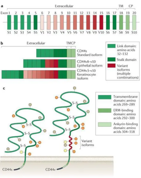
CD44 glycoproteins, which are encoded by a single gene [10], vary in size owing to N-glycosylation and O-glycosylation [8] and the insertion of alternatively spliced exon products in the extracellular domains of the molecule [10]. The smallest, standard or haematopoietic isoform (CD44s) is present on the membrane of most vertebrate cells [11]. CD44 has seven extracellular domains, a transmembrane domain and a cytoplasmic domain [12]. The cytoplasmic domain can be encoded
by exons 9 or 10 (Ref. [13]). Between domains 5 and 6, up to ten variant exon products can be inserted by alternative splicing [10] (Fig. 1).
The five amino-terminal exons encode a globular structure that contains two binding domains, the link domain (amino acids 32–132) and a basic motif outside the link domain (amino acids 150–158) to which HA binds [9]. Binding sites within the globular structure for collagen, laminin [14], fibronectin [15] and cell surface receptors such as CD62E and CD62L [16] have also been characterized. CD44 has two active binding sites for other glycosaminoglycans (GAGs)22. Four conserved cysteines are important for the stability of the extracellular domain, and two cysteines in the flanking region are important for correct folding of the link domain [17]. Between the N-terminal globular domain and the transmembrane domain there is a stretch of 46 amino acids that form a stalk-like structure [18]. This structure contains putative proteolytic cleavage sites and is heavily glycosylated [19]. This stretch can be enlarged by the insertion of variable exon products [20].
O-glycosylation and the cytoplasmic tail of CD44 can affect membrane subdomain localization and so influence the interaction of CD44 with HA [21].
Figure 1. (a) CD44 consists of several exons, some of which are constant region exons that are used in every CD44 mRNA and protein (green bars) and others are variant exons (red bars) that are used in the CD44 variant proteins and are selected by alternative splicing. (b) Examples of alternatively spliced CD44 proteins. (c)The CD44 protein is composed of an extracellular link domain, a stalk-like region in the extracellular domain close to the transmembrane region, where the variant exon products (red) are inserted, the transmembrane region (TM) and the cytoplasmic tail (CP). There are multiple sites for N-glycosylation (brown circles) and O- glycosylation (orange circles) and two active glycosaminoglycan (GAG)-binding sites (yellow circles), one located in the v3 exon product. The link module contains
cytoskeletal linker proteins, as well as for SRC kinases. ERM, ezrin, radixin and moesin; S–S, disulphide bonds. (Zöller, M. Nat. Rev. Cancer 2011, 11, 254–267) [22]
• CD44 as target receptor
As mentioned before CD44 has been identified as one of the most established and common biomarkers associated with CSCs that exhibit highly malignant and chemo-resistant properties in a variety of tumors. CD44 is also expressed on hemopoietic, epithelial, and neuronal cells at low levels and known to participate in a wide variety of cellular functions including regulation of cell adhesion, proliferation, migration, growth, survival, angiogenesis, differentiation, and matrix- cell signalling processes in collaboration with other cellular proteins [23]. Clinical studies have shown a positive correlation between expression of CD44 and the tumor biological behaviors such as prognosis, tumor genesis growth and metastasis [22]. These findings stress the importance of CD44 as a potential attractive receptor for therapeutic targeting especially in tumors over expressing CD44 [24].
Among different strategies, antibody-based cancer treatments represent the major anti-CSC approach [25]. It has been shown that anti-CD44 antibodies can inhibit tumor progression and induce differentiation or apoptosis of leukemic cells [26]. Other studies showed that activating anti-CD44 monoclonal antibody (MAb) induces CD44 signalling, which can cause apoptosis [27] and suppress leukemic CSCs [28]. Based on this concept, we cultured hybridoma Hermes-3 cells (HB- 9480) to produce anti-hCD44 MAb. Anti-hCD44 MAb functioned as ligand to target CD44. Furthermore, this ligand would be bound to liposome encapsulated gPTX targeting CD44 positive ovarian cancer cells.
Result and Discussion
Anti-hCD44 MAb obtained from conditioned medium culturing HB-9480 cells. HB-9480 cells were adapted from normal medium (RPMI 1640 medium supplemented 10% FBS) to PFHM II medium, a serum free medium to get optimum purity of produced MAb. We could confirm the purity of produced Anti-hCD44 MAb from Figure 2 B. Two bands were appeared on fraction 1-5, first band was about 50 kDa and second band was about 28 kDa. Those are showed as heavy chain and light chain from IgG2a of Anti-hCD44 MAb. Western blot result of conditioned medium of HB-9480 cells culture for 0 to 10 days in Figure 1A, confirmed Anti- hCD44 MAb as mouse IgG with comparable protein expression more than 100 ng in 10 days culture. Prior to the result, we cultured HB-9480 cells in the bioreactor for 10 days. We could obtain 4-5 mg/ml in 1.5 -2 ml protein from 50 ml conditioned medium cultured 10 days.
Next, we evaluated the protein expression levels of CD44 positive and negative cancer cell lines detected by obtained of Anti-hCD44 MAb by western blot assay.
This step could assessed whether our obtained MAb could detect or immuno-react to CD44 in cancer cells. We observed CD44 expression in the ovarian cancer cell lines SK-OV-3, OVCAR-3, and OVK18 (Figure 3A) and glioblastoma cell lines U251MG and U251MG-P1 (Figure 3B). Protein expression of CD44 detected by anti-hCD44 MAb, was found high in SK-OV-3, U251MG, U251MG-P1cells whereas was barely undetectable in OVCAR-3 and OVK18. The anti-hCD44 MAb showed the immunoreactive protein approximately at 85 kDa [29,30] which is attributed to the predominant isoform of CD44 known as the standard form. The presence of CD44+ within the SK-OV-3, U251MG, U251MG-P1cells were confirmed by flow cytometric analysis (Figure 3C). Since the expression of CD44 found consistent from western blot assay and flow cytometry assay, anti-hCD44
MAb was considered as suitable ligand to target CD44 in CD44 positive cancer cells.
Figure 2. Anti-hCD44 MAb preparation. (A) Western blot result of conditioned medium of HB-9480 cells culture for 0 to 10 days detected by goat anti-mouse IgG- HRP (Santa Cruz Biotechnology Inc., CA, USA), 100 ng of Mouse IgG as positive control. (B) CBB staining of SDS PAGE of anti-hCD44 MAb eluted from protein A column.
Figure 3. Anti-hCD44 MAb could detect CD44 protein expression in ovarian cancer and glioblastoma cells. Western blot analysis of human ovary cancer derived cells (A) and (B) human glioblastoma derived cells probed with anti-hCD44 MAb and human beta-actin antibody. (C) Cells contain CD44+ population analyzed by flow cytometry by staining for CD44. The margins CD44 for each cell line were set up by non-stained cells as the negative control shown at the bottom of each analysis.
After conjugation to liposome encapsulated gPTX, the expression of anti- hCD44 confirmed in Figure 4. Further immunoliposome based drug delivery to CD44 positive cancer cells could be confirmed using anti-hCD44 MAb obtained as the ligand.
Figure 4. Dot blotting analysis of liposomes conjugated to ligands. Liposomes containing approximately 6 µg of gPTX in 2 µL were blotted onto PVDF membrane and probed with peroxidase-labelled Protein A (KPL, USA) (red dashed circle indicate the positions of dots). The immunoreactivity indicates that anti-hCD44 MAb was conjugated to liposome.
Materials and Method
• Materials
RPMI 1640 medium and DMEM were obtained from Sigma-Aldrich (St Louis, MO, USA).
• Cells Culture and Experimental Animal
The human ovarian cancer cell lines SK-OV-3 cells (HTB-77, ATCC, VA), OVK18 cells (TKG 0323, Cell Bank, Tohoku University, Sendai, Japan), and U251MG-P1 was isolated from a xenograft tumor of human glioblastoma cell line U251MG cells in mouse were cultured in DMEM medium supplemented with 10% fetal bovine serum (FBS) (Thermo Fisher Scientific, Waltham, MA, USA), containing 100 U/mL penicillin (Nacalai tesque, Kyoto, Japan), and 100 μg/mL streptomycin and OVCAR-3 (HTB-161, ATCC, VA) were cultured in RPMI 1640 medium supplemented with 10% FBS containing 100 U/mL penicillin, and 100 μg/mL streptomycin.
• Preparation of anti-hCD44 MAb
To produce anti-hCD44 MAb, hybridoma Hermes-3 cells (HB-9480, ATCC, VA) cells were cultured using a bioreactor, miniPERM (SARSTEDT, Nümbrecht, Germany). Twenty million of the cells were suspended in 50 mL of PFHM-II (Gibco, NY, USA) medium and were transferred into production module. The production module was connected to nutrient module containing 350 mL of PFHM-II. The bioreactor was rotated for 10 days at 37˚C in 5% CO2. The medium in production module was then collected and centrifuged at 150 xg for 5 min at 4˚C to remove the cells. The supernatant was re-centrifuged at 10,000 xg for 5 min at 4˚C. The supernatant was then passed through 0.20 µm filter (Sartorius Stedim Biotech GmBH, Geottingen, Germany) to completely remove cell debris. Anti-
hCD44 MAb was then purified as follows. The supernatant was passed through a 0.5 mL of Protein A Sepharose (GE Healthcare, Uppsala, Sweden) equilibrated with PBS. After washing the column with PBS, anti-hCD44 MAb was eluted using 0.1 M sodium-acetic buffer at pH 2.6. Five hundred µL of each fraction was readily neutralized with 10 µL of 2 M sodium phosphate buffer, pH 8.0. The fraction containing anti-hCD44 MAb was detected by western blotting using polyclonal anti mouse IgG HRP (DAKO, Denmark) and the protein concentration was determined using a BCA assay kit (Pierce Biotechnology, Rockford, IL, USA).
• Expression of CD44 in Ovarian Cancer Cells line.
o Western blotting
Proteins following the SDS-PAGE were transferred to polyvinylidene difluoride (PVDF) membranes (Merck Millipore, Burlington, MA, USA). To detect CD44 epitope, the blot was probed using anti-hCD44 MAb (2 mg/mL, 1:2000) followed by polyclonal anti-mouse IgG HRP (1:4000) (DAKO, Denmark).
Quantitative assessment of relative intensity of the blots were analyzed using ImageJ. The actin immunoreact to anti-beta actin Rabbit MAb (1:1000, 4970S, Cell Signalling Technology, Inc., Beverly, MA, USA) was used as a normalization control.
o 3.4.2 Flow cytometry analysis
SK-OV-3, OVCAR-3, OVK18, U251MG, U251MG-P1 cells were harvested at logarithmic growth phase, followed by being re-suspended in 100ul PBS, stained with APC labelled mouse anti-human CD44 MAb (BD Science Pharmingen, San Diego, CA, USA) and analyzed by BD AccuriTM C6 plus flow cytometer (Becton
& Dickinson, Franklin Lakes, NJ, USA). Data of each experiment was analyzed using FlowJo software (FlowJo, LLC, Ashland, OR, USA).
References
1. Gallatin, W.M.; Weissman, I.L.; Butcher, E.C. A cell-surface molecule involved in organ-specific homing of lymphocytes. Nature 304, 30–4.
2. Günthert, U.; Hofmann, M.; Rudy, W.; Reber, S.; Zöller, M.; Hauβmann, I.;
Matzku, S.; Wenzel, A.; Ponta, H.; Herrlich, P. A new variant of glycoprotein CD44 confers metastatic potential to rat carcinoma cells. Cell 1991, 65, 13–24, doi:10.1016/0092-8674(91)90403-L.
3. Fábián, Á.; Barok, M.; Vereb, G.; Szöllősi, J. Die hard: Are cancer stem cells the Bruce Willises of tumor biology? Cytom. Part A 2009, 75A, 67–74, doi:10.1002/cyto.a.20690.
4. Sales, K.M.; Winslet, M.C.; Seifalian, A.M. Stem Cells and Cancer: An Overview. Stem Cell Rev. 2007, 3, 249–255, doi:10.1007/s12015-007-9002-0.
5. Allan, A.L.; Vantyghem, S.A.; Tuck, A.B.; Chambers, A.F. Tumor Dormancy and Cancer Stem Cells: Implications for the Biology and Treatment of Breast Cancer Metastasis. Breast Dis. 2007, 26, 87–98, doi:10.3233/BD-2007-26108.
6. Conway, A.E.; Lindgren, A.; Galic, Z.; Pyle, A.D.; Wu, H.; Zack, J.A.;
Pelligrini, M.; Teitell, M.A.; Clark, A.T. A Self-Renewal Program Controls the Expansion of Genetically Unstable Cancer Stem Cells in Pluripotent Stem Cell- Derived Tumors. Stem Cells 2009, 27, 18–28, doi:10.1634/stemcells.2008-0529.
7. Adams, J.M.; Strasser, A. Is Tumor Growth Sustained by Rare Cancer Stem Cells or Dominant Clones? Cancer Res. 2008, 68, 4018–4021, doi:10.1158/0008-5472.CAN-07-6334.
8. Stamenkovic, I.; Amiot, M.; Pesando, J.M.; Seed, B. A lymphocyte molecule implicated in lymph node homing is a member of the cartilage link protein
family. Cell 1989, 56, 1057–1062, doi:10.1016/0092-8674(89)90638-7.
9. Aruffo, A.; Stamenkovic, I.; Melnick, M.; Underhill, C.B.; Seed, B. CD44 is the principal cell surface receptor for hyaluronate. Cell 1990, 61, 1303–1313, doi:10.1016/0092-8674(90)90694-A.
10. Screaton, G.R.; Bell, M. V; Jackson, D.G.; Cornelis, F.B.; Gerth, U.; Bell, J.I.
Genomic structure of DNA encoding the lymphocyte homing receptor CD44 reveals at least 12 alternatively spliced exons. Proc. Natl. Acad. Sci. U. S. A.
1992, 89, 12160–4, doi:10.1073/PNAS.89.24.12160.
11. Naor, D.; Wallach-Dayan, S.B.; Zahalka, M.A.; Sionov, R.V. Involvement of CD44, a molecule with a thousand faces, in cancer dissemination. Semin.
Cancer Biol. 2008, 18, 260–267, doi:10.1016/J.SEMCANCER.2008.03.015.
12. Idzerda, R.L.; Carter, W.G.; Nottenburg, C.; Wayner, E.A.; Gallatin, W.M.; St John, T. Isolation and DNA sequence of a cDNA clone encoding a lymphocyte adhesion receptor for high endothelium. Proc. Natl. Acad. Sci. U. S. A. 1989, 86, 4659–63, doi:10.1073/PNAS.86.12.4659.
13. Goldstein, L.A.; Butcher, E.C. Identification of mRNA that encodes an alternative form of H-CAM(CD44) in lymphoid and nonlymphoid tissues.
Immunogenetics 1990, 32, 389–97.
14. Ishii, S.; Ford, R.; Thomas, P.; Nachman, A.; Steele, G.; Jessup, J.M. CD44 participates in the adhesion of human colorectal carcinoma cells to laminin and type IV collagen. Surg. Oncol. 1993, 2, 255–64.
15. Jalkanen, S.; Jalkanen, M. Lymphocyte CD44 binds the COOH-terminal heparin-binding domain of fibronectin. J. Cell Biol. 1992, 116, 817–25.
16. Konstantopoulos, K.; Thomas, S.N. Cancer Cells in Transit: The Vascular