U
lcerative colitis is an inflammatory bowel disease that causes inflammation in the digestive tract.As the name “ulcerative colitis” implies, it has long been believed that the inflammatory response is local- ized in the large intestine. However, studies conducted over the last 20 years have led to advances in our under- standing of the extent of the disease. Macroscopic and microscopic involvement of the stomach and duode- num in ulcerative colitis patients has been reported by
several research groups [1-7] as “gastroduodenitis asso- ciated with ulcerative colitis,” “ulcerative gastroduode- nal lesions,” or “ulcerative colitis-associated upper gas- trointestinal inflammation.” These lesions are reportedly observed as friable mucosa, granular mucosa, or mul- tiple aphthae during esophagogastroduodenoscopy [7].
Due to the increasing frequency of ulcerative colitis in developed countries since the mid-20th century, ulcerative colitis patients have been treated at various institutions ranging from tertiary care centers and com-
CopyrightⒸ 2018 by Okayama University Medical School.
http ://escholarship.lib.okayama-u.ac.jp/amo/
Original Article
Evaluation of the Upper Gastrointestinal Tract in Ulcerative Colitis Patients
Ryo Katoa, Masaya Iwamuroa*, Sakiko Hiraokaa, Shiho Takashimaa, Toshihiro Inokuchia, Masahiro Takaharaa, Yoshitaka Kondob,
Takehiro Tanakac, and Hiroyuki Okadaa
Departments of aGastroenterology and Hepatology, and bGastroenterological Surgery, Okayama University Graduate School of Medicine, Dentistry and Pharmaceutical Sciences,
cDepartment of Pathology, Okayama University Hospital, Okayama 700-8558, Japan
To analyze the clinical characteristics of patients with ulcerative colitis who have upper gastrointestinal lesions, we retrospectively reviewed the data of 216 patients with ulcerative colitis who underwent esophagogastroduo- denoscopy at our institute in April 2008-March 2016. We investigated the endoscopic features and compared the clinical characteristics between the patients with and without upper gastrointestinal lesions. Forty-two patients (19.4%) had upper gastrointestinal lesions, including multiple erosions (n=18), bamboo joint-like appearance (n=17), mucosa with white spots (n=4), friable mucosa (n=2), ulcer (n=1), and purulent depos- its within the mucosa (n=1) in the stomach and/or duodenum. Compared to the patients without upper gas- trointestinal lesions, those with upper gastrointestinal lesions showed significantly more frequent extraintesti- nal manifestations (19.0% vs. 8.0%, p<0.05) and a significant history of colectomy (33.3% vs. 12.1%, p<0.01).
There were no significant differences with regard to the sex ratio, age at esophagogastroduodenoscopy, gastro- intestinal symptoms, time since the diagnosis of ulcerative colitis, type of colitis at the initial diagnosis of ulcerative colitis, or gastric atrophy between the groups. In conclusion, gastroduodenal lesions were identified in 19.4% of the patients with ulcerative colitis. Esophagogastroduodenoscopy is particularly recommended for ulcerative colitis patients who show extraintestinal manifestations and for those who have undergone a colec- tomy.
Key words: ulcerative colitis, esophagogastroduodenoscopy, gastritis
Received June 6, 2017 ; accepted October 30, 2017.
*Corresponding author. Phone : +81-86-235-7218; Fax : +81-86-225-5991
E-mail : [email protected] (M. Iwamuro) Conflict of Interest Disclosures: No potential conflict of interest relevant to this article was reported.
munity hospitals to clinics. One of the concerns in treating ulcerative colitis is that the upper gastrointesti- nal involvement may be overlooked because of the lack of recognition of the gastroduodenal lesions by the attending physician. In addition, since the clinical backgrounds of patients with ulcerative colitis-associ- ated upper gastrointestinal inflammation have not been fully revealed, no criteria have been established to indi- cate which patients should undergo esophagogastrodu- odenoscopy.
Patients with ulcerative colitis-associated upper gas- trointestinal inflammation may require specific treat- ment for the gastroduodenal lesions. Hence, recogni- tion and detection of the upper gastrointestinal lesions are important. Here we describe 2 cases of ulcerative colitis and summarize the results of our investigation of the prevalence of upper gastrointestinal involvement detected by esophagogastroduodenoscopy. We also analyzed the macroscopic and microscopic features of the upper gastrointestinal lesions and patient character- istics.
Methods
A database search performed at the Department of Gastroenterology and Hepatology of our institute iden- tified 404 patients with ulcerative colitis who were treated at our institute between April 2008 and March 2016. Among them, 216 patients (53.5%) underwent one or more esophagogastroduodenoscopy examina- tions at our facility. Therefore, a total of 216 patients were enrolled in this study.
The presence or absence of esophageal, gastric, and duodenal lesions was macroscopically defined accord- ing to the results of the esophagogastroduodenoscopy examination(s). Atrophic gastritis, superficial gastritis, verrucous gastritis, single erosion, xanthoma, mucosal redness, candida esophagitis, esophageal hiatal hernia, hyperplastic polyp, fundic gland polyp, ulcer scar, reflux esophagitis, ectopic gastric mucosa, and varices were excluded from this study, since these lesions likely occur in no relation with ulcerative colitis. Verrucous gastritis is characterized by mucosal elevation with a central depression, which is usually multiple and arranged in chains or clusters in the antrum [8]. For the purposes of this paper, we defined all other lesions as upper gastrointestinal lesions because there is no con- sensus with regard to the definition of upper gastroin-
testinal involvement in ulcerative colitis to date [1].
Based on the presence of the upper gastrointestinal lesions, we divided the 216 patients into 2 groups: the patients with positive upper gastrointestinal lesions (the Lesion group) and the patients without upper gastroin- testinal lesions (the No-Lesions group). We retrospec- tively reviewed the patients’ endoscopic, radiological, biological, and pathological examination findings obtained from the clinical records of the patients. The patient information in the 2 groups was analyzed and compared with regard to sex, age at the esophagogas- troduodenoscopy, time (years) since the diagnosis of ulcerative colitis, the presence or absence of gastroin- testinal symptoms, the type of colitis (proctitis, procto- sigmoiditis, left-sided colitis, and pan-ulcerative coli- tis) at the initial diagnosis of ulcerative colitis, the presence or absence of extraintestinal manifestations, history of surgery for ulcerative colitis, and gastric atrophy. Two board-certified endoscopists blindly assessed the grade of gastric atrophy according to the Kimura and Takemoto grading classification [9]. In cases of a difference of opinion between 2 endoscopists, the grade was determined after discussion.
Pathological features were analyzed when biopsy sampling was performed during an esophagogastrodu- odenoscopy examination. Lesions with focal enhanced gastritis or with aggregates of neutrophils and sloughed epithelial cells within a partially ruptured gastrointesti- nal gland that resemble crypt abscesses were defined as ulcerative colitis-associated upper gastrointestinal inflam- mation [10].
Our statistical analyses of the groups were per- formed with t-tests, chi-square tests, and F-tests using JMP 12.0.1 software (SAS, Cary, NC, USA). P-values
<0.05 were considered significant.
The study design was approved by the Ethics Com- mittee of Okayama University Hospital (No. 1606-018), and it adhered to the Declaration of Helsinki.
Results
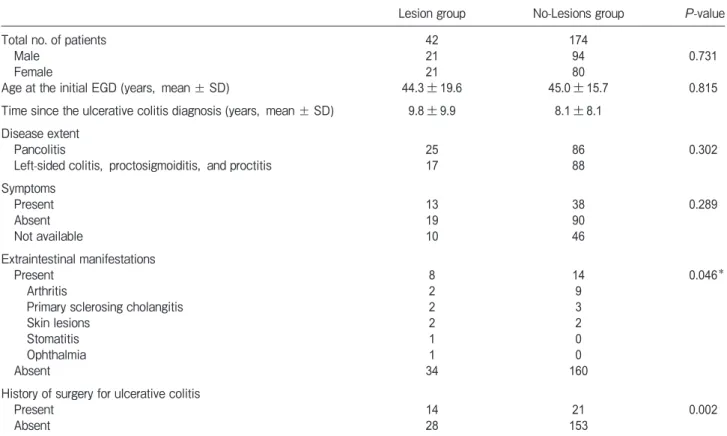
The patient backgrounds are summarized in Table 1.
Among the 216 patients who underwent esophagogas- troduodenoscopy, upper gastrointestinal lesions were identified in 42 (21 men and 21 women) patients (19.4%); these patients were classified as the Lesion group. The No-Lesions group was comprised of 174 patients (94 men, 80 women).
A comparison of the backgrounds of the patients in the 2 groups revealed that extraintestinal manifesta- tions were observed significantly more frequently in the Lesion group (19.0%) compared to the No-Lesions group (8.0%, p<0.05). The extraintestinal manifesta- tions in the Lesion group included arthritis (n=2), pri- mary sclerosing cholangitis (n=2), skin lesions (pyo- derma gangrenosum, n=2), stomatitis (n=1), and ophthalmia (scleritis, n=1). The extraintestinal mani- festations in the No-Lesions group were arthritis (n=9), primary sclerosing cholangitis (n=3), and skin lesions (n=2; one patient had erythema nodosum and the other had both pyoderma gangrenosum and erythema nodosum).
In addition, 33.3% of the patients in the Lesion group underwent surgery for ulcerative colitis, at a sig- nificantly higher rate than that in the No-Lesions group (12.1%, p<0.01). There were no significant differences between patients in the Lesion and No-Lesions groups with regard to sex ratio (males: 50.0% vs. 54.0%), age
at esophagogastroduodenoscopy (mean ± SD: 44.3±19.6 vs. 45.0±15.7 years), gastrointestinal symptoms (posi- tive: 40.6% vs. 29.7%), time since the diagnosis of ulcerative colitis (mean ± SD: 9.8±9.9 vs. 8.1±8.1 years), type of colitis at the initial diagnosis of ulcerative colitis (ratio of pan-ulcerative colitis: 59.5% vs. 49.4%), and gastric atrophy (ratio of C3, O1, O2, and O3: 14.4%
vs. 13.8%), respectively.
Macroscopic features of the upper gastrointestinal lesions were classified as multiple erosions (n=18), bamboo joint-like appearance (n=16), mucosa with white spots (n=4), friable mucosa (n=2), ulcer (n=1), and purulent deposits within the mucosa (n=1). The typical images of each endoscopic feature are shown in Fig.1. Table 2 shows the distribution of these features, which were observed in the stomach and duodenum.
No patient had macroscopic lesions in the esophagus.
One patient had mucosa with white spots along with a friable mucosa in the stomach. In 2 patients, the mac- roscopic features changed during the clinical course,
Table 1 Characteristics of the study subjects, 216 ulcerative colitis patients
Lesion group No-Lesions group P-value
Total no. of patients 42 174
Male 21 94 0.731
Female 21 80
Age at the initial EGD (years, mean ± SD) 44.3±19.6 45.0±15.7 0.815
Time since the ulcerative colitis diagnosis (years, mean ± SD) 9.8±9.9 8.1±8.1 Disease extent
Pancolitis 25 86 0.302
Left-sided colitis, proctosigmoiditis, and proctitis 17 88 Symptoms
Present 13 38 0.289
Absent 19 90
Not available 10 46
Extraintestinal manifestations
Present 8 14 0.046*
Arthritis 2 9
Primary sclerosing cholangitis 2 3
Skin lesions 2 2
Stomatitis 1 0
Ophthalmia 1 0
Absent 34 160
History of surgery for ulcerative colitis
Present 14 21 0.002
Absent 28 153
Lesion group: the patients with ulcerative colitis who had upper gastrointestinal lesions.
No-Lesions group: the patients without upper gastrointestinal lesions. *Present vs. absent.
EGD, esophagogastroduodenoscopy.
and are described below. All other patients showed a single type of macroscopic feature on esophagogastro- duodenoscopy. The detailed medication histories of the patients in the Lesion group were further investigated, revealing that during esophagogastroduodenoscopy, nonsteroidal anti-inflammatory drugs (NSAIDs) were prescribed for only one patient in whom an ulcer was identified.
Biopsy sampling was performed in 18 patients from the Lesion group. Focally enhanced gastritis was iden- tified in 3 patients; 2 patients had multiple erosions and
the third patient had bamboo joint-like appearance. No crypt abscess was found in any of the biopsied speci- mens. Consequently, the 3 patients were diagnosed with ulcerative colitis-associated upper gastrointestinal inflammation [9].
Among the 15 patients without focally enhanced gastritis, nonspecific inflammation within the mucosa and submucosa was identified in 11 patients, and an intact gastric mucosa lacking inflammation was observed in three patients. In these three patients, biopsy sampling was performed for the bamboo joint-
Table 2 Endoscopic features of the upper gastrointestinal lesions
Macroscopic feature No. of patients Stomach Duodenum Stomach and duodenum
Multiple erosions 18 13† 4* 1
Bamboo joint-like appearance 17 17* 0 0
Mucosa with white spots 4 2 2 1
Friable mucosa 2 1† 0 1
Ulcer 1 0 1 0
Purulent deposits within the mucosa 1 1 0 0
*Patient 1 presented with multiple erosions in the duodenum during the initial esophagogastroduodenoscopy (EGD), whereas the second EGD showed a bamboo joint-like appearance in the stomach.
†In Patient 2, gastric lesions were observed as mucosa with white spots at the initial EGD. The gastric lesions changed into multiple ero- sions at the second EGD.
A B C
D E F
Fig. 1 Typical images of macroscopic features of the upper gastrointestinal lesions in patients with ulcerative colitis. Esophago- gastroduodenoscopy revealed gastric multiple erosions (A), a bamboo joint-like appearance in the stomach (B), duodenal mucosa with white spots (C), duodenal friable mucosa (D), a duodenal ulcer (E), and purulent deposits within the gastric mucosa (F).
like appearance. In the remaining patient, since the samples were obtained from the purulent deposits within the mucosa, the pathologic features could not be evaluated due to the small volume of the sample.
Based on the results of an esophagogastroduode- noscopy examination, specific treatment for the upper gastrointestinal lesions was initiated in 2 patients. The clinical courses of these 2 patients are as follows.
Case 1. A 26-year-old Japanese woman was diag- nosed with ulcerative colitis. When she was 27 years old, subtotal proctocolectomy was performed based on the deterioration of colitis. At 32 years of age, the patient underwent esophagogastroduodenoscopy for the investigation of epigastralgia, which revealed multiple erosions in the duodenum (Fig.2A,B). Powdered mesalazine was administered to treat the duodenal lesions. When the patient was 35 years old, esophago- gastroduodenoscopy revealed resolution of the duode- nal erosions (Fig.2C) and a bamboo joint-like appear- ance was noted in the gastric cardia (Fig.2D); this appearance had not been identified during the initial esophagogastroduodenoscopy.
Case 2. A 14-year-old Japanese boy was diag- nosed with ulcerative colitis. He underwent subtotal proctocolectomy at 17 years of age. Esophagogastro- duodenoscopy performed at 19 years revealed white
spots in the gastric body (Fig.3A), whereas the gastric antrum seemed to be intact (Fig.3B). The patient was treated with powdered mesalazine. Esophagogastro- duodenoscopy performed 1 year later showed reddish mucosa in the gastric body (Fig.3C) and multiple ero- sions in the gastric antrum (Fig.3D,E).
Discussion
In this study, 42 of the 216 patients (19.4%) who underwent esophagogastroduodenoscopy had upper gastrointestinal lesions. Hori et al. prospectively per- formed esophagogastroduodenoscopy with biopsies on 250 patients with ulcerative colitis. They identified ulcerative colitis-associated upper gastrointestinal lesions, which were defined as friable mucosa, granular mucosa, or multiple aphthae, in 7.6% of the patients [7]. Abnormalities in macroscopic findings have been identified in 13-53% of patients with ulcerative colitis [7]. Horjus Talabur Horje et al. reported that no patient with ulcerative colitis had upper gastrointestinal lesions, whereas macroscopic abnormalities were found in 41%
of patients with Crohn’s disease during esophagogastro- duodenoscopy [11]. Such diversity of prevalence likely reflects the difference in the definition of upper gastro- intestinal lesions among the different studies. Several
A B
C D
Fig. 2 Endoscopy images of Patient 1.
Multiple erosions were initially observed in the duodenum (A,B). After the administration of mesalazine powder, the duodenal erosions resolved (C). A bamboo joint-like appearance appeared in the gastric cardia (D).
studies have revealed that a significant proportion of patients have macroscopic lesions in the upper gastro- intestinal tract, indicating the importance of esophago- gastroduodenoscopy examinations in the management of ulcerative colitis.
As we noted above, there is no consensus regarding the definition of upper gastrointestinal involvement in ulcerative colitis to date. In one prospective study, granular, erythematous mucosa, erosions, ulcers, nodular lesions and strictures were assessed as upper gastrointestinal lesions [11]. In a retrospective study, granular mucosa, erosion, mucosal friability, bleeding, and ulcers resembling colonic lesions, were classified as upper gastrointestinal lesions [12]. In the majority of studies, as well as in our present investigation, lesions were defined based on the macroscopic features.
Lin et al. pathologically investigated the upper gas- trointestinal mucosa and defined ulcerative colitis-asso- ciated upper gastrointestinal inflammation as lesions with focal enhanced gastritis or with aggregates of neu- trophils and sloughed epithelial cells within a partially ruptured gastrointestinal gland, resembling a crypt abscess [10]. This variation in the definitions reflects the difficulty of determining the lesions that actually occur
in relation to ulcerative colitis. In the present study, we included all mucosal alterations in the upper gastroin- testinal tract other than atrophic gastritis, superficial gastritis, verrucous gastritis, single erosion, xanthoma, mucosal redness, candida esophagitis, esophageal hia- tal hernia, hyperplastic polyp, fundic gland polyp, ulcer scar, reflux esophagitis, ectopic gastric mucosa, and varices. As a result, we identified multiple ero- sions, mucosa with white spots, friable mucosa, bam- boo joint-like appearance, purulent deposits within the mucosa, and ulcers. Among these, ulcer formation might occur as an adverse effect due to NSAID use. In contrast, mucosa with white spots and friable mucosa have been reported as findings specific to ulcerative colitis [7]. Multiple erosions are also reportedly indica- tive lesions of upper gastrointestinal involvement with ulcerative colitis, provided other disorders such as viral infections, collagen, allergic, autoimmune, and Crohn’s disease are excluded [7]. The significance of purulent deposits, which we observed in one patient, remains unknown, since the pathological analysis of the biop- sied specimen was not possible due to the small sample size.
The bamboo joint-like appearance of the gastric
A B
C D E
Fig. 3 Endoscopy images of Patient 2. White spots were observed in the gastric body (A), whereas the gastric antrum was intact (B).
The patient was treated with powdered mesalazine. The second esophagogastroduodenoscopy revealed reddish mucosa in the gastric body (C) and multiple erosions in the antrum (D,E).
mucosa (i.e., swollen longitudinal folds transversed by erosive fissures or linear furrows that are most fre- quently found at the gastric body and cardia) is a typical manifestation of Crohn’s disease. It is well known that 44-65% of patients with Crohn’s disease show a bamboo joint-like appearance of the gastric mucosa [13,14].
Recent reports on endoscopic gastroduodenal findings in patients with inflammatory bowel disease revealed that patients with ulcerative colitis have this feature [11,12,14]. In the present study, the bamboo joint-like appearance was found in 17 of the 216 patients (7.9%).
Our finding reinforces the notion that the bamboo joint-like appearance of the gastric mucosa can be seen in patients with ulcerative colitis as well as in those with Crohn’s disease.
In our study, extraintestinal manifestations were more frequently observed in the patients with positive upper gastrointestinal lesions. Moreover, the Lesion group included a higher percentage of patients who had undergone colectomy. The reported risks for develop- ing upper gastrointestinal lesions include more exten- sive colitis, a lower dose of prednisolone, the presence of pouchitis, post-colectomy status, and a longer post- operative period [7,11,16-22]. Although the precise mechanisms underlying the upper gastrointestinal lesions are unclear, several authors have speculated that immune response to the as-yet unidentified factors associated with ulcerative colitis may also be the cause, since patient characteristics that are suggestive of severe inflammation are reportedly related to gastroduodenal manifestations [1,2,5,23].
In the present study, the extent of colitis was not a significant factor in the development of upper gastroin- testinal lesions. However, the type of colitis was defined at the initial diagnosis of ulcerative colitis, and infor- mation regarding the disease extent at the time of esophagogastroduodenoscopy examination was not available, since esophagogastroduodenoscopy and colo- noscopy were performed at different time points in most of the patients. Although an evaluation of disease extent and activity may be useful to predict the presence of upper gastrointestinal lesions, further studies are required in this regard. Based on the results of the pres- ent study, we speculate that esophagogastroduodenos- copy is particularly recommended for patients with ulcerative colitis who show extraintestinal manifesta- tions and in those who have undergone colectomy.
Among the 188 patients with ulcerative colitis who had
never undergone esophagogastroduodenoscopy, 21 (11.2%) were in post-colectomy status. We thus intend to perform esophagogastroduodenoscopies in this patient population.
Pathologically, focally enhanced gastritis was identi- fied in 3 patients. This feature represents intense focal inflammation characterized by localized collections of lymphocytes, neutrophils, and macrophages in the lamina propria [10]. Focally enhanced gastritis was originally described in patients with Crohn’s disease by Oberhuber et al. in 1997 [24]. To date, this is the most common upper gastrointestinal inflammation in ulcer- ative colitis. Lin et al. reported that 29% of patients with ulcerative colitis had focally enhanced gastritis, whereas 9% of control patients had this feature. A 12-29% prev- alence of focally enhanced gastritis was reported in ulcerative colitis [10, 25-27]. Crypt abscess, a patho- logical hallmark of the large intestine in ulcerative coli- tis, can be identified in the upper gastrointestinal tract as well. Hori et al. reported that crypt abscesses were observed significantly more frequently in the gastrodu- odenal mucosa of patients with ulcerative colitis than in controls [7]. However, none of the patients in the pres- ent study had a crypt abscess. Nonspecific inflamma- tion within the mucosa and submucosa was also identi- fied in 11 patients without focally enhanced gastritis.
This observation is consistent with a report that non- specific inflammation in the upper gastrointestinal tract is observed in 60-90% of patients with ulcerative colitis [5].Upper gastrointestinal lesions in ulcerative colitis may be treated with steroids, leukocytapheresis, and/or peroral mesalazine powder [12]. Immunosuppression with cyclosporine, tacrolimus, or infliximab is likely to be the treatment of choice for upper gastrointestinal inflammation as well. In the present study, mesalazine powder was administered to 2 patients, and the macro- scopic features of the upper gastrointestinal lesions were found to have changed after the administration of this drug. One possible explanation is that mesalazine pow- der contributed to the altered macroscopic features.
Another hypothesis is that the patients’ morphology simply changed with time. Since the change in mor- phology was observed in only 2 patients, further stud- ies are required to determine the exact factor(s) that affect the morphology.
Our study has several limitations. It was a retro- spective analysis. Since esophagogastroduodenoscopy
was indicated in each patient by his or her attending physician, the timing of the examination during the course of ulcerative colitis varied. At our institution, we perform esophagogastroduodenoscopy for almost all patients with ulcerative colitis who have symptoms related to the upper gastrointestinal tract. Moreover, we usually recommend that patients with ulcerative colitis undergo esophagogastroduodenoscopy at least once. However, 188 of the 404 patients with ulcerative colitis (46.5%) in our study had never undergone an esophagogastroduodenoscopy examination. It is likely that the number of patients who underwent esophago- gastroduodenoscopy and the reason for esophagogas- troduodenoscopy might have affected the prevalence of the upper gastrointestinal lesions.
Second, our institute is a tertiary care center, where patients with ulcerative colitis who show relatively severe inflammation are referred from primary and sec- ondary care centers. Such a composition of patients might affect the prevalence of upper gastrointestinal lesions. Third, although Helicobacter pylori is a well- known pathogen that causes various mucosal alterations in the stomach, the status of infection was not exam- ined in this study, and only the presence of atrophic gastritis was endoscopically evaluated. Therefore, the influence of H. pylori on the gastroduodenal features could not be precisely assessed. It is noteworthy that several studies reported a negative association between H. pylori infection and ulcerative colitis [28-30].
Further studies with detailed information on H. pylori infection status are required to determine the roles of this organism in the pathogenesis, disease extent, severity, and upper gastrointestinal involvement of ulcerative colitis.
In conclusion, macroscopic lesions on esophago- gastroduodenoscopy were observed in 19.4% of the patients with ulcerative colitis. Esophagogastroduo- denoscopy is particularly recommended for patients with ulcerative colitis who show extraintestinal mani- festations and for patients who have undergone a colec- tomy.
References
1. Kovacs M, Muller KE, Arato A, Lakatos PL, Kovacs JB, Varkonyi A, Solyom E, Polgar M, Nemes E, Guthy I, Tokodi I, Toth G, Horvath A, Tarnok A, Tomsits E, Csoszanszky N, Balogh M, Vass N, Bodi P, Dezsofi A, Gardos L, Micskey E, Papp M, Szucs D, Cseh A, Molnar K, Szabo D and Veres G; Hungarian
IBD Registry Group (HUPIR): Diagnostic yield of upper endoscopy in paediatric patients with Crohnʼs disease and ulcerative colitis.
Subanalysis of the HUPIR registry. J Crohns Colitis (2012) 6: 86- 2. Tobin JM, Sinha B, Ramani P, Saleh AR and Murphy MS: Upper 94.
gastrointestinal mucosal disease in pediatric Crohn disease and ulcerative colitis: a blinded, controlled study. J Pediatr Gastro- enterol Nutr (2001) 32: 443-448.
3. Lemberg DA, Clarkson CM, Bohane TD and Day AS: Role of esophagogastroduodenoscopy in the initial assessment of children with inflammatory bowel disease. J Gastroenterol Hepatol (2005) 20: 1696-1700.
4. Castellaneta SP, Afzal NA, Greenberg M, Deere H, Davies S, Murch SH, Walker-Smith JA, Thomson M and Srivistrava A:
Diagnostic role of upper gastrointestinal endoscopy in pediatric inflammatory bowel disease. J Pediatr Gastroenterol Nutr (2004) 39: 257-261.
5. Abdullah BA, Gupta SK, Croffie JM, Pfefferkorn MD, Molleston JP, Corkins MR and Fitzgerald JF: The role of esophagogastrodu- odenoscopy in the initial evaluation of childhood inflammatory bowel disease: a 7-year study. J Pediatr Gastroenterol Nutr (2002) 35:
636-640.
6. Sawczenko A and Sandhu BK: Presenting features of inflammatory bowel disease in Great Britain and Ireland. Arch Dis Child (2003) 88: 995-1000.
7. Hori K, Ikeuchi H, Nakano H, Uchino M, Tomita T, Ohda Y, Hida N, Matsumoto T, Fukuda Y and Miwa H: Gastroduodenitis associated with ulcerative colitis. J Gastroenterol (2008) 43: 193- 8. Green PH, Fevre DI, Barrett PJ, Hunt JH, Gillespie PE and Nagy 201.
GS: Chronic erosive (verrucous) gastritis. A study of 108 patients.
Endoscopy (1977) 9:74-78.
9. Kimura K and Takemoto T: An endoscopic recognition of the atro- phic border and its significance in chronic gastritis. Endoscopy (1969) 3: 87-97.
10. Lin J, McKenna BJ and Appelman HD: Morphologic findings in upper gastrointestinal biopsies of patients with ulcerative colitis: a controlled study. Am J Surg Pathol (2010) 34: 1672-1677.
11. Horjus Talabur Horje CS, Meijer J, Rovers L, van Lochem EG, Groenen MJ and Wahab PJ: Prevalence of Upper Gastrointestinal Lesions at Primary Diagnosis in Adults with Inflammatory Bowel Disease. Inflamm Bowel Dis (2016) 22:1896-1901.
12. Hisabe T, Matsui T, Miyaoka M, Ninomiya K, Ishihara H, Nagahama T, Takaki Y, Hirai F, Ikeda K, Iwashita A, Higashi D and Futami K: Diagnosis and clinical course of ulcerative gastro- duodenal lesion associated with ulcerative colitis: possible rela- tionship with pouchitis. Dig Endosc (2010) 22:268-274.
13. Hirokawa M, Shimizu M, Terayama K, Tamai M, Takeda M, Iida M and Manabe T: Bamboo-joint-like appearance of the stomach: a histopathological study. APMIS (1999) 107: 951-956.
14. Kuriyama M, Kato J, Morimoto N, Fujimoto T, Okada H and Yamamoto K: Specific gastroduodenoscopic findings in Crohnʼs disease: Comparison with findings in patients with ulcerative coli- tis and gastroesophageal reflux disease. Dig Liver Dis (2008) 40:
468-475.
15. Fujiya M, Sakatani A, Dokoshi T, Tanaka K, Ando K, Ueno N, Gotoh T, Kashima S, Tominaga M, Inaba Y, Ito T, Moriichi K, Tanabe H, Ikuta K, Ohtake T, Yokota K, Watari J, Saitoh Y and Kohgo Y: A Bamboo Joint-Like Appearance is a Characteristic Finding in the Upper Gastrointestinal Tract of Crohnʼs Disease Patients: A Case-Control Study. Medicine (Baltimore) (2015) 94:
e1500.
16. Terashima S, Hoshino Y, Kanzaki N, Kogure M and Gotoh M:
Ulcerative duodenitis accompanying ulcerative colitis. J Clin Gastroenterol (2001) 32:172-175.
17. Valdez R, Appelman HD, Bronner MP and Greenson JK: Diffuse duodenitis associated with ulcerative colitis. Am J Surg Pathol (2000) 24:1407-1413.
18. Mitomi H, Atari E, Uesugi H, Nishiyama Y, Igarashi M, Arai N, Ihara A and Okayasu I: Distinctive diffuse duodenitis associated with ulcerative colitis. Dig Dis Sci (1997) 42: 684-693.
19. Rubenstein J, Sherif A, Appelman H and Chey WD: Ulcerative colitis associated enteritis: is ulcerative colitis always confined to the colon?: J Clin Gastroenterol (2004) 38: 46-51.
20. Annese V, Caruso N, Bisceglia M, Lombardi G, Clemente R, Modola G, Tardio B, Villani MR and Andriulli A: Fatal ulcerative panenteritis following colectomy in a patient with ulcerative colitis.
Dig Dis Sci (1999) 44:1189-1195.
21. Sasaki M, Okada K, Koyama S, Yoshioka U, Inoue H, Fujiyama Y and Bamba T: Ulcerative colitis complicated by gastroduodenal lesions. J Gastroenterol (1996) 31:585-589.
22. Kaufman SS, Vanderhoof JA, Young R, Perry D, Raynor SC and Mack DR: Gastroenteric inflammation in children with ulcerative colitis. Am J Gastroenterol (1997) 92: 1209-1212.
23. Berrebi D, Languepin J, Ferkdadji L, Foussat A, De Lagausie P, Paris R, Emilie D, Mougenot JF, Cezard JP, Navarro J and Peuchmaur M: Cytokines, chemokine receptors, and homing mol- ecule distribution in the rectum and stomach of pediatric patients with ulcerative colitis. J Pediatr Gastroenterol Nutr (2003) 37:
300-308.
24. Oberhuber G, Puspok A, Oesterreicher C, Novacek G, Zauner C, Burghuber M, Vogelsang H, Potzi R, Stolte M and Wrba F: Focally enhanced gastritis: a frequent type of gastritis in patients with Crohnʼs disease. Gastroenterology (1997) 112: 698-706.
25. Sharif F, McDermott M, Dillon M, Drumm B, Rowland M, Imrie C, Kelleher S, Harty S and Bourke B: Focally enhanced gastritis in children with Crohnʼs disease and ulcerative colitis. Am J Gastro- enterol (2002) 97: 1415-1420.
26. Parente F, Cucino C, Bollani S, Imbesi V, Maconi G, Bonetto S, Vago L and Bianchi Porro G: Focal gastric inflammatory infiltrates in inflammatory bowel diseases: prevalence, immunohistochemical characteristics, and diagnostic role. Am J Gastroenterol (2000) 95:
705-711.
27. Xin W and Greenson JK: The clinical significance of focally enhanced gastritis. Am J Surg Pathol (2004) 28:1347-1351.
28. Oliveira AG, das Graças Pimenta Sanna M, Rocha GA, Rocha AM, Santos A, Dani R, Marinho FP, Moreira LS, de Lourdes Abreu Ferrari M, Moura SB, Castro LP and Queiroz DM:
Helicobacter species in the intestinal mucosa of patients with ulcerative colitis. J Clin Microbiol (2004) 42: 384-386.
29. Parlak E, Ulker A, Dis¸ibeyaz S, Alkim C and Dagˇli U: There is no significant increase in the incidence of Helicobacter pylori infection in patients with inflammatory bowel disease in Turkey. J Clin Gastroenterol (2001) 33:87-88.
30. Jin X, Chen YP, Chen SH and Xiang Z: Association between Helicobacter Pylori infection and ulcerative colitis--a case control study from China. Int J Med Sci (2013) 10: 1479-1484.