Biochemical Analyses of Functional Metabolites in Allium:
Prospective Strategies for Improving Crop Stress Tolerance
ネギ属における機能性代謝物の生化学分析:
作物ストレス耐性の改変に関する将来戦略
Mostafa Abdelwahed Noureldein Abdelrahman
2015
Biochemical Analyses of Functional Metabolites in Allium:
Prospective Strategies for Improving Crop Stress Tolerance
ネギ属における機能性代謝物の生化学分析:
作物ストレス耐性の改変に関する将来戦略
By
Mostafa Abdelwahed Noureldein Abdelrahman
A Thesis Submitted as Part Fulfillment for a PhD in Agriculture
The United Graduate School of Agricultural Sciences Tottori University
JAPAN
2015
Major Supervisor Masayoshi Shigyo
Department of Biological and Environmental Sciences, Faculty of Agriculture, Yamaguchi University
Supervisor 1. Hisashi Tsujimoto
Center Division of Biological Production, Arid Land Research Center, Tottori University 2. Naoki Yamauchi
Department of Biological and Environmental Sciences, Faculty of Agriculture, Yamaguchi University
TABLE OF CONTENT
Page CHAPTER I. GNERAL INTRODUCTION………..………...1
CHAPTER II. METABOLITE PROFILING OF ORANMENTAL ALLIUM SPECIES SUBG. MELANOCROMMYUM AND NECTAROSCORDIUM USING CHEMICAL AND ANTIMICROBIAL ASSAYS
Introduction……….………..….9 Materials and Methods………...11 Results and discussion……..………16 CHAPTER III. AGINOSIDE SAPONIN, A POTENT ANTIFUNGAL COMPOUND, AND
SECONDARY METABOLITE ANALYSES FROM ALLIUM NIGRUM L.
Introduction………...………...26 Materials and Methods...27 Results and discussion………...32 CHAPTER IV. COMPARATIVE SAPONIN PROFILES IN DIFFERENT ALLIUM
GENOTYPES AND ITS PROSPECTIVE APPLICATIONS AS CHEMICAL MARKER FOR FUSARIUM BASAL ROT DISEASE RESISTANCE
Introduction……….45 Materials and Methods……….………...47 Results and Discussion………...50 CHAPTER V. COMPARTMENTATION AND LOCALIZATION OF BIOACTIVE
METABOLITES IN DIFFERENT ORGANS OF ALLIUM ROYLEI
Introduction……….64
Materials and Methods……….………...65
Results and Discussion………72
CHPTER VI. INTEGRATING TRANSCRIPTOME AND TARGET METABOLOME VARIABILITY IN DOUBLED HAPLOIDS OF ALLIUM CEPA FOR ABIOTIC STRESS PROSPECTING Introduction……….90
Materials and Methods………...92
Results……….95
Discussion………...107
CHAPTER VII. GENERAL DISCUESSION………...112
LITERATURE CITED………...118
LIST OF PAPERS RELATED TO THE THESIS………...143
ACKNOWLEDGMENT………..144
SUMMARY……….145
JAPANESE SUMMARY………...………..150
1
CHAPTER I. GNERAL INTRODUCTION
Onions (Allium cepa L.) are the third most economically important crop worldwide, with a total production of 85 million tons per year (FAOSTAT, 2013). Its consumption as a food or ethnomedicine is mainly attributed, respectively, to its nutritional and functional properties, including antiasthmatic, antichlolesterolemic, and antimicrobial properties (Mostafa et al., 2013;
Abdelrahman et al., 2014; Caruso et al., 2014). The Sumerians were the first to write about onions, dating back to 3500–3200 BC. In the Papyrus Ebers, which is based on old Egyptian tomb wall art, inscriptions, and carvings, we learn that onions and garlic played an important role in the Old Kingdom of ancient Egypt. They were used medicinally for indigestion and as diuretics, spasmolytics, and aphrodisiacs.
The genus Allium is an enormous genus (850 species) that is spread broadly across the northern hemisphere from the dry subtropics to the boreal zone; the one exception is A. dregeanum, which is located in South Africa (Kamenetsky and Rabinowitch, 2006) (Fig. 1). A region of exceptionally high species diversity stretches from the Mediterranean Basin to Central Asia, and a second, less sound, center of species diversity occurs in western North America (Kamenetsky and Rabinowitch, 2006). Allium species have adapted to diverse ecological niches, leading to the development of an astonishing number of different morphotypes. This is the main reason for the widely recognized difficulties in taxonomy and classification of Allium (Gregory et al., 1998).
Many plants of this genus have high economic significance, including vegetables [A. cepa (bulb onion and shallot), A. sativum (garlic), A. fistulosum (Japanese bunching onion), A. ampeloprasum (leek, kurrat, great-headed garlic, and pearl onion), A. schoenoprasum (chives), A. tuberosum (Chinese chives)] and ornamentals [(A. aflatunense, A. giganteum, A. karataviense)]
2
(Kamenetsky and Rabinowitch, 2006). In addition, about two dozen other Allium species are locally cultivated or collected as highly valued vegetables, seasonings, and/or medicinal plants (Hanelt, 2001; Fritsch and Friesen, 2002). However, knowledge about these species is incomplete.
The genus Allium is divided into various subgenera. The following figure (Fig. 2) shows the phylogenetic division of the genus Allium, according to Fritsch et al. (2010). The listed subgenera are also divided into numerous sections.Similar to many other plant crops, the possibility of gene introgressions from both parental species and close relatives is significantly reduced the farther they are from the center of evolution. Additionally, since their initial domestication, many immediate ancestors have either been lost or changed beyond recognition. Genetic shifts and drastic, unbalanced selection pressure by growers and breeders resulted in the loss of many traits important for modern agriculture; therefore genes of potentially useful characteristics were lost or are not readily available for crop improvement (Kamenetsky and Rabinowitch, 2006). The most characteristic constituents in Allium plants are organosulfur compounds, which are the most important substances in terms of both their chemotaxonomic value and biological activity (Rose et al., 2005). Organosulfur compounds are associated with a hot pungent taste and strong sulfur smell, and many health benefits of Allium species are attributed to these compounds (Fritsch and Keusgen, 2006). These substances are physiologically active and are used as antibiotic and antitumor agents (Kusterer and Keusgen, 2010). High levels of cysteine sulfoxides have also been shown to have antibacterial and antifungal properties that are probably beneficial for onions during extreme environmental conditions (Fritsch and Keusgen, 2006). The most important sulfur- containing substances are the amino acid cysteine and its derivatives, especially the S-substituted cysteine sulfoxides and the γ-glutamyl peptides.
3
Fig. 1 Scheme of geographical distribution of bulbous Allium species, based on floristic literature from several different regions according to Kamenetsky and Rabinowitch, (2006)
Equator 0°
4
Fig. 2 Phylogenetic tree of Allium species according to Fritsch et al. (2010) (left side) and inflorescences photos of different Allium species grown at Yamaguchi University greenhouse (right side)
A. flavum
A. christophii A. flavum A. roseum
A. roylei A. siculum
A. staminue
A. cepa L.
A. pharantherum
5
There are four basic representatives of cysteine sulfoxides: (+)-S-methyl-L-cysteine sulfoxide, (+)-Spropyl-L-cysteine sulfoxide, (+)-S-(2-propenyl)-L-cysteine sulfoxide, and (+)-S- (1-propenyl)-L-cysteine sulfoxide (Freeman and Whenham, 1975; Hashimoto et al., 1984). (Fig.
3). Differences between species and cultivars in flavour characteristics probably arise from variability in sulfur uptake and in its metabolism through the flavour biosynthetic pathway. Some Allium species exhibits a characteristic pattern of cysteine sulfoxide considerably different from that of garlic or onion, and their total amount may be higher that 1% of the bulb fresh weight (Fritsch, 2001).
Chemotaxonomy of more than 40 Allium species from various subgenera revealed at least seven different chemotypes of the aroma profiles and showed specific arrays of volatile sulfur compounds in the rhizomatous species (Storsberg et al., 2003; Kamenetsky and Rabinowitch, 2006). This classification can contribute to a better selection of wild species for breeding experiments aimed at purposeful improvement of aroma, taste, and pharmacological properties of interspecific Allium hybrids. However, the proposed classification into chemotypes does not agree with taxonomical or bio-morphological divisions within the genus (Kamenetsky and Rabinowitch, 2006). On the other hand, various researchers tend to attribute the potential pharmacological benefits of Allium plants to constituents other than sulfur compounds, such as steroidal saponins and polyphenolic compounds, especially flavonoids, as well as fructans, N-cynnamic amides, and antioxidative enzymes, considered to be equally important (Matsuura, 2001; Lanzotti, 2005;
Stajner et al., 2006; Lanzotti, 2012b).
6
Fig. 3 Chemical structure of the four major representative cysteine sulfoxides compounds in Allium species
(Isoalliin) (Methiin)
(+)-S-methyl-L-cysteine sulfoxide
(Alliin)
(+)-S-(2-propenyl)-L-cysteine sulfoxide
(Propiin)
(+)-S-(propyl)-L-cysteine sulfoxide
(+)-S-(1-propenyl)-L-cysteine sulfoxide
7
Allium species are a rich source of steroidal saponins with potential antifungal activity (Adao et al., 2011; Mostafa et al., 2013; Abdelrahman et al., 2014).Apart from the Amaryllidaceae family, steroidal saponins are widely distributed in other monocot families: Asparagaceae (Agave, Asparagus, Convallaria, Hosta, Nolina, Ornithogalum, Polygonatum, Sansevieria, Yucca), Costaceae (Costus), Dioscoreaceae (Dioscorea), Liliaceae (Lilium), Melanthiaceae (Paris), and Smilacaceae (Smilax). Interestingly, these compounds have been reported in some dicotyledonous angiosperms as well: Zygophyllaceae (Tribulus, Zygophyllum), Solanaceae (Solanum, Lycopersicon, Capsicum), Plantaginaceae (Digitalis), and Fabaceae (Trigonella) (Sobolewska et al., 2014).Several reports refer to pharmacological activities of steroidal saponins. Some of them showed promising antifungal, cytotoxic, anti-inflammatory, antithrombotic, and hypocholesterolemic effects (Sparg et al., 2004; Lanzotti, 2005). Steroidal sapogenins and saponins have been identified in more than 40 different Allium species so far. The earliest reports regarding Allium saponins date back to the 1970s and dealt with the identification of diosgenin in A. albidum (Kereselidze et al., 1970). Additional studies performed worldwide in the following years led to the isolation of a large number of new compounds (Sobolewska et al., 2014). Despite much research that has addressed the role of saponins as remarkable antifungal metabolites against different pathogens, few investigations have considered characterizing the distribution of total saponins within the different organs in Allium species (Abdelrahman et al., 2014). This information is very important, especially in vegetable crop research, for obtaining the most valuable plant material with optimum nutritional components for proper harvesting or breeding strategies. The prospects of saponin compounds as chemical markers for fungal disease resistance genotype
8
selection would be an interesting point for future onion genetic research (Abdelrahman et al., 2014).
With the development of genomic initiatives to outline genome and gene expression in the context of plant-environment interaction, metabolic profiling is required to better understand plant response mechanisms against various environmental stresses (Peremarti et al., 2014). MS-based metabolomics allow concomitant detection of several hundred metabolites as a snapshot of metabolomic phenomena (Sawada et al., 2009). Integrated metabolomics, genomics, and transcriptomics—referred to as omics technologies—play an important role in phytochemical genomics and crop breeding in sequenced plants (Saito and Matsuda, 2010).
In the present study, an approach for profiling targeted metabolites and RNAseq genotyping was adopted with a strong focus on saponin compounds and its biosynthesis genes as prospective chemically based genetic markers to 1) explore intraspecific variation within Allium species subg. Melanocrommyum and Nectaroscordum; 2) isolate, purify, and evaluate potential saponin compounds related to Fusarium disease resistance from A. nigrum; 3) isolate and purify several saponin compounds from shallots and their application as chemical markers for screening of Fusarium disease resistance candidates within different hybrid populations of shallots-bulb onions, F1 and F2 progeny; 4) explore the transcriptomic regulation of saponin biosynthesis genes and transcriptional factors by using a complete set of A. fistulosum-shallot monosomic additional lines; 5) evaluate the involvements of A. roylei metabolites in the phenotypic expression for disease resistance; and 6) investigate the transcriptome and target metabolome variability in doubled haploids of A. cepa for prospective stress responsiveness.
9
CHAPTER II. METABOLITE PROFILING OF ORANMENTAL ALLIUM SPECIES SUBG. MELANOCROMMYUM AND NECTAROSCORDUM USING CHEMICAL AND
ANTIMICROBIAL ASSAYS
Introduction
The genus Allium L., comprising over 800-850 species (Hanelt et al., 1992; Fritsch et al., 2010).
One of the large subgenera with in the genus is Melanocrommyum, which includes about 150 species distributed mainly in the Oriental-Turanic region, especially in the Turkestanic province (Fritsch, 1992a; Khassanov and Fritsch, 1994).The subgenus consists mostly of diploid perennial species with an extremely short developmental period (Fritsch, 1992a). This subgenus consists of xerophilous and heliophilous plants. Their preferred habitats are dry steppes, semi-desert and deserts and stony slops (Fritsch, 1993; Hanelt et al., 1992). Karyological analyses revealed uniform karyotypes without any clear species-specific or section-specific characteristics (Fritsch and Astanova, 1998). Despite the uniform karyotypes, genome size in Melanocrommyum is quite variable ranges from 26 to 50 pg for 2C DNA content (Ohri et al., 1998; Gurushidze et al., 2008).
Due to their large and often dense inflorescences and peculiar flower colors, some members of the subgenus are cultivated as ornamentals, including hybrids derived from a few wild species such as A. christophii, A. Karataviense, A. stipitatum and A. hollandicum (Friesen et al., 1997). These non- edible Allium species may represent a good source for many natural compounds with therapeutic qualities (Kamenetsky & Rabinowitch, 2006).
Cysteine sulfoxides are one of the major metabolites that characterize Allium genus and play a critical role in determining the smell and taste of these plants (Keusgen, 1999). A previous
10
phytochemical analyses for Menlanocrommyum subgenera revealed that these plants species have a low ACSO contents (Fritsch and Keusgen, 2006), and this explain the odorless smell for most of the plants belongs to this subgenera. Polyphenols, anthocyanin, flavonoids, quercetin, kaempferol and their glycosides have been reported in Allium genus (Crozier et al., 1997; Fossen et al., 1997).
These active metabolites have been reported to have antimicrobial and antioxidant properties, which is highly potential for the human health as well as for plant protection (Challier et al., 1998;
Yin and Cheng, 1998; Stajner and Varga, 2003). Triterpene saponins are widely distributed in nature and typical constituents of dicotyledonous, while steroidal saponins are less distributed and usually found in many monocotyledonous, especially Dioscoreaceae, Agavaceae and Liliaceae.
Allium plants belong to this last family and are known to possess steroidal saponins (Lanzotti, 2006). Saponins are a major family of secondary metabolites generally considered to be produced by plants to counteract pathogens (Osbourn, 1996; Francis et al., 2002; Sparg et al., 2004). Besides their role in plant defense, saponins are of growing interest for drug research as they are active constituents of several folk medicines and provide valuable pharmacological properties (Augustin et al., 2011). Saponins antifungal activity is attributed to their ability to form complexes with sterols and results in an increase of fungal membrane permeability and leakage of cell contents (Morrissey and Osbourn, 1999). Saponin accumulation is also known to be influenced by several environmental factors such as nutrient and water availability (Szakiel et al., 2011). Saponins are valuable compounds to humans due to their use in pharmacy industry, cosmetics, agriculture and food market (Tanaka et al., 1996; Cheeke 1998; Skene and Sutton, 2006; Sun et al., 2009).
Therefore, significant efforts have been made in the qualitative and quantitative analyses of saponins in different plant species (Sen et al., 1998; Ward, 2000; Pecetti et al., 2006). In this chapter we describe the biochemical characterization of some bioactive compounds such as
11
S-alk(en)yl-L-cysteine sulfoxide (CSO), polyphenol , total sugar content and crude saponin content in the fresh bulbs of different Allium species from Melanocrommyum and Nectaroscordeum subgenera. Moreover, antimicrobial bioassay for the crude saponin extract against wide range of fungal pathogen was conducted.
Materials and methods
Plant material
Fresh bulbs from different Allium species subgenus Melanocrommyum and Nectaroscordum from Netherlands (Baltus) were used in this study (Fig. 4).
Determination of S-alk (en)yl-L-cysteine sulfoxide
Bulb fresh weight (5 g) was measured and microwaved for two minutes around 500W. The cooked tissue was gently weighted after heating to calculate the amount of water lost by heating (Y gm), (fresh weight X + water lost by heat Y) for the same amount of tissue dry weight was homogenized in a mortar pestle tissue. The homogenate was transferred to a centrifuge tube, and centrifuged at 4000 rpm for 10 minutes at room temperature. The supernatant was collected using micropipette and keep at -20 ºC. 300 µl was collected in Eppendorf tubes and centrifuged at 15000 rpm for 2 minutes. Supernatant was diluted 10X using 0.005% TFA buffer (Trifluroacetic acid) and filtrated by using 0.45 µm syringe-type filter (HCL-Disk3, Kanto Chemical Co., Inc., Tokyo, Japan). A 60 µl filtered sample was injected into a high performance liquid chromatography (HPLC) system and quantified. The HPLC system include pump, a Degasser, a Column oven, a diode array detector set to 220 nm, a data collection system (EZchrom Elite TM , Hitachi high-Technologies Corporation, Tokyo, Japan) SS-1251 column (4.6 mm i.d. x 250 mm long, Senshu Scientific Co.,
12
Fig. 4 Bulbs (left) and inflorescences (right) of A. nigrum (N), A.aflatunense (AF), A. atropurpureum (AT), A. Mount Everest (M), A. christophii (C), A. schubertii (S), A. gladiator
(G), A. oreophilum (O) and A. siculum (SI). Scale bar = 1.5 cm
13
Ltd., Japan). The solvent was 0.005% TFA and flowed for 15 minutes at a flow rate 0.6 ml/min.
Standard compounds were synthesized at Somatech Center (House Food Corporation, Japan). A series of standard were dissolved in distilled water and analyzed as described above.
Determination of total polyphenol
Total polyphenol measurement was performed according to the Folin-Denis colorimetric method (Folin and Denis, 1915). Two ml of sample Ethanol 70% extraction was first mixed with 5 volumes of water. Then one ml of this extract was mixed with one ml of Folin-Ciocalteau's reagent. After 10 min, one ml of 10% saturated sodium bicarbonate was added and mixed well. After one hour the absorbance of the mixed samples was measured by a spectrophotometer at 530 nm. Catechin was used as the standard to which the samples were compared.
Determination of total reduced sugar content
Sugar from fresh bulbs (5g) was extracted with hot 74% ethanol (final concentration 70%) for 15 minutes. The extract was filtered through filter paper 90 mm (ADVANCTEC). Five ml of the extract was filtered by a Sep-Pac C18 cartridge (waters) to remove pigments, the first two ml is eliminated and the other three ml is filtered with 0.45µm filter (ADVANCETEC). The filtrate was analyzed by HPLC analysis according to Yamashita et al. (1993). For HPLC analysis; a Hitachi Model L-2130 pump and L-7490 RI detector were used. Sugars were separated on LiChrospher NH2 column (MERCK), 4X250 mm using Acetonitrile:water solvent (85:15 v/v). The flow rate was 0.8 ml/min and the injection volume 20 µl and analysis time 30 minutes. Identification of fructose, sucrose and glucose was based on retention time using standards (0.5% fructose, sucrose, and glucose).
14 Saponin extraction
Bulbs were hand-cut, air-dried at room temperature without exposure to light giving a dry weight (A. christophii, A. schubertii, A. mount everest, A. aflatunense, A. atropurpureum, A. gladiator, A. nigrum, A. siculum, A. oreophilum 21.0, 11.5, 20.0, 8.7, 10.0, 13.5, 8.3, 4.3, and 2.5 g, respectively). The dry bulbs were exhaustively extracted, at room temperature, with the following solvents in this order (100 ml n-Hexane, the mixture was sonicated for 15 minutes and keep in room temperature for one day. The defatted bulbs materials were extracted with 100 ml 100 % methanol (MeOH), the mixture was sonicated for 15 minutes and keep in room temperature for one day, each step was repeated three times and filtered. The MeOH extract was partitioned using 100 ml n-Butanol and H2O (1:1, v/v) in order to remove sugar compounds using separation funnel this step was repeated three times. The butanol organic layer was filtered and then concentrated under vacuum dried giving a crude extract.
Determination of total saponin content
Total saponin content in the all species under study was determined spectrophotometrically according to (Ebrahimzadeh and Niknam, 1998). 500 µl of crude saponin extract solution was diluted with 500 µl of H2O. one ml of the diluted sample was mixed with one ml of (0.7% Vaniline- 60% H2SO4, v/v) reagent. After that mixture was incubated in water path at 60ºC for one hour it was followed by ice cooling for 10 minutes, the color will be changed into brown and sample is ready to measure spectrophotometrically at 473 nm using Hitachi, Model U-2001. The same process was submitted for control preparation and saponin standard (Disogenin, Merck).
Determination of saponin concentration was based on average value of absorbance at each concentration of standard.
15
Crude saponin polymorphism using thin layer chromatography
A common simple, inexpensive and fast method for the separation of saponin compounds from mixture is thin layer chromatography (TLC). A small amount of the extract (10-20 µl) is applied approximately at 1.5 cm from the bottom of a thin layer of the TLC plate. The TLC plate placed in a glass container with solvent CHCl3: MeOH: H2O (7:3:1, v/v/v) filled to approximately one cm from the bottom. The solvent will move to the top of the TLC plate as result of capillary action.
Since each compound of saponin extract in the matrix will have a unique way of interacting with the matrix and the solvent, some saponin compounds will move faster on the top of the TLC than others.
Antifungal activity
Antifungal activity of the crude saponin extract from different ornamental Allium species were tested on soil-borne pathogens (Fusarium oxysporum f. sp. cepae, F. oxysporum f. sp. spinaciae, F. oxysporum f. sp. radicis-lycopersici, F. oxysporum f. sp. fragariae, F. oxysporum f. sp.
lagenariae, F. oxysporum f. sp. lycopersici and F. verticillioides) and air-borne pathogen Colletotrichum graminicola and Colletotrichum gloeosporioides). Antifungal activity was assessed by the disc diffusion method. Plates of 9 cm were filled with 20 ml of potato dextrose agar media (PDA), after solidification a 5 mm disc of selected agar fungal growth was inoculated in the center of the plate media surface. A 5 mm disc of agar was removed by sterilized cork-porer at 1.5 cm from the Center of the fungal inoculation and three different concentrations 500, 1000 and 1500 μg/ml of crude saponin solution were added to the wells. Plates were incubated at 25°C and after 96 h the inhibition zone was measured in compare with control.
16 Results and discussion
S-alk(en)yl-L-cysteine sulfoxides (ACSOs) content in ornamental Allium species
The quantitative and qualitative differences in flavor precursor content result in the different flavors of many of the Allium spp., particularly the presence or absence of PeCSO which is responsible for the lachrymatory effect of onions and AlCSO which produces the characteristic taste of garlic (Lancaster and Shaw 1989). The examined plants in this study are belonging to subgenus Melanocrommyum except for A. siculum which belongs to Nectaroscordum section, according to the modern Allium taxonomy (Gregory et al., 1998) and two ornamental species A.
Mount Everest and A. glaidator. The S-alk(en)yl-L-cysteine sulfoxides were separated with baseline resolution in this method. MeCSO was the first separated (retention time 6.4 min), followed by AlCSO (7.4 min) and PeCSO (9.4 min). Among the all plants examined, MeCSO was the most abundant organo-sulfur compound. The highest MeCSO content was recorded in A.
gladiator (1.73 mg/g FW), A. siculum (1.77 mg/g FW) and A. Mount Everest (0.31 mg/g FW).
Only very low traces of AlCSO and PeCSO were detected in the all tested plants except for A.
siculum which show a detectable amount of AlCSO (0.7 mg/g FW) and PeCSO (0.1 mg/g FW) (Figs. 5 and 6). These findings are in accordance with previous investigations (Freeman and Whenham, 1975; Fritsch and Keusgen, 2006) who found that cysteine sulfoxides content were below 0.1% in the species of bulbous subgenera Melanocrommyum, where most of the plants materials in this study belong. Furthermore, the detectable amount of cysteine sulfoxides in A.
siculum was previously reported by (Lancaster and Shaw 1989) who found that A. siculum contains MeCSO as the major flavor precursor with minor amounts of PeCSO. A. gladiator and A. Mount Everest which show a high MeCSO content are commercial ornamental plant originated from A.
17
stipitatum and this later one have been reported to have a strong unpleasant smell, and its MeCSO content was high (Fritsch and Keusgen, 2006). Moreover, most of these plants have no special smell except for A. siculum which make this plant species is potential to be involved in the Allium breeding programs to improve the aroma and taste of the hybrid Allium.
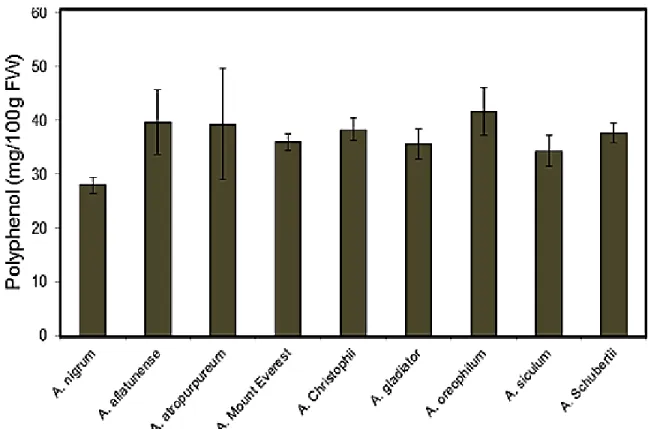
Total polyphenol content
Phytochemicals, particularly polyphenols from natural sources such as vegetables and fruits have gained popularity due to their protective properties against several chronic diseases such as cancer and cardiovascular diseases (Temple, 2000). Most of the polyphenol studies on Allium have
been focused on edible Allium species such as garlic (A. staivum), onion (A. cepa), shallot (A. cepa L. Aggregatum group) and leek (A. porrum). However, data on the polyphenol content in
other Allium species are certainly lacking (Dziri et al., 2012). Total phenol compounds (TP), as determined by Folin Ciocalteu method, are reported as Catechol equivalents by reference to standard curve. There was no much variance in the TP content among the plants examined in this study. The total phenolic contents ranged from 28 to 41.6 mg/100g FW (Fig. 7). A. oreophilum showed the highest TP content (41.6 mg/100g FW) and A. nigrum was the lowest (28 mg/100g FW). The TP content of these ornamental Allium species expressed in equivalent catechol was in general lower than garlic (61.8 mg/100 g FW) and shallot who has the highest phenolic content (114.7 mg/100 g) among the bulb onion verities tested by (Lanzotti, 2006), and higher than that of onion (31.0 mg/100 g FW) as reported by (Kaur and Kapoor, 2002). Previous reports regarding the quantification of TP in other Allium species showed quantitative differences that seem to depend on plant species/cultivar or variety origin and plant organ (Yang et al., 2004; Prakash et al., 2007).
18
Fig. 5 Representative chromatograms of MeCSO (1), AlCSO (2) and PeCSO (3) in: (A) standard, (B) A. siculum and (C) A. nigrum
Fig. 6 Total content of MeCSO, AlCSO and PeCSO in different ornamental Allium species. Values are means + SE (n=2)
Response detected 220nm
19
Fig. 7 Total polyphenol content mg/100g FW in different ornamental Allium species. Values are means + SE (n=2)
20 Total reduced sugar content
Carbohydrates are the most abundant class of chemical compounds in Allium species and include glucose, fructose and sucrose, together with a series of oligosaccharides, the fructans (Darbyshire and Steer, 1990; Kamenetsky and Rabinowitch, 2006). The water soluble carbohydrates content in the examined Allium species ranged from 7.5 to 19 mg/g FW (Fig. 8).
Only sucrose was represented on the chromatogram however, glucose and fructose was not detected. The highest sucrose content was determined in A. oreophilum (19 mg/g FW) and the lowest content was determined in A. schubertii (7.5 mg/g FW). The total reduced sugars contents in the investigated plants showed low sugars content in compare with the edible one. Total sugar content in A. cepa was ranged from 13.5% to 71.8% in Nigerian cultivars (Ketiku, 1975). A previous record for total reduced sugars content in A. cepa was 55 mg/g DW according to (Abrameto et al., 2010). Three sugars (glucose, fructose and sucrose) were identified in all the onion samples examined by (Galdon et al., 2009), and the total sugar content ranged from 2.3 to 3.79%. These findings are higher than the total sugar in our data. The wide range variability of the carbohydrates content with in the different Allium species could be influenced by the differentiation of the genomic levels. A previous study by (McCallum et al., 2006), revealed a one significant QTL in chromosome 8 of intraspecific mapping population ‘BYG15-23’ x
‘AC43’using a complete molecular marker map and this locus, provisionally named Frc, may account for the major phenotypic differences in bulb carbohydrate content.
21
Fig. 8 Total reducing sugars content (mg/g FW) in different ornamental Allium species. Values are means + SE (n=2)
22 Total saponin content
Saponins are a large group of glycosides, widely distributed in the plant kingdom. They are believed to form the main constituents of many plant drugs and folk medicines (Estrada et al., 2000). The content of saponins with in plants is quite variable and it’s mainly reflects the physiological status of the plant, which mainly depend on the stage of the growth and development (Szakiel et al., 2011). Total saponin content among the examined plants revealed variable qualitative and quantitative saponin content (Fig. 9). TLC analysis of the crude saponin from A.
schubertii showed a remarkable saponin profile in compare with other TLC profiles from other extracts. Moreover, the quantitative spectral analysis of the total saponin content was almost in correlation with the TLC figure where high saponin content was detected in A. schubertii and lowest content was detected in A. Mount Everest. The overall conclusion from these outputs, that each plant has its unique saponin profile which can be translated into a specific bioactivity and this will be precious tools for better selection of wild Allium species to be used in the breeding program to improve disease resistance in edible Allium. Furthermore, previous reports have showed that even though among the same species the saponin content could be variable and this variability of saponin content could be influenced by the surrounding environment. The local geoclimate, seasonal changes, external conditions such as light, temperature, humidity and soil fertility as well as cultivation techniques affect both quantitative amount and qualitative composition of saponins (Augustin et al., 2011; Szakiel et al., 2011). Significant quantitative and qualitative differences in saponin content depending on the site of growth have been reported (Lim et al., 2005).
23 Crude Saponin assay
Antifungal activity of the crude saponin extract (500, 1000 and 1500 ppm) from each plant was tested on nine different fungal pathogens. Seven soil-born pathogen (Fusarium oxysporum f.sp.
cepae, F. oxysporum f. sp. spinaciae, F. oxysporum f. sp. radicis-lycopersici, F. oxysporum f. sp.
fragariae, F. oxysporum f. sp. lagenariae, F. oxysporum f. sp. lycopersici and F. verticillioides), and two air pathogen Colletotrichum graminicola and Colletotrichum gloeosporioides). All the crude saponin extracts significantly showed concentration-dependent antifungal activity except for the crude saponin from A. siculum which doesn’t revealed a strong antifungal activity (Fig. 11).
Relatively higher antifungal activities against all fungus strains were observed in A. schubertii, A.
atropurpurm, and A. nigrum in compare with other plants extracts. C. graminicola and C.
gloeosporioides were highly sensitive to the crude saponin in compare with Fusarium strains. The high antifungal activity of the crude saponin from the ornamental Allium species suggested that these bioactive metabolites may act as chemical barriers to protect the plant against pathogen attack.
Our finding is matching with many previous reports about the antifungal activity of saponin compounds (Zimmer et al., 1967; Osbourn, 1996; Nicol et al., 2002; Lanzotti et al., 2012a and b).
Moreover, the different Fusarium sensitivity to the variant crude saponin may be related to the different nature of the aglycone in each extract. The correlation coefficient between the total saponin content and antifungal activity revealed a positive but week correlation (Fig. 11). The scatter plot of the metabolite signature in each genotype was illustrated by principal components analyses (Fig. 10). PC1 show the highest significant of variance with 45.87%, mainly dominated by ACSO and saponin, and PC2 18.06 % with sucrose and saponin as a major component. Further biochemical analysis will be conducted to isolate and identify the pure compounds responsible for this bioactivity.
24
Fig. 9 Total saponin contents (I) and TLC profiles (II) of crude extracts in A. nigrum (N), A. aflatunense (AF), A. atropurpureum (AT), A. Mount Everest (M), A. christophii (C), A. schubertii (S), A. gladiator (G), A. oreophilum (O) and A. siculum (SI).
25
Fig 11 Percentages of antifungal inhibition at 500, 1000 and 1500 µg/ml of the crude saponins against different fungal strains (A) and correlation coefficient between total saponin content (x- axis) and antifungal activity (y-axis) (B). Values are means + SE (n=3)
Fig. 10 PCA of the metabolite profile in different ornamental Allium species
Fungal growth inhibition %
(A) (B)
26
CHAPTER III. AGINOSIDE SAPONIN, A POTENT ANTIFUNGAL COMPOUND, AND SECONDARY METABOLITE ANALYSES FROM ALLIUM NIGRUM L.
Introduction
Plants during their life cycles produce a diverse array of secondary bioactive metabolites, many of which are involved in plant defense (Dixon, 2001). Based on this concept, the phytochemical diversity of antimicrobial compounds includes saponins, phenolics, and indoles. Saponins are a major family of glycosides with triterpenoid or steroidal aglycone and show remarkable antifungal activities widespread among many plant species (Osbourn, 1996; Barile et al., 2007; Lanzotti et al., 2012a). Triterpene saponins are the most common saponins, occurring mainly in dicotyledonous angiosperms, while steroidal saponins are less widely distributed and are present exclusively in monocotyledonous families (Bruneton, 1995). Allium spp. possess antibacterial and antifungal activities and contain powerful sulfur and other phenolic compounds which hold potential for future research (Griffiths et al., 2002). Allium plants represent the most economically important and representative genus of the Alliaceae. They are also a rich source of steroidal saponins with potential antifungal activity (Adao et al., 2011). Different Allium species, such as bulb onion (Allium cepa L.), garlic (Allium sativum L.), shallot (A. cepa L. Aggregatum group), chive (Allium schoenoprasum L.), and leek (Allium porrum L.), have been used in folk medicine and food for a long time (Fattorusso et al., 2000). Allium nigrum L. is an ornamental plant of the Alliaceae family subgenera Melanocrommyum, first described by Linnaeus in 1762. According to an ethnobotanical reference in Sicily, this species was used as a food spice (Lentini and Venza, 2007). Aginoside, a spirostane saponin, was first isolated from bulbs of Allium giganteum
27
(Kawashima et al., 1993) and recently was reported in A. nigrum (Jabrane et al., 2011). To date, most pharmacological and phytochemical studies regarding aginoside have focused on clinical pathogens and cancer cell lines. However, the antifungal activity of the aginoside compound against phytopathogens has not been investigated. In this study, a phytochemical analysis of A.
nigrum was carried out to evaluate the potential of A. nigrum for use in plant breeding programs to improve disease resistance and health properties in other Allium species. In addition, A. nigrum is a natural source of agrochemicals that can be used to manage pests and fungi. We determined the content of cysteine sulfoxides, total polyphenols, and total saponins in different organs of A.
nigrum by using high-performance liquid chromatography (HPLC) and spectral techniques.
Furthermore, we isolated, quantified, and assessed the antifungal activity of the aginoside compound in a wide range of phytopathogens
Materials and Methods
General experimental procedures
Optical rotations were taken with a JASCO DIP-1000 automatic digital polarimeter. 1H and 13C NMR spectra were recorded in a pyridine-d5 solution on a JEOL ECA 500 (500 and 125 MHz, respectively) spectrometer. Chemical shifts are reported in parts per million (ppm) and J values in Hz using pyridine-d5 for 1H NMR (7.20 ppm) and 13C NMR (123.5 ppm) as an internal standard.
The HR-ESI-MS was recorded with a JEOL JMS-T100LP spectrometer (Tokyo, Japan). Isolation of the compound was carried out using glass column chromatography (3 cm x 60 cm) on silica gel C-300 Wakogel (Wako, Tokyo, Japan). Thin-layer chromatography (TLC) was performed on
28
precoated silica gel plates (60 F254; Merck, KgaA, Darmstadt, Germany). The saponin profile was detected by spraying with a 5 ml p-anisaldehyde reagent.
Plant materials
Bulbs of wild A. nigrum L. were obtained from the Netherlands (Baltus Co., Vaassen). They were grown in a greenhouse (23 + 2°C) at Yamaguchi University, Japan, from October 2010 and harvested in May 2011.
Extraction and isolation
Fresh root–bulb basal stem of A. nigrum (80 g) was hand-cut and air-dried at room temperature (22°C), and the final dry weight (30 g) was obtained and used for this study. The dry weight was exhaustively extracted at room temperature with the following solvents: n-Hexane and 70%
MeOH. Each solvent extraction step was conducted for 1 day and repeated three times with 30 min of sonication and filtering. The MeOH extract was dried in a rotary evaporator with a vacuum pump (v-700; BUCHI, Rotavapor R-3) and then partitioned between BuOH and H2O (1:1, v/v).
The BuOH layer was filtered and then concentrated under vacuum, giving a saponins crude extract (1.69 g). The aliquot of the crude extract was chromatographed by C300 silica gel column chromatography (3 cm X 60 cm; AG Tokyo, Japan). The column was developed using a gradient solvent system starting with CHCl3, CHCl3–MeOH (9:1–1:9), MeOH, and MeOH–H2O (9:1–7:3) as eluents to give 10 fractions after the evaporation of solvents (AN1–AN10). Fractions were re- chromatographed using TLC silica gel plates (60 F254; Merck KgaA, Darmstadt, Germany). The chromatogram was developed with chloroform:methanol:water (30:15:2.5, v/v/v). In a preliminary NMR study of the eluted fractions, AN8 gave a pure compound (13 mg).
29
Determination of aginoside content in different organs
Different concentrations (6, 12, 30, and 36 mg) of purified aginoside compound isolated from the root–bulb basal stem and crude saponin extract from the root, bulb, and leaves were subjected to TLC, as mentioned earlier. Quantification of the TLC band intensity of the purified aginoside compound and crude extract was detected using Image J version 1.42Q/Java 1.6.0_10 image analysis. The aginoside content in the crude extract of each organ was calculated by comparison to the purified aginoside calibration curve.
Extraction and analysis of S-alk(en)yl-L-cysteine sulfoxides
The A. nigrum bulb, leaf, and root fresh weight (2.5 g) were water extracted according to the method of Masamura et al. (2011). The extract was diluted 10 times using a 0.005% trifluroacetic acid buffer (TFA) and filtrated using a 0.45 mm syringe-type filter (HCL-Disk3; Kanto Chemical Co., Inc., Tokyo, Japan). A 60 ml filtered sample was injected into a high performance liquid chromatography (HPLC) system and quantified. The HPLC system includes a pump, a degasser, a column oven, a diode array detector set to 220 nm, a data collection system (EZchrom Elite TM;
Hitachi High-Technologies Corporation, Tokyo, Japan), and an AQUASIL SS-1251-120 column (4.6 mm i.d. 250 mm long; Senshu Scientific Co., Ltd., Japan). The solvent, 0.005% TFA, flowed for 15 min at a flow rate of 0.6 ml/min. Standard compounds were synthesized at the Somatech Center (House Food Corporation, Japan).
Determination of total phenol content
Two grams of leaf, bulb, and root tissue was extracted by 70% EtOH according to the method of Hang et al. (2004). The extracted materials were adequately diluted with water, and the total phenolic (TP) contents were determined by the Folin–Ciocalteau method (Folin and Denis, 1915).
30
The polyphenol contents were quantified spectrophotometrically at 530 nm (Hitachi, Model U- 2001, Tokyo, Japan).
Determination of total saponin content
Dried root, bulb, and leaf powder (160 mg) of A. nigrum was extracted with 70% MeOH, as previously described. The total saponin content was determined spectrophotometrically at 473 nm in the crude extract from each organ according to Ebrahimzadeh and Niknam (1998) using 0.7%
Vanillin-60% H2SO4 reagent and a saponin standard serial dilution (Disogenin, Merck, Germany).
Biological assays
The antifungal activity of crude saponins, CSO, and TP was tested on seven soil-borne pathogens (F. oxysporum f. sp. cepae, F. oxysporum f. sp. spinaciae, F. oxysporum f. sp. radicis-lycopersici, F. oxysporum f. sp. fragariae, F. oxysporum f. sp. lagenariae, F. oxysporum f. sp. lycopersici, and F. verticillioides) and one air-borne pathogen (C. gloeosporioides). Pathogens were obtained from the Laboratory of Plant Molecular Pathology, Faculty of Agriculture, Yamaguchi University, Japan. The antifungal activity was evaluated with the agar plate diffusion method using 3.2 cm Perspex plates of potato dextrose agar (PDA). Crude saponin, CSO, and TP were added to obtain three final concentrations (500, 1000, and 1500 ppm), and the plates were inoculated with a 5 mm plug containing the fungi grown on PDA for five days. The plates were incubated at 25°C, and the fungal radical growth was measured after one week. The antifungal activity of the aginoside
compound was assessed against three soil-borne pathogens (F. oxysporum f. sp. cepae, F. oxysporum f. sp. radicis-lycopersici, and F. verticillioides) and two airborne pathogens (B. squamosa and C. gloeosporioides). Plates of 3.2 cm of PDA added to the aginoside at four
final concentrations (10, 100, 200, and 400 ppm) were inoculated with a 5 mm plug containing the
31
fungi grown on PDA for five days. The plates were incubated at 25°C, and the fungal radial growth was measured after one week. The antifungal activity was assessed by the in vitro spore germination test as described by Lorito et al. (1996). A suspension of 103 spores of B. cinerea and C. gloeosporioides was prepared in 100 ml of potato dextrose broth (PDB) separately. Aginoside was added to obtain three final concentrations (10, 50, and 100 ppm). One hundred microliters of each solution was placed in an ELISA 96-well plate (Nunclon, Denmark) and incubated at 25°C.
The number of germinated spores and the hyphal length were measured after 24 h using ELISA (Emax, S/N 9807, USA) at 620 nm. Tomato and strawberry leaves were detached from 4- to 6- week-old greenhouse plants and sprayed with aginoside at three concentrations (10, 50, and 100 ppm). Treated and non-treated tomato and strawberry leaves were inoculated with 5 mm mycelial agar discs of B. cinerea and C. gloeosporioides, respectively. A disc was placed in the middle of each leaflet, which was then placed in a closed plastic container on wet filter paper to maintain 70% humidity. The containers were placed in an incubator at 22°C for 10 days. Lesion development was recorded daily.
Statistical analysis
All experiments consisted of three replications. Values are expressed as the mean + standard error (SE) and analysis of variance (ANOVA) calculated using SPSS, Inc. 16.0. The significant effects of treatments were determined by the magnitude of the P-value (P < 0.05). Treatment means were separated by the Tukey’s Honestly Significant Differences (HSD) test.
32 Results and discussion
S-alk(en)yl-L-cysteine sulfoxides
Methiin (MeCSO) was first separated at a retention time of 6.4 min, followed by alliin (AlCSO) at 7.4 min and isoalliin (PeCSO) at 9.4 min, as shown in a previous study (Masamura et al., 2011).
Only a small amount of total cysteine sulfoxide (0.367 mg/g FW) was detected in A. nigrum (Fig.
12A). These findings are in agreement with previous investigations in which the total cysteine sulfoxide contents were below 0.1% in proportion to the fresh weight in the examined Allium species of bulbous subgenera Melanocrommyum (Fritsch and Keusgen, 2006). Among different plant organs, the highest MeCSO content was detected in the bulb (0.17 mg/g FW), whereas a trace amount of MeCSO was detected in the leaf (0.10 mg/g FW) and root (0.08 mg/g FW). Our data agree with those obtained by Kusterer et al. (2011), who reported methiin as the predominant CSO in all investigated samples of a wild Allium species subgen. Melanocrommyum, with the highest amount found in Allium suworowii (0.6% FW) and the lowest in Allium rosenorum (0.01%
FW). AlCSO was detected only in the bulb (0.011 mg/g FW). On the other hand, this compound was not detected in the leaf or root. PeCSO was not found in any plant organs. These findings contradict the previous reports by Fritsch and Keusgen (2006), who found detectable amounts of PeCSO in all investigated Allium species subgen. Melanocrommyum. The low level of cysteine sulfoxides in A. nigrum is the reason many Allium species among the Melanocrommyum lack a specific smell (Rabinowitch and Currah, 2002).
33 Total polyphenol
The total phenolic contents were highest in the leaf organ (116.05 mg CE/100 g FW), followed by the root (65.09 mg CE/100 g FW). The lowest amount of TP (33.29 mg CE/100 g FW) was detected in the bulb (Fig. 12B). This organ-dependent compartmentation of TP was recently reported for A. roseum (736.65 and 749.54 mg CE/100 g of DW) in flowers and leaves, respectively, the richest organs in terms of TP (Najjaa et al., 2011). Nencini et al. (2011) reported higher TP contents (1.23, 1.22, and 1.36 mg GAE/g) in the leaves than in the bulbs (0.25, 0.39, and 0.73 mg GAE/g) of A. neapolitanum Cyr., A. subhirsutum L., and A. sativum L., respectively.
From a quantitative viewpoint, it is difficult to compare our findings with those of previous reports because of the variations in the conditions, extraction solvent used, and TP assays. Most reports regarding the quantification of TP in other Allium species show quantitative differences that seem to depend on the plant species/ cultivar or variety origin and plant organ (Prakash et al., 2007;
Dziri et al., 2012). Exposure to UV-B radiation and light intensity could be a reason for the higher accumulation of polyphenol compounds in the leaves than in the bulbs, which, from a biological viewpoint, have a protective role against environmental abiotic stress (Wang and Feri, 2011). The interesting finding in our results is the detectable amount of polyphenols in the root organ. The antimicrobial activity of the polyphenol compounds to protect a plant against pathogens could explain this accumulation. Many previous studies have reported the antimicrobial activity of polyphenols as efficient protection against pathogens and predators (Popa et al., 2002; Wang and Feri, 2011).
34 Total saponin content
Saponins accumulated in the vegetative organs of A. nigrum, with a higher content in the roots (19.38 mg/g DW) and lower contents in the bulbs (15.65 mg/g DW) and leaves (10.48 mg/g DW).
These results show a dynamic variance trend of total saponins among different organs (Fig. 12C).
Such organ repartition, with higher saponin accumulation in the root organs than in the aerial parts (stem and leaves), was previously reported by Teng et al. (2009). Szakiel et al. (2011) reported that the saponin content of plants is variable and may be affected by the surrounding environment, growth, and developmental stage. In addition, the biological activities of saponins (Sparg et al., 2004) could be one of the parameters influencing saponin distribution among different organs in A. nigrum.
35
Fig. 12 Total ACSOs (A), polyphenol (B), and total saponins content (C) in root, leaf and bulb organs of A. nigrum. a Values are means of three independent replicates. b P < 0.05, bars represent standard errors. Means followed by the same letter(s) within the column are not significantly different according to Tukey’s HSD
0 20 40 60 80 100 120 140
Total polyphenol (mg CE/100 g fw)
0 0.05 0.1 0.15 0.2 0.25
ACSOs content (mg/g fw)
0 5 10 15 20 25
Root Bulb leaf
Saponin (mg/g dw)
A
B
C
36 Characterization of the isolated compound
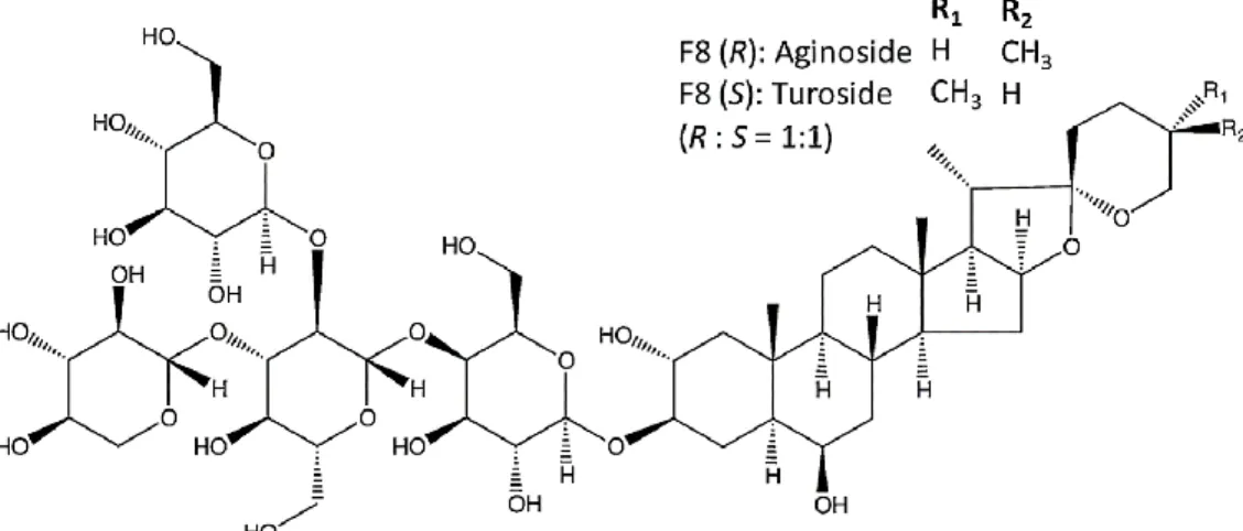
The BuOH root–bulb basal stem extract was subjected to TLC (CHCl3/MeOH/H2O, 30:15:2.5, v/v/v) and was shown to contain saponins. It was initially separated by column chromatography with a gradient solvent system starting with CHCl3, CHCl3/MeOH (9:1–1:9), MeOH, and MeOH/H2O (9:1–7:3), yielding a partially purified fraction AN8 (20 mg). This fraction was further purified by TLC, affording a pure aginoside compound (13 mg), which was recently isolated from A. nigrum (Jabrane et al., 2011). The structure of the aginoside (AN8) was elucidated by 600 MHz NMR analyses, including 2D NMR experiments, and compared with literature data (Fig. 13). The aginoside compound, isolated as an amorphous white solid power in high yield, showed a molecular formula of C50H82O24, deduced by high-resolution FABMS measurements and confirmed by 13C NMR data: HR-ESI-MS (positive-ion mode): m/z 1089.5027 [M+Na]+ (calcd.
for 1089.5094): 1H NMR (pyridine-d5, 500 MHz) d: 5.54 (1H, d, J = 8.0 Hz, terminal-Glc-H-1), 5.14 (1H, d, J = 7.5 Hz, Xyl-H-1), 5.10 (1H, d, J = 8.0 Hz, inner-Glc-H-1), 4.93 (1H, d, J = 8.0 Hz, Gal-H-1), 3.97 (1/2H, m, H-26(S)), 3.54 (1/2H, br d, J = 8.1 Hz, H- 26(R)), 3.44 (1/2H, t, J = 11.5 Hz, H-26(R)), 3.32 (1/2H, d, J = 10.9 Hz, H-26(S)), 1.16 (3H, s, H-19), 1.07 (3H, d, J = 6.9 Hz, H-21), 0.99 (3/2H, d, J = 6.9 Hz, H-27(S)), 0.75 (3/2H, s, H-18(R)), 0.74 (3/2H, s, H-18(S)), 0.62 (3/2H, d, J = 5.7 Hz,H-27(R)); 13C NMR (pyridine-d5, 125 MHz) data (Table 1). The aginoside content has a variable distribution within different organs in A. nigrum (Fig. 14).The highest accumulation (2.9 mg/g DW) was detected in the root, followed by the bulb (2.34 mg/g DW), and the lowest was recorded in the leaves (1.57 mg/g DW). Huhman et al. (2005) similarly reported that soyasapogenol was mainly accumulated in the root organ (>32% of the total saponin content) compared with the leaf and stem of Medicago truncatula.
37
Tabel 1. 13 C NMR spectroscopic data of the aglycone and sugar moieties of aginoside (S/R) in pyridione-d, in ppm
Pos. AN8 (S)
Turoside
AN8®
Aginoside
Pos.
C-1 2 3 4 5 6 7 8 9 10 11 12 13 14 15 16 17 18 19 20 21 22 23 24 25 26 27
46.3 69.9 83.6 31.5 47.0 69.3 39.4 29.3 53.7 36.3 20.7 39.8 40.2 55.5 31.1 80.6 62.2 15.9 16.4 41.8 14.2 108.8
25.6 25.4 26.8 64.5 15.6
46.3 69.9 83.6 31.5 47.0 69.3 39.4 29.3 53.7 36.3 20.7 39.8 40.2 55.5 31.1 80.6 62.2 15.9 16.4 41.8 14.4 109.3
30.9 28.5 29.9 66.4 16.7
gal-1 2 3 4 5 6 i-glc-1
2 3 4 5 6 t-glc-1
2 3 4 5 6 Xyl-1
2 3 4 5
102.2 71.8 74.6 78.8 75.0 60.3 103.6
80.2 86.4 69.3 76.7 61.9 103.7
75.0 77.2 69.0 77.6 62.0 104.0
74.2 77.6 70.6 66.4
38
Fig. 14 Aginoside content detected in different plant organs. Quantification of TLC band intensity of purified aginoside compound and crude extract was detected using Image J version 1.42Q/Java 1.6.0_10 image analysis. The right y-axis represents mean relative hybridization intensity of three independent replicates expressed in arbitrary unit. Left y-axis represents concentration (mg/g DW)
Fig. 13 Chemical structure of aginoside
39 Phytochemical bioactivity
The crude saponins showed the following rank of antifungal activity depending on the applied concentration and the tested fungal species (Fig. 15). Fusarium oxysporum f. sp. lycopersici had the highest resistance to the crude saponin of all tested concentrations, whereas Colletotrichum gloeosporioides was the most susceptible pathogen. The potential antifungal activity of the crude saponins in the root organs of A. nigrum supports our hypothesis that this bioactive metabolite plays a major role in the plant’s defense mechanism, which explains the high accumulation of saponins in the root organs. The antifungal activity of the three dominant CSO compounds, MeCSO, AlCSO, and PeCSO, at different concentrations (500, 1000, and 1500 ppm) revealed no inhibition for MeCSO and PeCSO, whereas AlCSO at 1500 ppm recorded a maximum inhibition of 16% against C. gloeosporioides and F. oxysporum strains (10%). These results are in agreement with those of Coley-Smith et al. (1987), who reported that a high CSO content is directly associated with a high disease incidence in Allium species. Yamada and Azuma (1977) confirmed that the
antifungal activity of thiosulfinates was linked to allicin, which was effective against C. cryptococcus; however, allicin showed less inhibition against Aspergillus. Furthermore,
Yoshida et al. (1998) suggested that the antimicrobial activity of the cystine sulfoxide group could be ranked as follows: AlCSO > MeCSO > PrCSO. A fair to moderate fungal inhibition of phenolic extract from the leaves was noticed among tested concentrations (500, 1000, and 1500 ppm) against phytopathogens; the highest inhibition of 35% was recorded in C. gloeosporioides at 1500 ppm. Similar findings of concentrations dependent on the antifungal activity of phenolic compounds were reported in onion and garlic extracts (Benkeblia et al., 2005). Various phenolic compounds are known to exhibit antifungal activity, as reviewed by Cushnie and Lamb (2005).
40
Fig. 15 Antifungal activity against FOC (Fusarium oxysporum f. sp. cepae), FOS (F. oxysporum f. sp. spinaciae), FORL (F. oxysporum f. sp. radicis-lycopersici), FOF (F. oxysporum f. sp.
fragariae), FOL (F. oxysporum f. sp. lagenariae), FOLY (F. oxysporum f. sp. lycopersici), FV (Fusarium verticillioides) and CG (Colletotrichum gloeosporioides) of crude saponins isolated from A. nigrum at three final concentrations (500, 1000 and 1500 ppm). Data are inhibition of fungal growth in percentage compared to control (= 0). a Values are means of three independent replicates. b P < 0.05, bars represent standard errors. Means followed by the same letter(s) within the column are not significantly different according to Tukey’s HSD.
b b
c
c c
a
c
b
c d
bc
c
a
bc
a
b
b
0 10 20 30 40 50 60 70 80
CG FV FOC FORL FOS FOLY FOF FOL
Inhibition of fungal growth (%) 500 ppm
1000 ppm 1500 ppm
41
Further investigation may be necessary to examine the presence of organo sulfur and phenolic compounds in other Allium plants and to clarify the contribution of these two metabolites to antifungal activity. Aginoside compounds showed significant (P < 0.05) antifungal activity (Fig.
16). The 400 ppm of aginoside completely inhibited the fungal growth of C. gloeosporioides, F. verticillioides, and B. squamosa and partially suppressed that of F. oxysporum f. sp. cepae and F. oxysporum f. sp. radicis-lycopersici. The potent antifungal activity displayed by aginoside suggests that this compound, either alone or in addition to others, may act as a chemical barrier to protect plants against fungal attacks. Our results are in agreement with those of Jabrane et al.
(2011), who reported aginoside to be the most active spirostane saponin against colon cancer cell lines in comparison with other steroidal saponins from A. nigrum. In other research, Sata et al.
(1998) stated that aginoside displayed in vitro cytotoxicity against P388 leukemic cells at 2.1 mg/
ml and antifungal activity against Mortierella ramanniana at 10 mg/disk. Moreover, Coleman et al. (2010) reported the significant antifungal activity of aginoside against the clinical fungal pathogen Candida albicans at MIC (47.0 mg/ml).
Our results regarding the antifungal activity of aginoside against F. oxysporum f. sp.
Lycopersici are consistent with the data obtained by Lanzotti et al. (2012b), who found high resistance of FOL against three identified saponin compounds , cepasoide A, B and C isolated from the bulbs of A. cepa at three different concentrations (10, 50, and 200 ppm). Many fungi attack plants by producing enzymes that degrade saponins into non-toxic molecules (Morrissey and Osbourn, 1999). Most probably, the relatively high tolerance of Fusarium oxysporum to aginoside could be explained by this suggestion. The in vitro spore germination test, together with different aginoside concentrations (10, 50, 100 ppm), showed a high inhibition of spore germination in all tested pathogens (B. cinerea and C. goleosporioides). A relatively high
42
suppression of spore germination (< 40%) was detected at two different concentrations, 50 and 100 ppm (Fig. 17). The results for B. cinerea and C. goleosporioides are in agreement with those of the in vitro assay (Fig. 18). Fungal infection expansion and black necrotic lesions were observed in the control (untreated leaves). In contrast, lesion expansion was totally inhibited in infected leaves treated with a concentration of 100 ppm.
Fig. 16 Examination of aginoside-antifungal activities against C. gloeosporioides (CG), B. squamosa (BSQ), F. verticillioides (FV), F. oxysporum f. sp. cepae (FOC), F. oxysporum f. sp.
radices lycopersici (FORL), at four final concentrations (10, 100, 200 and 400 ppm). Data are inhibition of fungal growth in percentage compared to control (= 0). a Values are means of three independent replicates. b P < 0.05; bars represent standard errors. Means followed by the same letter(s) within the column are not significantly different according to Tukey’s HSD
a a
a a
b a
b b b
c
a b
c c
d
a b
c c c
0 20 40 60 80 100 120
CG BSQ FV FOC FORL
Inhibition of fungal growth (%)
10 ppm 100ppm 200ppm 400ppm
43
Fig. 17 Examination of Aginoside activity against spore germination of B. cinerea and C. gloeosporioides at three different concentrations 10, 50 and 100 ppm (A). Light microscopic
examination of aginoside (Ag) treated and untreated on spore inhibition of C. gloeosporioides and B. cinerea at 200 µg (B). Spore staining using Evan blue. a Values are means of three independent replicates. b P < 0.05; bars represent standard errors. Means followed by the same letter(s) within the column are not significantly different according to Tukey’s HSD
A
a a
b
b
c
c d
d
0 20 40 60 80 100 120
BC GC
Spore germination (%)
Control 10 ppm 50 ppm 100 ppm
B
Spore germination %
44
Fig. 18 Antifungal activities of the Aginoside (10, 50 and 100 ppm) upon artificial infection of B. cinerea on tomato leaves (A) and C. gloeosporioides on strawberry leaves (B) for 7 days after infection. (C) Leaf lesion symptoms (%)
45
CHAPTER IV. COMPARATIVE SAPONIN PROFILES IN DIFFERENT ALLIUM GENOTYPES AND THEIR PROSPECTIVE APPLICATIONS AS CHEMICAL
MARKERS FOR FUSARIUM BASAL ROT DISEASE RESISTANCE
Introduction
Steroidal saponins are broadly disseminated among monocots, including the Amaryllidaceae family in which the Allium genus is currently classified. Apart from sulfur compounds, these are important biologically active compounds that are considered to be responsible for the observed activity of Allium species (Sobolewska et al., 2014). Numerous reports refer to the pharmacological activities of steroidal saponins. Some showed promising antifungal, cytotoxic, anti-inflammatory, antithrombotic, and hypocholesterolemic effects (Sparg et al., 2004; Lanzotti, 2005; Sobolewska et al., 2014). The first chemical survey of saponins from the genus Allium was published by Kravets in 1990; this was followed by an update by Lanzotti in 2005 (Kravets et al., 1990; Lanzotti, 2005). The approach for structurally elucidating saponins is mainly based on the use of spectroscopic techniques, including HRFABMS and advanced 2D NMR experiments, thus keeping the material for further biological assays (Mostaf et al., 2013).
Chemical methods are only applied for the stereostructure elucidation of sugar moieties. 2D NMR methods have the enormous advantage in overcoming the overlapping of a large number of signals in the 1D NMR spectra of saponins and in determining: the aglycone structure, the number of sugar residues (including their nature and their configuration), the glycosylation site, and the interglycosidic linkages (Lanzotti, 2005, 2012a). Surprisingly only a few studies have investigated