緒 言 Hemichoreaの原因となる疾患は,脳血管障害,免疫疾患, 腫瘍性疾患,代謝性疾患などがあげられるが1)2),それらの うち脳血管障害は頻度も高く重要である3).近年内頸動脈狭 窄に伴う脳血流障害が原因で生じたと考えられるhemichorea 症例が東アジアを中心に報告されている4)~12).このたび我々 は,対側の内頸動脈狭窄を伴い,頸動脈内膜剝離術(carotid endarterectomy; CEA)にて改善した hemichorea の 1 例を経験 したので報告する. 症 例 症例:71 歳,男性 主訴:顔面左側と左手足の異常な動き 既往歴:後縦靭帯骨化症. 家族歴:父が脳梗塞.家族に類症なし. 内服薬:なし. 現病歴:2014 年 11 月中旬,自動車運転中に数秒間持続す る左下肢の不随意にねじれるような動きが出現した.その 後同様の症状が日に数回の頻度で見られるようになった.11 月下旬からは左上肢を不随意にねじるような動き,顔面左側 にしかめっ面をするような動きが出現した.12 月初頭から症 状の頻度が急速に増加し,左上下肢及び顔面左側の症状が昼 夜を問わず持続するようになったため,精査加療目的に入院 した. 入院時現象:身長 160 cm,体重 65 kg,血圧 141/79 mmHg, 脈拍 54/ 分・整,体温 36.5°C.一般身体所見に特記すべき異 常所見は認められなかった.神経学的には左上下肢全体をく ねらせるような比較的なめらかで非律動的な不随意な動きを 認め,左眼を開閉させる・口唇を突き出す・左口角を上外側 方向へ引っ張る,舌をねじるようなといった不随意運動が見 られた.意識清明で高次脳機能に異常は見られず,脳神経も 顔面の不随意運動以外に異常は見られなかった.四肢の筋力 低下は無く,深部腱反射に異常は認められず,感覚障害も認 められなかった. 検査所見:血液検査では血算に異常なく,凝固系も正常で あった.一般生化学検査では腎・肝機能に異常なく,脂質代 謝はトリグリセリド 145 mg/dl,LDL コレステロール 120 mg/dl, HDLコレステロール 89 mg/dl,糖代謝は HbA1c 5.7%であっ た.抗核抗体,リウマトイド因子,抗 ds-DNA 抗体は陰性で あった.心電図では正常洞調律であった. 画像所見:頭部 MRI では右側脳室後角後方と右前頭葉皮 質下に拡散強調画像で点状の高信号域を認めたが,T2強調画 像・FLAIR 画像では陳旧性梗塞巣や慢性虚血性変化は認めら れなかった.頭蓋内MRAでは有意狭窄や途絶は見られなかっ たが,右内頸動脈(internal carotid artery; ICA)から中大脳動 脈(middle cerebral artery; MCA)にかけて描出が不明瞭で あり,頸部 MRA では右 ICA 分岐部近傍に高度狭窄が見ら れ,頭部 MRA での描出不良は右 ICA 狭窄が原因と考えられた (Fig. 1).頸動脈エコーでは右 ICA に European Carotid Surgery
Trial(ECST)法で 82%・North American Symptomatic Carotid Endarterectomy Trial(NASCET)法で 73%の高度狭窄を認め, 収縮期最大血流速度 404 cm/s・拡張末期血流速度 211 cm/s と
右内頸動脈狭窄により hemichorea を生じた 1 例
六車 一樹
1)元田 敦子
1)杉本 太路
1)北村 健
1)*
要旨: 症例は 71 歳男性.2014 年 11 月中旬から左半身の hemichorea が出現した.頭部 MRI 拡散強調画像で右 側脳室後角後方と右前頭葉皮質下に点状の高信号域を認めた.右内頸動脈起始部に高度狭窄を認め,脳血流 SPECT では右大脳半球にびまん性の血流低下を認めた.2015 年 1 月に右内頸動脈の頸動脈内膜剝離術を施行し た後,hemichorea は消失し,右大脳半球の血流は改善した.Hemichorea は基底核領域の梗塞が原因となること は以前から知られているが,本症例のように内頸動脈狭窄を有するが基底核領域に梗塞を伴わずに内頸動脈領域 全般の血流低下を認める症例が,近年東アジアを中心に報告されている. (臨床神経 2019;59:509-514) Key words: 片側舞踏運動,内頸動脈狭窄,頸動脈内膜剝離術 *Corresponding author: 中国労災病院脳神経内科〔〒 734-8551 広島県広島市南区霞 1 丁目 2-3〕 1)中国労災病院脳神経内科(Received January 25, 2019; Accepted May 30, 2019; Published online in J-STAGE on July 23, 2019) doi: 10.5692/clinicalneurol.cn-001276
流速上昇が見られた(Fig. 2).
Xenon-CTでは安静時の cerebral blood flow(CBF)に左右 差は見られなかったが,acetazolamide 負荷後,右前頭葉・側 頭葉・基底核領域の血流予備能低下が見られた.脳 single photon emission computed tomography(SPECT)検査(123I-IMP
定性)でも右前頭葉,側頭葉及び頭頂葉の皮質領域,右基底 核領域の血流低下を認めた(Fig. 3). 臨床経過:不随意運動に対してハロペリドール 2 mg/ 日の 内服を開始したが効果は乏しかった.クロピドグレル 75 mg/ 日, アスピリン 100 mg/ 日,ロスバスタチン 5 mg/ 日の内服を行っ たところ,不随意運動の頻度は著明に減少し,数秒間の不随 意運動が一日に数回見られる程度となった.2015 年 1 月中旬 に右 CEA を行ったところ,直後から不随意運動は完全に消失 しその後再発することはなかった.CEA 後に脳血流 SPECT を再検したところ,右大脳半球に見られていた血流低下が改 善していた(Fig. 4). 考 察 本症例は hemichorea を生じた対側の ICA 狭窄を伴い,亜 急性に左半身の hemichorea を発症した.抗血小板剤およびス タチンの内服により症状が改善し,さらに右 ICA の CEA 後 に脳血流の改善とともに症状が消失したこと,右内頸動脈狭 窄以外に hemichorea の原因となりえる疾患が認められな かったことから,右内頸動脈狭窄による血流低下が原因で hemichoreaが生じたものと考えられた.なお,本症例は臥位 で不随意運動が消失せず立位で顕著な増悪がなかったこと, 症状が連続的で長期間持続していることなどから本症例の症 候は limb shaking ではなく hemichorea と考えた.しかし,limb
shakingの病態には脳血流の低下が関与していると考えられ ることから13),本症例が limb shaking と類似した病態である 可能性は否定しえないと考えられる.頭部 MRI にて右側脳室 後角後方と右前頭葉皮質下に梗塞が認められたが,病巣が微 小であることや,基底核など hemichorea の原因となり得る部 位とは一致しないことから,これらが hemichorea の原因では ないと考えられた. Hemichoreaの原因としては,基底核を障害する脳血管障害や 脳腫瘍,多発性硬化症や,Sydenhamʼs chorea などの免疫疾患, 傍腫瘍症候群,非ケトン性高血糖症などが知られている1)2).
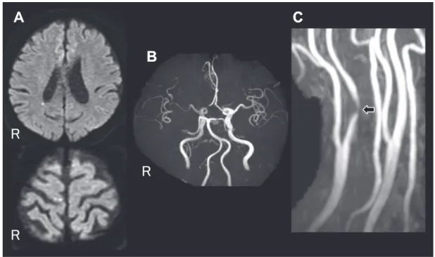
Fig. 1 MR images on admission.
A: Axial diffusion weighted images showing small hyper intense area in white matter near right cerebral ventricle posterior horn and right frontal lobe. B: Cerebral MR angiography image showing hypoperfusion of the right internal carotid artery and the right middle cerebral artery. C: Cervical MR angiography image showing right internal carotid artery stenosis (arrow).
Fig. 2 Carotid ultrasonography image of the right internal carotid artery showing stenosis with low echoic plaque.
脳血管障害後に発症した hemichorea は全体の 0.54%14)と稀 である.しかし,Dewey らは hemichorea を生じた 14 例の うち 7 例は脳血管障害が原因であったと報告しており15), hemichoreaの原因として脳血管障害は重要である. Choreaの機序は一般的に一次運動野と尾状核,被核,淡蒼 球内節・外節,視床下核,黒質細網部といった基底核の神経 核から形成される神経ネットワークである大脳皮質大脳基底 核ループ(Alexander-Crutcher-DeLong のループ)が障害され ることで生じると考えられている1). 脳血管障害に伴う hemichorea の責任病巣は,健側の視床下 核などの基底核病変が多く報告されている14)~17).しかし Piccoloらは脳梗塞が原因となった chorea 症例 20 例のうち基 底核,視床に原因病変があったのは 2 例のみであったとして いる3).分水嶺領域や11),運動を統制する役割である皮質や 皮質下の病変18)が原因となるとの報告もあり,様々な領域に よる報告がある. Hemichoreaの原因となる脳血管障害として脳梗塞,脳出血 以外にも頸部・頭蓋内の主幹動脈狭窄による血流低下があ り,中大脳動脈や内頸動脈狭窄やもやもや病の症例が報告さ れている2).ICA 狭窄に伴った hemichorea 症例は,近年日本 や韓国など東アジアを中心に本症例を合わせて 13 症例(男性 10例,女性 3 例.平均年齢 70.5 ± 8.0)の報告が見られる4)~12) (Table 1).これは,頭蓋内主幹動脈狭窄がアジア人に多いこ と19)も関連している可能性がある.なお,同じくアジア人に 多いことが知られているもやもや病も hemichorea を生じるこ とが報告されている20)が,この 13 例には含めなかった.小堺 らは血管性 hemichorea に関する論文の中で,内頸動脈終末部 に限局した狭窄と豊富な側副血行路を有し若年者に多いもや もや病と,アテローム硬化を基礎に持つ症例とでは脳循環動 態が異なる7)と考察しており,本論文でもその考えを踏襲した.
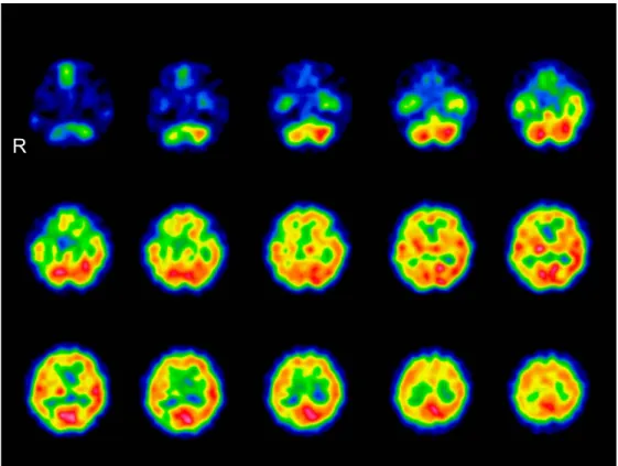
Fig. 3 Single photon emission computed tomography scan image on admission. Single photon emission computed tomography scan image (123
I-IMP qualitative) showing reduction in cerebral blood flow of the right frontal lobe cortex, the temporal lobe cortex, the parietal lobe cortex, and the basal ganglia.
Fig. 4 123I-IMP single photon emission computed tomography (SPECT) scan before and after carotid endarterectomy.
A: 123I-IMP SPECT scan before carotid endarterectomy. B: 123I-IMP SPECT scan after carotid endarterectomy showing the improvement of cerebral blood flow.
これらのうち脳血流 SPECT 検査がなされた症例は 7 症例
あり6)8)10)~12),全例で健側の大脳半球に血流低下を認めた.
CEAあるいは頸動脈ステント留置術(carotid artery stenting; CAS)を行った結果全例で症状が消失しており,このことか
らも内頸動脈狭窄による血流低下が hemichorea の原因と推 察される.
内頸動脈狭窄を伴った hemichorea 症例 10 例のうちでは基 底核に脳梗塞を認める症例は 2 例のみであった.放線冠や分 Table 1 Cases of hemichorea induced by ICA stenosis.
Case Age/ Sex of stenosisLocation Location of infarction Blood reduction Treatment Therapeutic effect Blood flow after treatment Waterston JA,
19904)
60/M Rt ICA Rt centrum semiovale. ND ND ND ND
Fukui T, 19935)
75/M Bil ICA Rt fronto-parietal subcorti-cal white matter and centrum semiovale.
Right frontal convex-ity and the centrum semiovale.
ND ND ND
Shimizu T, 20016)
72/M Bil ICA T2WI hyper intense area in bilateral cerebrum white matter and basal ganglia.
Bilateral cerebral hemisphere.
Bil CEA Hemichrea disappeared.
ND
Kosakai A, 200027)
48/M Lt ICA Small DWI hyper intense area in watershed of bilateral cerebral parasagit-tal section and subcortical white matter of lt ACA area.
Rt ICA area. Antiplatelet and anticoagulant drugs
Hemichorea improved in few days but did not disappeared.
ND
Morigaki R, 20068)
75/M Rt ICA Small T2WI hyper intense area in bilateral corona radiate. Right cerebral hemisphere. CEA Hemichrea disappeared in 24 months.. Improved after treatment. 77/F Rt ICA Small T2WI hyper intense
area in right head of caudate nucleus, bilateral basal ganglia, and semioval center. Right cerebral hemisphere. CEA Hemichrea disappeared in 18 months. Improved after treatment. Galea I, 20089)
72/F Lt ICA No infarction. ND CEA Hemichrea
disappeared in 6 months.
ND
72/M Lt ICA Left posterior parietal gray matter.
ND CEA Hemichrea
disappeared in 2 weeks.
ND
73/M Lt ICA Left anterior parietal lobe. ND CEA Hemichrea disappeared immediately.
ND
Pareés I, 201110)
81/M Lt ICA No infarction. Left thalamus and putamen. CAS Hemichrea disappeared in 3 months. Improved after treatment. Kim DW, 201311)
67/F Rt ICA Small DWI hyper intense area in right frontal lobe.
Subcortical area of right cerebellar hemisphere (do not include basal ganglia).
CAS Hemichrea disappeared. ND Kodera Y, 201512) 73/M Lt ICA/ Rt CCA
No infarction. Right cerebellar hemisphere including basal ganglia. CAS Hemichrea disappeared in 5 months. Improved after treatment. This case 71/M Rt ICA Small DWI hyper intense
area in white matter near right cerebral ventricle posterior horn and right frontal lobe. Right cerebellar hemisphere including basal ganglia. CEA and antiplatelet drugs Hemichrea disappeared immediately. Improved after treatment.
Bil = bilateral; Rt = right; Lt = left; ICA = internal carotid artery; CCA = common carotid artery; ACA = anterior cerebral artery; T2WI = T2-weighted image; DWI = diffusion weighted image; CEA = carotid endarterectomy; CAS = carotid artery stenting; ND = no data.
大脳皮質大脳基底核ループの理論では,線条体の投射 ニューロンが淡蒼球外節,視床下核,淡蒼球内節・黒質網様 体と経由し,視床や大脳皮質を抑制する negative feedback loop
が存在し,不必要な運動を抑制する働きをしている21)22). Choreaは間接路のいずれかの部位の障害により生じうると 推察されるが16),これらの経路において興奮性の神経投射を 行う視床下核などの神経核に梗塞がなくとも chorea が生じ ている.MRI で信号変化が生じない程度の基底核の虚血が生 じて基底核の機能を障害している23),運動皮質から線条体へ 投射される経路が梗塞や血流低下で障害されている8),血流 低下が血液脳関門に影響して脳細胞が代謝異常などにたいし て脆弱になっている3)といった機序が hemichorea の原因と推 測されている. 本症例では右 ICA 高度狭窄により基底核をふくめ右大脳半 球に広範な血流低下を認めており,基底核の血流低下や運 動皮質から線条体へ投射される経路の血流低下が negative
feedback loopを阻害し hemichorea を生じたと推察される.
結 語 内頸動脈狭窄に伴う hemichorea の症例を経験した.外科的 血行再建術などの内頸動脈狭窄に対する治療により脳血流が 改善し,症状も消失しており,内頸動脈狭窄による脳血流低 下が hemichorea の原因と考えられた.脳血管障害による hemichoreaの病態を理解するうえで貴重な症例と考え,報告 した. 本報告の要旨は,第 96 回日本神経学会中国・四国地方会で発表し, 会長推薦演題に選ばれた. ※著者全員に本論文に関連し,開示すべき COI 状態にある企業,組 織,団体はいずれも有りません. 文 献
1) Cardoso F, Seppi K, Mair KJ, et al. Seminar on choreas. Lancet Neurol 2006;5:589-602.
2) Zijlmans JC. Vascular chorea in adults and children. Handb Clin Neurol 2011;100:261-270.
3) Piccolo I, Defanti CA, Soliveri P, et al. Cause and course in a series of patients with sporadic chorea. J Neurol 2003;250: 429-435.
4) Waterston JA, Brown MM, Butler P, et al. Small deep cerebral infarcts associated with occlusive internal carotid artery disease. A hemodynamic phenomenon? Arch Neurol 1990;47:953-957. 5) Fukui T, Hasegawa Y, Seriyama S, et al.
Hemiballism-hemichorea induced by subcortical ischemia. Can J Neurol Sci 1993;20:324-328.
6) Shimizu T, Hiroki M, Yamaoka Y, et al. Alternating paroxysmal
arteryおよび内頸動脈閉塞を伴った血管性ヘミヒョレアの
1例―低灌流による皮質下白質病変の病的意義について―.
JMDD 2002;12:93-100.
8) Morigaki R, Uno M, Suzue A, et al. Hemichorea due to hemodynamic ischemia associated with extracranial carotid artery stenosis. Report of two cases. J Neurosurg 2006;105: 142-147.
9) Galea I, Norwood F, Phillips MJ, et al. Pearls & Oy-sters: resolution of hemichorea following endarterectomy for severe carotid stenosis. Neurology 2008;71:e80-82.
10) Pareés I, Pujadas F, Hernández-Vara J, et al. Reversible hemichorea associated with extracranial carotid artery stenosis. J Neurol Sci 2011;300:185-186.
11) Kim DW, Ko Y, Jang SH, et al. Acute hemichorea as an unusual presentation of internal carotid artery stenosis. J Mov Disord 2013;6:17-20.
12) 小寺佑佳,中山 平,湯谷佐知子ら.舞踏様運動をきたし頸 動脈ステント治療が奏功した 73 歳男性例.臨床神経 2015; 55:356-359.
13) Ali S, Khan MA, Khealani B. Limb-shaking transient ischemic attacks: case report and review of literature. BMC Neurol 2006;6:5.
14) Chung SJ, Im JH, Lee MC, et al. Hemichorea after stroke: clinical-radiological correlation. J Neurol 2004;251:725-729. 15) Dewey RB Jr, Jankovic J. Hemiballism-hemichorea. Clinical and
pharmacologic findings in 21 patients. Arch Neurol 1989;46: 862-867.
16) Fukui T, Hasegawa Y, Seriyama S, et al. Hemiballism-hemichorea induced by subcortical ischemia. Can J Neurol Sci 1993;20:324-328.
17) Alarcón F, Zijlmans JC, Dueñas G. Post-stroke movement disorders: report of 56 patients. J Neurol Neurosurg Psychiatry 2004;75:1568-1574.
18) Umeh CC, Nichols P, Rosenthal LS, et al. Dual treatment of hemichorea-hemiballismus syndrome with tetrabenazine and chemodenervation. Tremor Other Hyperkinet Mov 2012;2. 19) Inzitari D, Hachinski VC, Taylor DW, et al. Racial differences in
the anterior circulation in cerebrovascular disease. How much can be explained by risk factors? Arch Neurol 1990;47:1080-1084.
20) Baik JS, Lee MS. Movement disorders associated with moyamoya disease: a report of 4 new cases and a review of literatures. Mov Disord 2010;25:1482-1486.
21) Penney JB Jr, Young AB. Striatal in homogeneities and basal ganglia function. Mov Disord 1986;1:3-15.
22) Albin RL, Young AB, Penney JB. The functional anatomy of basal ganglia disorders. Trends Neurosci 1989;12:366-375. 23) Mizushima N, Park-Matsumoto YC, Amakawa T. A case of
hemichorea-hemiballism associated with parietal lobe infarction. Eur Neurol 1997;37:65-66.
Abstract