Regime of gene silencing: Efficient siRNA
delivery into cancer cells using nanocapsules
著者
Archana Mukundrao Raichur
学位授与大学
東洋大学
取得学位
博士
学位の分野
バイオ・ナノサイエンス融合
報告番号
32663甲第387号
学位授与年月日
2015-09-25
URL
http://id.nii.ac.jp/1060/00008442/
Doctor’s Thesis
Regime of gene silencing: Efficient siRNA delivery into
cancer cells using nanocapsules
Archana Mukundrao Raichur
4R10120001
Doctor Course
Course of Bio-Nano Science Fusion
Graduate School of Interdisciplinary New Science
Toyo University, Japan
Doctor’s Thesis
Regime of gene silencing: Efficient siRNA delivery into
cancer cells using nanocapsules
Archana Mukundrao Raichur
4R10120001
Doctor Course
Course of Bio-Nano Science Fusion
Graduate School of Interdisciplinary New Science
Toyo University, Japan
December 2014
PREFACE
RNA interference (RNAi) is a gene regulation mechanism initiated by RNA molecules that enables sequence-specific gene silencing by promoting degradation of specific mRNAs. Molecular therapy using small interfering RNA (siRNA) has shown great therapeutic potential for diseases caused by abnormal gene overexpression or mutation. The major challenges faced in application of siRNA therapeutics include the stability and effective delivery of siRNA in vivo. Important progress in nanotechnology has led to the development of efficient siRNA delivery systems. Polymeric nanoparticles being biodegradable and biocompatible are used widely in the field of siRNA delivery. PLGA [poly (lactic- co-glycolic) acid] is United States Food and Drug Administration approved and possesses good physiochemical properties that are highly accessible for surface modifications. Nanoparticles synthesized from PLGA have generated increasing interest in the field of medical therapeutics and clinical trials.
This thesis entitled “Regime of Gene Silencing: Efficient siRNA delivery
using Nanocapsules” is divided into six chapters that deals with the synthesis,
characterization and biological application of siRNA encapsulated hollow PLGA nanocapsules.
In chapter one, we present the techniques used in siRNA delivery in RNAi therapeutics. Nanomedicine is giving rise to the advanced nano-biomedical techniques and has shown successful results in gene delivery. Gene therapy using siRNA has demonstrated therapeutic potential for diseases caused by over-expression of disease-associated genes. In this chapter, we present a brief history on the techniques used in siRNA delivery.
In chapter two, the theoretical details of the techniques and instrumentation to characterize the nanomaterials and its biological properties are discussed. The basic understanding and analysis of the physiochemical properties of nanomaterials are essential to acquire insight into the unique properties of nano-scale materials.
In chapter three, we discuss the importance of siRNA delivery in cancer cells by using different types of nanomaterials and their applications in targeted delivery. The basic understanding of gene delivery and RNAi therapeutics are discussed in this
chapter. siRNA delivery is a reliable and effective tool that can be used in the treatment of many diseases. We also discuss the application of polymeric nanoparticles in siRNA delivery.
In chapter four, we discuss the formulation of highly efficient and biocompatible PLGA hollow nanocapsules. Our work proposes a novel route to prepare hollow PLGA NPs (HNPs), which showed increased drug-encapsulation and release efficiency. Simple emulsion solvent evaporation technique was adopted to synthesize nano-hollow shells. We report the synthesis of PLGA hollow nanoparticles, which released the drug in controllable manner that can be used as nano vechicle to deliver gene and drug in cell organelles.
In chapter five, we have presented the use of hollow PLGA nano-capsules for the delivery of siRNA into the cancer cells by using highly efficient cell penetrating targeting moieties. siRNA was encapsulated in PLGA hollow nanoparticles. We found enhanced anti-proliferation and down regulation of MYC proto-oncogene expression in cancer cells on treatment with siRNA encapsulated functionalized PLGA nanoparticles.
Contents
!Chapter Title Page
1 History of Techniques used in siRNA delivery in the field of nanomedicine
1
1.1. Introduction 2
1.2. Techniques for siRNA delivery 4
1.2.1. Barriers to systemic siRNA delivery 5 1.3. Layer by layer formulation [LbL assembly] 6
1.3.1. Introduction 6
1.3.2. Structural aspects 8
1.3.3. Polyelectrolyte assembly for deposition of film
through interaction and driving forces 9 1.3.4. Polymer particles for LbL techniques 10
1.3.5. Other nanoparticles 12
1.4. Encapsulation techniques 14
1.4.1. Introduction 14
1.4.2. Polymers used for encapsulation of siRNA 16 1.4.3. Other nanoparticles used for siRNA delivery 16
1.5. PEGylation 17
1.5.1. Introduction 17
1.5.2. Y-shaped PEGylation reagents 18
1.5.3. Highly branched PEGylation reagents 19
1.6. Comparison of above techniques 19
1.7. Summary and outlook 20
1.8. References 22
2 Techniques for Characterization of
Nanomaterials 31
2.1. Introduction 32
2.2. Microscopic Techniques 33
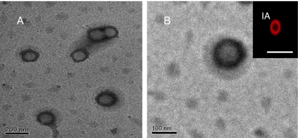
2.2.1. Transmission Electron Microscopy 33
2.2.2. Scanning Electron Microscopy 35
2.2.3. Confocal Microscopy 38
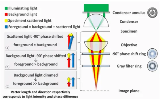
2.2.4. Phase Contrast Microscopy 39
2.3. Spectroscopic Techniques 41
2.3.1. Ultraviolet Visible Spectroscopic analysis 41 2.3.2. X-ray Photoelectron Spectroscopy 43
2.3.3. Energy Dispersive X-ray 44
2.4. Other Instruments 46
2.4.1. Zeta sizer 46
2.4.2. Particle Size Analyzer 47
2.5. Molecular Biology Techniques 49
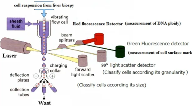
2.5.1. Flow cytometry (FCM) 49
2.5.2. Quantitative Real-Time Reverse Transcriptase
Polymerase Chain Reaction (qRT-PCR) 51
2.5.3. Agarose gel electrophoresis 53
2.6. Conclusion 54
3. Importance of Gene therapy and use of
polymeric nanoparticles in siRNA delivery 55
3.1. Introduction 56
3.2. Importance of Gene Therapy 56
3.3. Advantages of siRNA delivery 57
3.4. siRNA Delivery using Nanoparticles 59
3.5. Criteria of siRNA delivery system for cancer
therapy 60
3.6. Potential siRNA delivery systems for cancer therapy
62 3.7. Chemical modifications of anti-cancer siRNA 63 3.8. Polymer nanoparticles synthesized for anti-cancer
siRNA delivery system
64
3.9. Conclusions and future prospects 66
3.10. References 67
4. Hollow Polymeric (PLGA) Nano-capsules synthesis by solvent emulsion evaporation method for enhanced drug encapsulation and release efficiency
71
4.1. Introduction 72
4.2. Experimental 74
4.2.1. Materials and methods 74
4.2.2. Hollow NP synthesis (HNPs) 75
4.2.3. Non-hollow Nanoparticle Preparation (non HNPs) 75
4.2.4. Characterization of NPs 76
4.2.5. Drug encapsulation and loading 77
4.2.6. Drug release from NPs 78
4.3. Results and Discussion 79
4.3.1. Scanning electron microscopy (SEM) 80 4.3.2. Transmission Electron Microscopy (TEM) 81 4.3.3. Drug encapsulation and release efficiency 85
4.4. Conclusion 90
4.5. References 91
5. Strategist PLGA Nano-capsules to deliver siRNA for Inhibition of Carcinoma and
Neuroblastoma cell lines by knockdown of MYC proto-oncogene using CPP and PNA
95
5.1. Introduction 97
5.2. Materials and methods 99
5.2.1. Materials 99
5.2.2. Synthesis of PLGA Hollow nanoparticle
preparation (HNPs) 100
5.2.3. siRNA duplex preparation 100
5.2.4. Preparation of siRNA encapsulated PLGA nanoparticles
102
5.2.5. Release of siRNA 103
5.2.6. Preparation of Functionalized PLGA HNPs 104 5.2.7. Preparation of fluorescently labeled NPs 105
5.2.8. Characterization of NPs 106
5.2.9. Analysis of Cell Proliferation 106
5.2.10. Flow cytometry 108
5.2.11. Cellular uptake of nanoparticles 108
5.2.12. RNAi Treatment. 110
5.2.13. Relative gene expression studies using qRT-PCR 110
5.3. Results 111
5.3.1. Nanoparticle Characterization 111
5.3.2. siRNA encapsulation efficiency 112
5.3.3. siRNA release efficiency 112
5.3.4. Characterization of functionalized NPs 114 5.3.5. Effect of nanoparticles on cell 116 5.3.6. Cellular and nuclear uptake of PAPHNPs and
TPAPHNPs 118
5.3.7. Effect of NPs on gene expression 127
5.4. Conclusion 129
5.5. References 130
6 Conclusion 135
Acknowledgement i
List of Publications iii
Conference v Abbreviations vii !
!
!
! !!
!
!
!
!
!
!
!
!
!
!
Doctor’s Thesis
Regime of gene silencing: Efficient siRNA delivery into
cancer cells using nanocapsules
Archana Mukundrao Raichur
4R10120001
Doctor Course
Course of Bio-Nano Science Fusion
Graduate School of Interdisciplinary New Science
Toyo University, Japan
December 2014
! !
Chapter 1
History of Techniques used in siRNA
delivery in the field of nanomedicine
Abstract
Nanomedicine is giving rise to advanced nano-biomedical techniques and producing successful results in gene delivery. The potential ability of small interfering RNA (siRNA) to silence abnormal gene over-expression created a new horizon in the field of gene therapy. The major hindrances in the application of siRNA therapeutics are safety of gene, stability and effective delivery of siRNA in in vivo to reach the targeted cells. In this chapter, we present a brief history on the techniques used so far in siRNA delivery. We also mentioned about the future aspects of designing nanoparticles for effective siRNA delivery.
1.1. Introduction
Nanomedicine is a rapidly developing field in science giving satisfactory and promising results in drug and gene delivery. Until now, nanoparticles (NPs) played a major role in drug delivery. They are also utilized vastly in the field of gene delivery; especially in RNAi (RNA interference) therapeutics [1]. Various types of NPs are administered and have been proved efficacious in gene delivery; however the research is still in progress.
Organic and inorganic NPs, especially those, which are cationic in nature, show enhanced results in drug and gene delivery. NPs based targeted gene therapy aims to shuttle gene to the site-specific target so as to deliver effective concentrations of therapeutic drugs [2]. Despite many cellular and tissue-level hurdles, non-viral deliveries of NP-based approaches have been developed and hold great potential to transform medicine effectively [1, 3]. NPs have played a major role in gene targeting and delivery methods, giving the results for down-regulation of certain overexpressed gene in the diseased cells. siRNA treatment in non-viral gene delivery includes the insertion of siRNA using various types of NPs [4].
siRNA are small interfering RNA segments that are 23-29 nucleotides long and are categorized in non-coding genes. These siRNAs have the ability to silence the expression of almost all genes present in eukaryotic cells hypothetically. In practice, many diseases have been treated with high success rate. Therefore, siRNA treatment is gaining attention among researchers. RNAi treatment is giving effective results however there are some barriers that siRNA faces when administered inside the bloodstream and cell [4-6].
The major drawbacks using siRNA-based gene therapies without using any vectors are (i) low cytosol uptake, (ii) degradation of gene by enzymes, nucleases and (iii) rapid clearance through kidney, spleen, liver following systemic administration [5, 6]. The negative charge of siRNA fragments prevents active intracellular delivery. Cell membrane also has a negatively charged backbone and higher molecular weight, leading to electrostatic repulsion. Therefore, in this dilemma the NPs play a critical role in transportation of siRNA [4, 7]. Nanotechnology along with gene delivery is a reliable method that results in siRNA delivery in the reduction of multiplication of cancer cells by cleaving messenger RNA (mRNA) & breaking down the transcription resulting in no protein synthesis. NPs, which are inert, cationic, relatively moderate size (ranging from 60~300 nm) and having a large surface area are used and have shown promising results in siRNA delivery [4-12].
So far NPs have been an excellent vector for gene delivery, however the major barriers faced in the complete delivery of siRNA are (i) immune clearance through the liver and spleen, (ii) permeation through the endothelium into target tissues, (iii) penetration through the tissue and renal clearance, (iv) endosomal escape in target cells, (v) diffusion through the cytoplasm and (vi) entry into the nucleus. Also, the bio distribution of NPs varies according to the route of transport. The cellular uptake of the NPs carrying siRNA is unequal, leading lower effect and thereby increase in dose of the siRNA-NPs complex becomes necessary [13, 14].
In this chapter we focused on the techniques that are used to formulate NPs for targeted delivery of siRNA. We have emphasized on the upcoming techniques used so far in fabrication of different kinds of NPs and the effect in
targeted delivery. These different techniques involved in siRNA delivery give a clear picture for inventing novel ideas for delivering the drug completely in the cells in addition to bypassing the existing barriers.
1.2. Techniques for siRNA delivery
In the past decade siRNA delivery has been researched thoroughly. Transfection, electroporation and viral gene transfer were the delivery methods used earlier for gene delivery. However, due to emergence of nanotechnology the transfection of siRNA became easy and reliable [15]. Many NPs specially carbon nanotubes, lipoplexes, polymer complexes were widely used in siRNA transfection as a delivery vectors [16, 17]. Selection of siRNA delivery method depends on three aspects namely efficiency of siRNA, toxicity and optimization of the activity of the siRNA or siRNA-nano-conjugates. siRNA treated to a certain cell population must have the potency of target-gene knockdown at a specific siRNA concentration and systemic administration. The highly efficient delivery method depends on the rate of conjugation between siRNA and nano-complexes. The efficient delivery must target and affect the targeted gene with minimum amount of siRNA. Fine-tuning of experimentation is required for acquiring the target-gene down-regulation [16, 18]. Even though drugs are directly accessible to many tissues and can reach the tissues through systemic administration with the help of delivery agents in bloodstream, overdose and side effects of drugs are still persisting.
Till now techniques like encapsulation, conjugation (chemical reaction between targeting moieties and siRNA) was studied. Recent developments in
siRNA delivery techniques include layer-by-layer assembly (LbL) assembly and PEGylation on the NPs.
1.2.1. Barriers to systemic siRNA delivery
RNAi is a biological pathway in eukaryotic cells and is considered to be the fundamental pathway that cleaves complementary mRNA in sequence-specific manner. The lengthy double stranded RNA is introduced into the cytoplasm that triggers RNAi and further cleaved into siRNA (21–23 nucleotides long) [19]. Further, the siRNA recognizes complementary mRNA and is cleaved by the Dicer complex. Practically the siRNA is directly inserted into nucleus. In practice, siRNA can be synthetically produced and then directly introduced into the nucleus of the cell. When inserted appropriately the designed siRNA targets the gene, following this RNAi machinery activates and silences that gene in the body, giving it a broader therapeutic potential than specific small-molecule drugs. Even though the siRNA are capable of knocking down the disease-causing gene including cancer, siRNA needs to overcome certain barriers in vivo. Safe and effective delivery of siRNA is still a hurdle. Certain reliable and efficient systems must be developed for complete delivery of siRNA. While ‘naked’, chemically modified siRNA have shown efficacy in certain physiological settings [20]. Some siRNA get cleared from the renal and hepatic system very easily. This is because naked siRNA is subject to degradation by enzymes, and is too large (~14 kDa) and negatively charged to cross-cellular membranes. The issue of non-toxic and effective delivery is a key challenge and serves a barrier between siRNA interference usage, technology and its therapeutic application [4, 21].
cytoplasm and further the nucleus. Post-injection, the siRNA nano complex must navigate the circulatory system of the body while avoiding kidney filtration, uptake by phagocytes, aggregation with serum proteins, and enzymatic degradation by endogenous nucleases. Phagocytosis is one of the greatest immunological barriers found in extracellular matrix of tissues. Phagocytic cells such as macrophages and monocytes remove foreign material from body [22]. Unfortunately, phagocytes are efficient in eliminating therapeutic nano-complexes and macromolecules from the body, and therefore certain reliable techniques must be adopted to avoid opsonization, enzymatic infection when designing gene delivery vehicles [22, 23].
Efficient technique and different types of nanomaterials must be utilized for siRNA deliveries that have high loading capacity with maximum release efficiency. siRNA must be designed and also coupled with NPs in such a way that siRNA should not get cleaved in bloodstream. Also the technique must be accessible and should not affect the activity and longevity of siRNA [24-36]. Further, we have discussed about certain techniques used now a days that are studied in siRNA delivery.
1.3. Layer by layer formulation [LbL assembly]
1.3.1. Introduction
The designing and fabrication of nano carriers for the siRNA delivery has gained interest recently because of their clinical applications. Decher et al were the first group that introduced the LbL assembly method [38]. This assembly has attracted extensive attention in the field of siRNA delivery. Its
advantages in biomedical applications like easy preparation, versatility, and capability of assembling high loadings of different types of bio-structures in layers helps in formulating different types of NPs [39]. Fine control over the NP structure and robustness of the bio-moieties under provided physiological conditions enhance the activity of the vectors used. NPs used in gene delivery form ideal nano-vector. In this situation neutral moieties and positively charged biomolecules like antibodies, cell-penetrating peptides become necessary to be used in targeting application. Therefore, the LbL assembly helps to accumulate and control the size and structure that facilitates the targeted and safe delivery of the gene inside the specific cell [40-42].
Elbakry A et al [43] focused on the siRNA delivery using modified Gold NPs (AuNPs) with active and relevant siRNA molecules in LbL approach and cellular delivery. AuNPs are widely used in this type of delivery system because of its remarkable advantages like straightforward synthesis, easy surface modification, availability in different sizes and high biocompatibility with cells and tissues. Normally in LbL assembly technique the NPs are first addressed to surface modification by binding it mercaptoundecanoic acid, polyethylene glycol (PEG) and polyethyleneimine (PEI) [44-46]. Further, these surface-modified NPs are subjected to bind the subsequent layers of siRNA and polyelectrolytes with final completion of positively charged moiety around. This increase in layers can be controlled. The size of NPs could be controlled for suitable delivery of siRNA [41, 47].
To obtain a well-defined siRNA coated (layered) NPs, two oppositely charged polyelectrolytes are adsorbed on one another so as to keep the characteristics of the siRNA intact keeping siRNA safe from external forces. In
the LbL technique charge-reversible copolymers are used to improve the endosome escape capacity. Thus LbL technique improves the efficiency of transfecting the siRNA across the cell membrane [48-50]. The other main feature of LbL technique is that the film formulated is non-erodible regulatory membrane for release of siRNA to escape from the serum nucleases during delivery. These LbL assemblies show two significant approaches, namely the structural aspect and film deposition method between the interaction and driving forces during the film assembly [51, 52].
1.3.2. Structural aspect
The fabrication of multilayer on a substratum by consecutive adsorption of polymeric polyanions and polycations, polyelectrolytes controls the shape and size of functionalized NPs. It is obvious that the simplest lattice is a structure of multiple polyanion /polycation layers. This architecture built by very flexible cationic and anionic polyelectrolytes, like poly (allylamine hydrochloride) (PAH) and poly-styrenesulfonic (PSS) acid leads to the formation of one-dimensional structures along the layer. Further, if this film gets a strong hold on a substratum, then the strength of the whole moiety increases giving a structural integrity [53, 54].
More rigid ionic building blocks could be envisioned to reduce layer overlap. For example, creating a difference in the adsorption layer lipid bi-layers can be incorporated in between two anionic and cationic adsorbed moieties for the longevity of the siRNA incorporated in the NPs. In LbL assembly, the periodic laminating of organic and inorganic compounds gives a well-defined sandwiched multilayered film with distinguished super-lattices, which can be easily characterized by X-ray analysis, Zeta-potential analysis.
Size distribution is analyzed using a dynamic light scattering technique [55]. In these Nano composites, both the experimental and theoretical energy transfer efficiencies were correlated to show that the morphology of films are precisely on the molecular length scale, and that the polycation layer, once adsorbed hardly gets eroded by external forces [53, 56].
1.3.3. Polyelectrolyte assembly for deposition of film through
interaction and driving forces
In a solution, there are electrostatic interactions between oppositely charged polyelectrolytes. These oppositely charged polyelectrolytes readily get adsorbed because of their specific nature as a thin film deposition. There is an enthalpic contribution between point charges on the oppositely charged polyelectrolyte chains, and entropic contribution due to conformational changes [55, 56]. These charges on the polyelectrolyte chain are surrounded by counter ions from the aqueous solution.
Usually the balance between enthalpic and entropic contribution changes with the salt concentration in the solution. The electrostatic forces are higher in high salt concentration than that of low salt concentration. This enthalpic complexion driven by entropy complex in the salt concentration depends on the nature of electrolyte used in solution [55]. Temperature also plays a great role in this film assembly. It is found that the growth of the polyelectrolyte films turns to exponential when the concentration of the supporting electrolyte is increased. Thus the step-by-step assembly in the formation of film depends on the external parameters like salt concentration, temperature and the co-solvents used in the experiment [54-56].
Although there are advantages of layer-by-layer assembly techniques, there are certain limitations that are faced in gene delivery when very nano-scaled particles are used. The major disadvantage is water insoluble substances are used that is difficult for transportation of the gene through blood capillaries. Sometimes water-soluble moieties are modified with lengthy alkyl chains (either covalently or non-covalently) to adsorb on the nanocarrier [57].
In the synthesis of functionalized NPs where LbL technique is used, the interfacial width of rough surfaces does not give any well-defined scattering length gradients. But it depends on the spread and the distance between the layers. The internal organization in the LbL technique can be decayed monotonically away from the substrate. To overcome these limitations an alternate assembly technique is needed so as to deliver gene in the targeted region. The LbL technique may give a firm assembly; however, there are many other techniques like Encapsulation, Conjugation and PEGylation techniques that are used widely for gene delivery. The mechanisms of the LbL structure have proven successful and well developed only with polymeric ions and gold NPs [58, 59].
1.3.4. Polymer particles for LbL techniques
Polymer NPs are used for siRNA delivery because of their affinity towards holding siRNA and their proper delivery in cells. Positively charged polymer NPs have the potential ability to bind and condense large nucleic acids into NPs. They serve as efficient gene delivery agents with siRNA. LbL assembled polyelectrolyte multilayers (PEMs) were engineered to tune surface properties to modulate the cell adhesion on a surface of NPs [60].
Dimitrova M et al [61] demonstrated a fabrication of cell-degradable multilayered polyelectrolyte films (MPF) with PEI-siRNA complexes targeting cell- infection. MPFs were monitored for synthesizing the layered films using Quartz Crystal Microbalance (QCM). To form this PEI-siRNA complex, positively charged layer complex, adsorption technique of layering alternative precursor films were used [61, 62]. Electrolytes namely lysine and poly-glutamic acid contributed in supporting the film assembly. The film thickness of the polymer bilayers was found to be 150nm. The adsorption of PEI-siRNA increased till 10nm thickness. After the treatment to the cells the amount of siRNA released from culture medium was less for newly formed MPFs. These MPFs suggested the passive diffusion and spontaneous release of siRNA from films till the first deposited layer. These films showed efficient inhibition of nucleic acid replication using the delivered siRNA [61]. However the transfection ability of siRNA was limited.
Transfection was assessed in another framework where siRNA particularly for human PKR gene (protein kinase RNA-activated) was analyzed. A typical method to deliver small interfering RNA (siRNA) that capitalizes on a forward transfection method was adopted to increase siRNA transfection. A multilayer assembly mediated forward transfection (MFT) method separates the substrate-mediated delivery from the cell. Hence, pH responsive LbL assembled multilayer was used as the delivery platform. Micro-contact printing technique (µCP) pattern was utilized as transfection method. 25-kDa linear polyethylenimine (LPEI) was optimized as the siRNA transfection reagent through NPs. MFT efficiency ≥ 60% was achieved for LPEI-siRNA NPs in siRNA delivery system. Thus cationic polymers with siRNA complexes have
shown potential for application in the systemic assembly of siRNA layers on NPs for successful siRNA delivery. [63]
1.3.5 Other nanoparticles
AuNPs are the emerging NPs that are widely used in the nano-gene delivery systems. AuNPs being inert in nature and easy to customize, are coated with biodegradable polymers and served as the idealistic nano-carriers for siRNA delivery. Deng et al generated a nano-layered siRNA loadable LbL film onto functionalized NPs. They developed a substratum that was tailored to achieve highest loading of siRNA and other biomolecules, secured delivery, stability of siRNA in serum and tumor targeting [64]. They demonstrated the ability of AuNPs as an ideal gene carrier in MDA-MB-468 to silence GFP overexpression. This idea of creating a platform for siRNA suggests the reliability of LbL technique used in gene delivery.
Tan YF et al demonstrated the efficient delivery of siRNA using LbL NPs. The feasibility of encapsulating SPARC-siRNA using LbL NPs with poly (L-arginine) (Arg) and dextran as polyelectrolytes showed cellular binding and uptake of LbL NPs as well as siRNA delivery in FibroGRO cells. siRNA molecules were efficiently coated onto hydroxyapatite NPs [65].
Shutao Guo synthesized charge-reversal functional AuNPs using LbL assembly technique and delivered RNA (siRNA) and plasmid DNA into cancer cells. Cyanine 5-siRNA/polyethyleneimine/ cis-aconitic anhydride functionalized poly-allylamine/ polyethyleneimine/ 11-mercaptoundecanoic acid-gold NPs (cy5-siRNA/ PEI/ PAH-Cit/ PEI/ MUA-AuNP) complexes showed cytoplasmic uptake of cy5-siRNA [66]. Charge-reversal co-polymer has the property of pH-dependent shift charge nature between positive and negative.
The charge-reversal copolymers are used to improve the nucleic acid delivery efficiency by enhancing endosome escape capacity [67, 68].
Elbakry A et al therefore used LbL technique to obtain mono-dispersed AuNPs and deposited the oppositely charged polyelectrolyte thin films on the solid surfaces. AuNPs form an excellent tool for studying the size and surface properties that influence the portal entry into the cells [69]. Cystamine modified AuNPs were functionalized with hyaluronic acid (AuCM)/ siRNA/ polyethyleneimine (PEI)/ hyaluronic acid (HA) for intracellular delivery of siRNA by HA receptor mediated endocytosis. The HA in the outer layer of the AuCM/ siRNA/ PEI/ HA complex contributed to the stability and target specific gene silencing. This complex therefore, had a capacity of target specificity and ultimately gene knockdown. The layers one by one react within the cell and then the siRNA remains safe from the endosomic reactions when administered through blood circulation. Hence LbL assembly technique is widely used in siRNA delivery system and still the work is in progress with great demand and applications.
Recently, Jaganathan H et al reported the presence of tumor initiating cells (TICs) that are more lethal than the existed cancer cells [70]. These breast cancer cells are drug resistant and reappear even it is treated at initial stage. Hence the TICs were treated with siRNA that silenced Signal Tranducer and Activator of Transcription 3 (STAT 3) expression levels in MDA-MB cancer cells. They used LbL assembly technique and layered siRNA on Au tagged with poly-L-lysine. This clearly suggests the deposition of different bio-moieties on the surface of Au. Three layers were introduced on Au substratum and siRNA was delivered into the cytosol. The release of siRNA and poly-L-lysine was
measured spectrometrically at definite time intervals and accurate absorbance. Therefore in this study, LbL assembly serves as a reliable technique in delivering siRNA in TICs also. This technique could be used for siRNA delivery in cancer cells too.
1.4. Encapsulation Technique
1.4.1. Introduction
In the field of RNAi therapeutics it is known that siRNA reacts with cytoplasmic proteins and faces renal clearance inside the cell. Also, this mechanism includes first interaction with cell membrane, endosomal escape and finally complete escape from vesicle, so that siRNA gets delivered in the cytoplasm as mentioned earlier. Moreover, siRNA cannot be injected intravenously into the bloodstream as plasmatic nucleases degrade it. To overcome this consequence complete encapsulation of siRNA and its safe delivery is required [71].
Encapsulation is encasing the cargo material inside a capsule. Encapsulation of siRNA was widely adopted method using polymeric NPs, lipid NPs, liposomes, tri-block polymeric NPs and hybrid NPs were widely used giving highest encapsulation efficiency (80-90%) of siRNA. These NPs hardly interact with cytoplasmic proteins. siRNA are physically encapsulated in NPs and hence are safe. Therefore NPs face and escape endocytosis and deliver siRNA in cytosol [71, 72].
Cun D et al [73] determined the effect of PLGA (poly lactic co-glycolic acid) used during encapsulation of siRNA in synthesizing siRNA encapsulated PLGA NPs. They reported that formation of siRNA/ PLGA NPs depend on the various factors like (i) volume ratio between the inner water phase and the oil
phase, (ii) the PLGA concentration, (iii) the sonication time, (iv) the siRNA molecules, (v) charge on the molecule and the amount of acetylated bovine serum albumin (Ac-BSA) in the inner water phase that affects the encapsulation efficiency. The encapsulation efficiency finally depends upon the capacity of the molecule to get physically encapsulated in the NPs without having any effect on the activity of siRNA. Lipids substituted with PEI, which have low molecular weight, were used for siRNA delivery in MDA-MB-231 human breast cancer cell lines. The 50 to 120 fold of survivin down regulation was due to siRNA, which was hydrophobically encapsulated with modified PEI in lipid NPs. Toxicity of PEI was evaluated and found that the siRNA delivery was effective and safe [73-76].
Composite nano fibers of PLGA prepared by electro spinning were subjected to siRNA delivery. The higher integrity of encapsulated siRNA/ chitosan polyplex released siRNA to inhibit 50% of over expressed endogenous enhanced green fluorescent protein (EGFP) in H1299 cells. Hybrid NPs composed of human IgG and poloxamer 188 were efficiently used for siRNA gene delivery in A549 cells [77]. siRNA encapsulated NPs elicit immune response avoiding renal clearance and nuclease degradation that silenced the mutated KRAS gene successfully and siRNA/ NPs therapy was implemented. The important parameter that affects the encapsulation efficiency is the distribution of material that is dispersed in NPs and the siRNA distribution inside the NPs. Even the smallest volume of solidification of dispersed phase and stability of siRNA can affect the siRNA encapsulation efficiency. In order to prevent gene loss and gene migration the equal distribution of siRNA in NPs is essential. Also the optimized dispersed phase and continuous phase ratio in
the NPs determines the encapsulation efficiency and its controlled release efficiency [77, 78].
1.4.2. Polymers used for encapsulation of siRNA
Polymers are used widely in research area for siRNA delivery. Polymer encapsulated siRNA NPs or polymer-coated NPs are not only used in in vitro, but are successful in clinical trials too. Tagami et al [79] recently reported siRNA delivery using PEGylated cationic liposomes. They found that post-second dose injection; whole siRNA gets degraded due to the production of anti-PEG IgM [79]. Therefore, the technique of siRNA encapsulation was introduced as PEGylated lipid nanocarrier and “wrapsome” was invented. “Wrapsomes” are composed of siRNA and cationic lipofection complex in the center, which was completely enveloped with a neutral lipid bilayer and hydrophilic polymers [80]. Colombo S et al demonstrated the siRNA release kinetics of lipid-polymer hybrid NPs. Also the ratio of siRNA and PLGA used for synthesizing the NPs affects the encapsulation of siRNA. The encapsulation of siRNA depends on the surface reorganization of NPs. siRNA in its physical form gets encapsulated when the PLGA is low [81].
1.4.3. Other nanoparticles used for siRNA delivery
Nowadays magnetic NPs, including super-paramagnetic iron oxide NPs (SPIONs) and magnetic iron tetroxide particles are used as nanotheranostics for gene delivery [82]. The large surface area of SPIONs makes their functional modification feasible, enabling the conjugation of targeting molecules, siRNA, and imaging agents for enhancing siRNA delivery [83]. Z Medarova et al reported the synthesis and characterization of a new dual-purpose probe for the simultaneous non-invasive imaging and delivery of siRNAs to tumors [84]. Jing
Chen performed experiment on radioiodine labeling of human vascular endothelial growth factor (131I-hVEGF) siRNA by the Bolton-Hunter method. It is simple and reliable, and improved technique to give high yield. The dual functional properties of I-hVEGF siRNA and magnetic NPs in tumor therapy increased the down regulation of VEGF. Imaging of NPs provided an attractive system where real time monitoring of gene delivery, gene therapy can be integrated. Therefore, the combined therapy of 131I-hVEGF siRNA/ SPIONs might be a future treatment option against carcinoma[85]. Lu W et al [86] used near-IR (NIR) light–inducible therapy for NF-κB down regulation and coined the mode of action as “photo-thermal transfection”. They attached folate receptors on hollow gold nanospheres and showed pulsatile release of siRNA using NIR irradiation [86].
1.5. PEGylation
1.5.1. Introduction
The PEGylation technique is widely adopted and preferred method of adhering highly stabilized polymers onto NPs. PEGylation simply refers to the decoration of a particle surface by the covalently grafting, adsorbing of PEG chains. PEGylation helps in maintaining the stability of cargo material inside the NPs by preventing proteolytic and enzymatic degradation [87-92]. Recent studies demonstrated the rejection ability of targeting moieties like proteins on PEGylated surfaces. It was possible to understand due to use of freeze-fracture transmission electron microscopy (TEM) [89].
Owing to the siRNA resistivity with the NPs, many of PEG-containing polymers have conjugated with NPs and proved their ability to impart specificity and integrity towards siRNA and preserve it till it is delivered. The
repeating units of PEG and polypropylene glycol contribute in the functionalization of NPs. Because of these chemical structures they are also known as polyethylene oxide (PEO) and polypropylene oxide (PPO) chains. The PEG, a PEO extension on the NPs becomes conjugated to the targeting moieties. These functionalized NPs are used for treatment of cancer cells [92-95].
Nowadays using multi-arm-PEGs, PEG-dendrimers, PEGs, star-PEG-co-block polymers conjugation technique have been a practice in delivery of siRNA in the cancer treatment and genetic diseases [90]. This technique actually is the process of covalent attachment of PEG-polymer chains to a NP specially inert or cationic in nature. PEGylation is basically a chemical reaction with a derivative of PEG with the target molecule and ultimately attached to a nanovector [90, 96-98]. PEGylation of the NPs improved the ability of the vector to cross the barrier like endosomal activity, lysosomal activity [99]. Multi-arm PEGs that were conjointly attached layers of PEG having a functional group at the end. One of the arms is attached to NP and other arms are free. This helps the multi-arm PEG to stabilize and the site becomes active for the adsorption of targeting molecules. These PEG-arms are resistant to serum protein giving siRNA protection from external forces and provides secured mode of delivery [100, 101].
1.5.2. Y-shaped PEGylation reagents
Functional Y-shaped PEGs have received growing interest due to some reports indicating improved biological efficacy compared to the analogous therapeutics in which a linear conjugating PEG was used. 1-(1H, 1H, 2H, 2H-perfluorodecyloxy)-3-(3, 6, 9- trioxadecyloxy)-propan-2-yl acrylate was
synthesized for the gene delivery purpose. Sometimes, conjugated three blocks namely hydrophobic, hydrophilic and a polymerizable vinyl group using epichlorohydrin as a conjugating agent are widely used. This technique is cost effective and has simple synthesizing process, hence could be utilized in large scale. Therefore the Y-shaped PEGylation reagents having specific functional groups that cannot have adverse effect on the activity of siRNA were used widely for siRNA conjugation and delivery system [102]. The macromolecular structure of the conjugating polymer has proven to be crucial for the improved properties of the corresponding bio-conjugates, compared to linear PEG, branched PEG chains [103].
1.5.3. Highly branched PEGylation reagents
Comb-shaped polymers with one specific site of attachment represent a new type of PEGylation agent. One such polymer is POLY PEG, where PEG ‘teeth’ are linked to a methacrylate backbone via an ester bond. POLY PEG has more degrees of freedom than linear PEG, enabling structure optimization. Conventional PEGylation agents are prepared by ring-opening polymerization of ethylene oxide, usually initiated by an alkoxide derived from an appropriate alcohol [102, 104].
1.6. Comparison of above techniques
In the past decade, it is an observation that PEGylation and encapsulation techniques are widely adopted for siRNA delivery. Even though the techniques are reliable the siRNA targeting and site-specificity is still a hurdle. Although the encapsulation of siRNA is maximum, the pulsatile release and instant release from the nano-vectors is still under research. The
encapsulation method was adopted so as to maintain the siRNA integrity and ultimately have safe delivery.
However, encapsulation of siRNA is found to be nearly 100% efficient, but delivery is still low [105]. In the method of PEGylation, the long chain of polymers had low fixable ability. There are chances that the polymer chain increases the total weight of the nanovector and thereby leading to low targeting of the cells. The PEGylation of nanovector faces, the effect of nuclease and serum degradation. Owing to large molecular weight the accumulation of the nanovector is also observed specially in in vivo application. The large molecular weight of the PEGylated nano vectors decreases the mobility inside the blood stream and therefore the siRNA delivery is hindered [106, 107].
The LbL technique that is the new approach in the scientific world is quite durable and reliable. It is observed that the neutral moiety as the substratum does not cause any adverse effect on the siRNA activity. As the layers can be controlled and siRNA can be released as per the necessity inside the cell, LbL technique can be used as the ideal technique. The layers generated by siRNA and targeting moieties do not react with each other due to electrostatic stabilization. The siRNA activity remains independent of the reacting activity with targeting moieties and is physically adsorbed on the nano vector [108]. Therefore LbL technique is gaining great demand in siRNA delivery field.
1.7. Summary and outlook
In this chapter, we discussed about the different techniques used widely in siRNA delivery. Of the above techniques, LbL technique is widely accepted and researchers are keenly interested in investigating the effect of siRNA
delivery and the target sites in cancer cells. A detailed review of NPs used for siRNA delivery with LbL technique is mentioned with their application.
Recent developments in biological drugs have broadened the scope of therapeutic targets against the human diseases. These emerging drugs, which include non-coding genes along with NPs, demonstrate successful gene delivery when compared to the weak activity of siRNA delivery alone. Also, it is reported that siRNA/ nano-complex delivery have shown promising results in clinical trials. Further to overcome certain barriers faced in siRNA delivery, improved synthetic carrier and chemical modification of RNA are used. Systemic delivery of siRNA is a necessary aspect so that unmodified siRNA do not tend to accumulate in the kidneys, liver, spleen like vital organs. The siRNA therapeutic relies on filtering organs of the reticulo-endothelial system. Application of siRNA delivery requires a synthetic carrier decorated with specific targeting moieties. The major advantage of siRNA nano-carriers is their ability to engineer tissue specificity. Hence, to overcome these limitations the biodegradable polymers having a highest encapsulation efficiency and pulsatile release efficiency should be employed for efficient and tissue-specific delivery. Highly advanced techniques are necessary to fully harness the power of RNA interference and increase the flexibility of RNA-based therapeutics. Engineered NPs, such as peptide nucleic acid siRNA (PNA-siRNA) chimeras, peptide-siRNA, ligand-siRNA and transferrin-decorated NPs, will continue to improve the delivery of RNA drugs. The widely adopted technique could be the LbL assembly as it involves the physical adsorption of siRNA rather any chemical activity. When nano vectors are taken into consideration, due to their size, accessibility to deliver siRNA is comparatively faster than the naked siRNA
alone. Therefore, the future prospects of RNA-based nano-carriers will require efficient technique to maximize gene potency while minimizing off target toxicity and immunogenicity. Also the proper administration technique is essential that will cure the diseases.
1.8. References
[1] Lee, J. M ,Yoon T. J, and. Cho Y. S,2013, Recent developments in NPs-based siRNA delivery for cancer therapy,Biomed Res Int, vol. 2013, p. 782041.
[2] Ozpolat,B A.. Sood A. K and. Lopez-Berestein G, 2010,Nanomedicine based approaches for the delivery of siRNA in cancer, J Intern Med, vol. 267, pp. 44-53.
[3] Cheng F. F.,. Chen, W. Hu, L. H. Chen, G. Miao, H. T.. Li, C. Z et al., 2013, Highly dispersible PEGylated graphene/Au composites as gene delivery vector and potential cancer therapeutic agent, Journal of Materials Chemistry B, vol. 1, pp. 4956-4962.
[4] Whitehead, K. A. Langer R., and. Anderson, D. G 2009, Knocking down barriers: advances in siRNA delivery, Nat Rev Drug Discov, vol. 8, pp. 129-38.
[5] Reischl D and. ZimmerA,2009, Drug delivery of siRNA therapeutics: potentials and limits of nanosystems,Nanomedicine, vol. 5, pp. 8-20. [6] Wang,J Lu,Z Wientjes, M. G and. Au, J. L 2010,Delivery of siRNA
therapeutics: barriers and carriers," AAPS J, vol. 12, pp. 492-503.
[7] Thakur,A Fitzpatrick, S. Zaman A.,. Kugathasan, K. Muirhead B,. Hortelano G, et al.,2012, Strategies for ocular siRNA delivery: Potential and limitations of non-viral nanocarriers,J Biol Eng, vol. 6, p. 7.
[8] McCarroll J and. Kavallaris, M 2012, NPs delivery of siRNA as a novel therapeutic for human disease, Nucleus, vol. 7, p. 75.
[9] Rana T. M, 2007, Illuminating the silence: understanding the structure and function P. S of small RNAs,Nat Rev Mol Cell Biol, vol. 8, pp. 23-36.
[10] Lee R. C and. Ambros V,2001, An extensive class of small RNAs in Caenorhabditis elegans,Science, vol. 294, pp. 862-4.
[11] Lagos-Quintana M., Rauhut R., Lendeckel W, and. Tuschl T, 2001, Identification of novel genes coding for small expressed RNAs, Science, vol. 294, pp. 853-8.
[12] Wightman B.,. Ha I, and. Ruvkun G,1993, Posttranscriptional regulation of the heterochronic gene lin-14 by lin-4 mediates temporal pattern formation in C. elegans, Cell, vol. 75, pp. 855-862.
[13] Barua S.and. Mitragotri S,2014, Challenges associated with Penetration of NPs across Cell and Tissue Barriers: A Review of Current Status and Future Prospects, Nano Today, vol. 9, pp. 223-243.
[14] Daniel de Paula et.al;2007, Hydrophobization and bioconjugation for enhanced siRNA delivery and targeting, RNA, Vol. 13, No. 4.
[16] Liu Z,. Sun, X. Nakayama-Ratchford N, and. Dai H,2007, Supramolecular chemistry on water-soluble carbon nanotubes for drug loading and delivery, ACS Nano, vol. 1, pp. 50-6.
[17] Urban-Klein B.,. Werth S, Abuharbeid S.,. Czubayko, F and. Aigner A, 2005, RNAi-mediated gene-targeting through systemic application of polyethylenimine (PEI)-complexed siRNA in vivo, Gene Ther, vol. 12, pp. 461-6.
[18] Low,L S. Henne W. A, and. Doorneweerd D. D, 2008, Discovery and development of folic-acid-based receptor targeting for imaging and therapy of cancer and inflammatory diseases, Acc Chem Res, vol. 41, pp. 120-9.
[19] Gregory J. Hannon, 2002, RNA interference, Nature, vol. 418,pp 244-251.
[20] Morrissey,., Lockridge D. V,., Shaw J. A,., Blanchard L,., Jensen K, , Breen, K. Hartsough W.,,., Machemer K,., Radka L,., Jadhav S,. et al. V 2005, Potent and persistent in vivo anti-HBV activity of chemically modified siRNAs., Nat. Biotechnology. Vol. 23, pp 1002- 1007.
[21] Zabner, J., Fasbender, A. J., Moninger, T., Poellinger, K. A. and Welsh, M. J.,1995, Cellular and molecular barriers to gene transfer by a cationic lipid., J. Biol. Chem. vol. 270, pp18997-19007.
[22] Monika Dominska, Derek M. Dykxhoorn, 2010, Breaking down the barriers: siRNA delivery and endosome escape, Journal of Cell Science, vol. 123, pp 1183-1189.
[23] McNamara J. O, 2nd. Andrechek, E. R,. Wang, Y. Viles, K. D. Rempel, R. E. Gilboa E, et al., 2006, Cell type-specific delivery of siRNAs with aptamer-siRNA chimeras,Nat Biotechnol, vol. 24, pp. 1005-15.
[24] Jonathan E. Zuckerman1,Chung Hang J. Choi1 , Han Han, and Mark E. Davis, 2012, Polycation-siRNA NPs can disassemble at the kidney glomerular basement membrane, PNAS ,vol. 109 ,no. 8 . vol. 3137– 3142. Feb.2012.
[25] Roh Y. H,. Lee J. B,. Shopsowitz, K. E Dreaden E. C.,. Morton S. W,. Poon, Z et al.,2014, Layer-by-layer assembled antisense DNA microsponge particles for efficient delivery of cancer therapeutics,ACS Nano, vol. 8, pp. 9767-80,
[26] Shen J.,. Kim H. C, Mu C., Gentile E., Mai J.,. Wolfram J, et al., 2014, Multifunctional gold nanorods for siRNA gene silencing and photothermal therapy,Adv Healthc Mater, vol. 3, pp. 1629-37.
[27] Dreaden E. C,. Morton S. W,. Shopsowitz, K. EChoi J. H.,. Deng Z. J,. Cho N. J, et al., 2014, Bimodal tumor-targeting from microenvironment responsive hyaluronan layer-by-layer (LbL) NPs, ACS Nano, vol. 8, pp. 8374-82.
[28] Wang Y. C,. Yang C. B,. Hu R,. Toh H. T,. Liu X,. Lin G. M, et al., 2015, Assembling Mn:ZnSe quantum dots-siRNA nanoplexes for gene silencing in tumor cells,Biomaterials Science, vol. 3, pp. 192-202.
[29] Ganas C,. Weiss A,. Nazarenus M,. Rosler S,. Kissel T,. Gil P. R, et al., 2014, Biodegradable capsules as non-viral vectors for in vitro delivery of PEI/siRNA polyplexes for efficient gene silencing, Journal of Controlled Release, vol. 196, pp. 132-138.
[30] Chen Y.,. Gu H,. Zhang D. S,. Li, F. Liu T, and. Xia W,2014, Highly effective inhibition of lung cancer growth and metastasis by systemic
delivery of siRNA via multimodal mesoporous silica-based nanocarrier, Biomaterials, vol. 35, pp. 10058-69.
[31] Liao Z. X.,. Peng S. F,. Chiu Y. L,. Hsiao C. W,. Liu H. Y, Lim W. H., et al., 2014, Enhancement of efficiency of chitosan-based complexes for gene transfection with poly(gamma-glutamic acid) by augmenting their cellular uptake and intracellular unpackage, Journal of Controlled Release, vol. 193, pp. 304-315.
[32] Li Y. Y,. Zheng S. Q,. Liang X. L,. Jin Y. S,. Wu Y. D, Bai H. C., et al., 2014, Doping Hydroxylated Cationic Lipid into PEGylated Cerasome Boosts in Vivo siRNA Transfection Efficacy, Bioconjugate Chemistry, vol. 25, pp. 2055-2066.
[33] Godinho B. M. D. C, Ogier, J. R. Quinlan A.,. Darcy R,. Griffin B. T, Cryan, J. F. et al.,2014, PEGylated cyclodextrins as novel siRNA nanosystems: Correlations between polyethylene glycol length and NPstability, International Journal of Pharmaceutics, vol. 473, pp. 105-112.
[34] Forbes D. C and. Peppas N. A, 2014, Polymeric nanocarriers for siRNA delivery to murine macrophages, Macromol Biosci, vol. 14, pp. 1096-105.
[35] Leus N. G,. Morselt H. W,. Zwiers P. J, Kowalski P. S.,. Ruiters M. H, Molema G., et al., 2014, VCAM-1 specific PEGylated SAINT-based lipoplexes deliver siRNA to activated endothelium in vivo but do not attenuate target gene expression, International journal of pharmaceutics, vol. 469, pp. 121-131.
[36] Zhu H., Dong C.,. Dong H Ren, T.,. Wen X, Su J., et al., 2014, Cleavable PEGylation and hydrophobic histidylation of polylysine for siRNA delivery and tumor gene therapy, ACS Appl Mater Interfaces, vol. 6, pp. 10393-407.
[37] Wen Y. T.,. Zhang Z. X, and. L J i,2014, Highly Efficient Multifunctional Supramolecular Gene Carrier System Self-Assembled from Redox-Sensitive and Zwitterionic Polymer Blocks, Advanced Functional Materials, vol. 24, pp. 3874-3884.
[38] Decher G., Eckle M., Schmitt J., and. Struth B, 1998, Layer-by-layer assembled multicomposite films, Current Opinion in Colloid & Interface Science, vol. 3, pp. 32-39.
[39] Wood K. C., Chuang H. F.,. Batten R. D. Lynn, D. M, and Hammond P. T., 2006, Controlling interlayer diffusion to achieve sustained, multiagent delivery from layer-by-layer thin films, Proc Natl Acad Sci U S A, vol. 103, pp. 10207-12.
[40] Lee K. G, Hong J.,. Wang K. W, Heo N. S.,. Kim H do,. Lee S. Y, et al., 2012, In vitro biosynthesis of metal NPs in microdroplets,ACS Nano, vol. 6, pp. 6998-7008.
[41] Jewell C. M.and. Lynn, D. M 2008, Multilayered polyelectrolyte assemblies as platforms for the delivery of DNA and other nucleic acid-based therapeutics,Advanced Drug Delivery Reviews, vol. 60, pp. 979-999.
[42] Garza J. M,. Jessel, N. Ladam G,. Dupray V,. Mulle S r,. Stoltz J. F, et al., 2005, Polyelectrolyte multilayers and degradable polymer layers as multicompartment films, Langmuir, vol. 21, pp. 12372-12377.
[43] Elbakry A., Wurster E. C.,. Zaky A,. Liebl R, Schindler E.. Bauer-Kreise l, P, et al., 2012, Layer-by-Layer Coated Gold NPs: Size-Dependent Delivery of DNA into Cells,Small, vol. 8, pp. 3847-3856.
[44] Chuang H. F., Smith, R. C. and. Hammond P. T, 2008, Polyelectrolyte multilayers for tunable release of antibiotics, Biomacromolecules, vol. 9, pp. 1660-8.
[45] Fisman D. N, Reilly D. T.,. Karchmer A. W, and. Goldie S. J, 2001, Clinical effectiveness and cost-effectiveness of 2 management strategies for infected total hip arthroplasty in the elderly, Clin Infect Dis, vol. 32, pp. 419-30.
[46] Buchholz H. W,. Elson R. A, and. Heiner K t, 1984, Antibiotic-loaded acrylic cement: current concepts, Clin Orthop Relat Res, vol. 190, pp. 96-108.
[47] Moskowitz J. S.,. Blaisse M. R,. Samuel R. E,. Hsu H.-P, Harris M. B.,. Martin, S. D et al., 2010, The effectiveness of the controlled release of gentamicin from polyelectrolyte multilayers in the treatment of Staphylococcus aureus infection in a rabbit bone model,Biomaterials, vol. 31, pp. 6019-6030.
[48] Hammond P. T., 2012, Building biomedical materials layer-by-layer, Materials Today, vol. 15, pp. 196-206.
[49] Smith R. C, Riollano M.,. Leung A, and. Hammond P. T, 2009, Layer-by-layer platform technology for small-molecule delivery, Angew Chem Int Ed Engl, vol. 48, pp. 8974-7.
[50] Giuliano F.and. Warner, T. D 1999, Ex vivo assay to determine the cyclooxygenase selectivity of non-steroidal anti-inflammatory drugs, Br J Pharmacol, vol. 126, pp. 1824-30.
[51] Hales K and. Pochan D. J, 2006, Using polyelectrolyte block copolymers to tune nanostructure assembly, Current Opinion in Colloid & Interface Science, vol. 11, pp. 330-336.
[52] Cho J.,. Hong J,. Cha r K, and Caruso F., 2006, Nanoporous block copolymer micelle/micelle multilayer films with dual optical properties, J Am Chem Soc, vol. 128, pp. 9935-42.
[53] Volodkin D, Arntz Y.,. Schaaf, P. Moehwald H. Voegel, J. C, andBall V., 2008, Composite multilayered biocompatible polyelectrolyte films with intact liposomes: stability and temperature triggered dye release, Soft Matter, vol. 4, pp. 122-130.
[54] Ma N.,. Wang Y. P, Wang Z. Q., and Zhang X., 2006, Polymer micelles as building blocks for the incorporation of azobenzene: Enhancing the photochromic properties in layer-by-layer films, Langmuir, vol. 22, pp. 3906-3909.
[55] Marc Michel, Vale ́rie Toniazzo, David Ruch,Vincent Ball.2012, Deposition Mechanisms in Layer-by-Layer or Step-by-Step Deposition Methods: From Elastic and Impermeable Films to Soft Membranes with Ion Exchange Propertie Materials Science Volume 13 Article ID 701695, 13 pages.
[56] Advincula R, Aust E, Meyer W, Knoll W:1996, In-situ investigations of polymer self-assembly solution adsorption by surface plasmon spectroscopy. Langmuir , 12:3536-3540.
[57] Kirstein S:,1995, White-light electroluminescence from self-assembled Q-CdSe/PPV multilayer structures.Adv Mater, vol 9: pp802-805.
[58] Katsuhiko Ariga, Yuri Lvov et al.,1997, Assembling Alternate Dye-Polyion Molecular Films by Electrostatic Layer-by-Layer Adsorption. J.
Am. Chem. Soc. vol. 119,pp 2224-2231.
[59] Izquierdo A,. Ono S. S,. Voegel J. C,. Schaaf P, and Decher G., 2005, Dipping versus spraying: exploring the deposition conditions for speeding up layer-by-layer assembly, Langmuir, vol. 21, pp. 7558-67. [60] Christopher J. Detze,2011, Polyelectrolyte Multilayers in Tissue
Engineering, TISSUE ENGINEERING: Part B Volume 17, Number 2, pp 101-113.
[61] Dimitrova M.,. Affolter C, Meyer F.,. Nguyen I,. Richard, C. D Schuste r. G, et al., 2008, Sustained delivery of siRNAs targeting viral infection by cell-degradable multilayered polyelectrolyte films, Proc Natl Acad Sci U S A, vol. 105, pp. 16320-5.
[62] Yokota T, et al.,2003, Inhibition of intracellular hepatitis C virus replication by synthetic and vector-derived small interfering RNAs.EMBO Rep 4:602–608.
[63] Sumit Mehrotra et.al, 2009, Multilayer mediated forward and patterned siRNA transfection using linear-PEI at extended N/P ratios, Acta Biomater., vol. 5(5), pp. 1474–1488. 2009.
[64] Deng D,. Yan C,. Pan X, Mahfouz M.,. Wang J,. Zhu J. K, et al., 2012, Structural basis for sequence-specific recognition of DNA by TAL effectors, Science, vol. 335, pp. 720-3.
[65] Yang Fei Tan , Raghavendra C. Mundargi , Min Hui Averil Chen , Jacqueline Lessig ,Björn Neu ,. Venkatraman Subbu S ,Tina T. Wong,2014, Layer-by-Layer NPs as an Efficient siRNA Delivery Vehicle for SPARC Silencing, Small.vol.10, No. 9, pp1790–1798. [66] Shutao Guo et.al. 2010, Enhanced Gene Delivery and siRNA Silencing
by Gold NPs Coated with Charge-Reversal Polyelectrolyte, ACS Nano. VOL. 4 , NO. 9, pp. 5505–5511.
[66] Inoue, H.; Sato, K.; Anzai, J.,2005, Disintegration of Layer-byLayer Assemblies Composed of 2-Iminobiotin-Labeled Poly(Ethyleneimine) and Avidin.Biomacromolecules ,vol. 6, 27 –29. 44.
[67] Lee, Y.; Miyata, K.; Oba, M.; Ishii, T.; Fukushima, S.; Han, M.; Koyama, H.; Nishiyama, N.; Kataoka, K.2008, Charge-Conversion Ternary Polyplex with Endosome Disruption Moiety: A Technique for Efficient and Safe Gene Delivery. Angew. Chem., Int. Ed., 47, pp 5163– 5166.
[68] Xu, P. S.; Van Kirk, E. A.; Zhan, Y. H.; Murdoch, W. J.; Radosz, M.; Shen, Y. Q. 2007, Targeted Charge-Reversal NPs for Nuclear Drug Delivery. Angew. Chem., Int. Ed. 46, 4999–5002.
[69] Elbakry A, Zaky A., Liebl R.,. Rachel R,. Goepferich A, and Breunig M., 2009, Layer-by-layer assembled gold NPs for siRNA delivery, Nano Lett, vol. 9, pp. 2059-64.
[70] Jaganathan H,. Mitra S, Srinivasan S.,. Dave B, and. Godin B, 2014, Design and in vitro evaluation of layer by layer siRNA nanovectors targeting breast tumor initiating cells, PLoS One, vol. 9, p. e91986. [71] White P. J, 2008, Barriers to successful delivery of short interfering
RNA after systemic administration, Clin Exp Pharmacol Physiol, vol. 35, pp. 1371-6..
[72] Joshua McCarroll,2012, NPs delivery of siRNA as a novel therapeutic for human disease, Australian biochemist, vol 43 no 3 pp 09-20.
[73] Cun D,. Jensen D. K,. Maltesen M. J,. Bunker M. Whiteside, P,. Scurr D, et al., 2010. High loading efficiency and sustained release of siRNA encapsulated in PLGA NPs: quality by design optimization and characterization, Eur J Pharm Biopharm, vol. 77, pp. 26-35.
[74] Montazeri Aliabadi, H.; Landry, B.; Bahadur, R. K.; Neamnark, A.; Suwantong, O.; Uludag, H. 2011. Impact of Lipid Substitution on Assembly and Delivery of siRNA by Cationic Polymers. Macromol. Biosci, 11, 662–672
[75] Neamnark, A.; Suwantong, O.; Bahadur, R. K.; Hsu, C. Y.; Supaphol, P.; Uludag, H. 2009, Aliphatic lipid substitution on 2 kDa polyethylenimine improves plasmid delivery and transgene expression. Mol. Pharmaceutics 2009, 6, 1798–1815. Correction: 7, 618.
[76] Montazeri Aliabadi H.,. Landry B,. Mahdipoor, P and Uludag H., 2011, Induction of apoptosis by survivin silencing through siRNA delivery in a human breast cancer cell line,Molecular pharmaceutics, vol. 8, pp. 1821-1830.
[77] Lakshmikuttyamma A.,2014, Stable and Efficient Transfection of siRNA for Mutated KRAS Silencing Using Novel Hybrid NPs, Mol. Pharmaceutics.vol. 11, pp 4415−4424.
[78] Caruso F,. Caruso R. A, and. Mohwald H, 1998, Nanoengineering of inorganic and hybrid hollow spheres by colloidal templating, Science, vol. 282, pp. 1111-4.
[79] Tagami T, Uehara Y.,. Moriyosh i N, Ishida T., and Kiwada H., 2011, Anti-PEG IgM production by siRNA encapsulated in a PEGylated lipid nanocarrier is dependent on the sequence of the siRNA, Journal of Controlled Release, vol. 151, pp. 149-154.
[80] Nobuhiro Yagi et.al;2009, A NPsystem Specifically Designed to Deliver Short Interfering RNA Inhibits Tumor Growth In vivo, Cancer Res., vol. 69: (16).pp6531-39 .
[81] Colombo S.,. Cun D,. Remaut, K. Bunker M Zhang, J.. Martin-Bertelsen, B, et al., 2015, Mechanistic profiling of the siRNA delivery dynamics of lipid–polymer hybrid NPs,Journal of Controlled Release, vol. 201, pp. 22-31.
[82] Pankhurst, J.. Connolly Q. A,. Jones S. K, and Dobson J., 2003, Applications of magnetic NPs in biomedicine, Journal of Physics D-Applied Physics, vol. 36, pp. R167-R181.
[83] Willard M. A,. Kurihara, L. K. Carpenter E. E,. Calvin S, and. Harris V. G, 2004, Chemically prepared magnetic NPs, International Materials Reviews, vol. 49, pp. 125-170.
[84] Medarova Z. Pham, W., Farrar C. Petkova, V., and Moore, A. 2007, In
vivo imaging of siRNA delivery and silencing in tumors,Nature
medicine, vol. 13, pp. 372-377.
[85] Chen J., Zhu S.,. Tong, J. Li, F. Chen, Y. Han, et al., 2014, Superparamagnetic iron oxide L NPs mediated 131I-hVEGF siRNA inhibits hepatocellular carcinoma tumor growth in nude mice, BMC cancer, vol. 14, p. 114.
[86] Lu W.,. Zhang G,. Zhang R,. Flores L. G, 2nd, Huang Q.,. Gelovani J. G, et al., 2010, Tumor site-specific silencing of NF-kappaB p65 by targeted
hollow gold nanosphere-mediated photothermal transfection, Cancer Res, vol. 70, pp. 3177-88.
[87] Davis F. F, 2002, Commentary - The origin of pegnology, Advanced Drug Delivery Reviews, vol. 54, pp. 457-458.
[88] Chen H,. Paholak H,. Ito M,. Sansanaphongpricha K,. Qian W Che, Y., et al., 2013, Living' PEGylation on gold NPs to optimize cancer cell uptake by controlling targeting ligand and charge densities,Nanotechnology, vol. 24, p. 355101.
[89] Owens D. E, 3rd and. Peppas N. A,2006, Opsonization, biodistribution, and pharmacokinetics of polymeric NPs, Int J Pharm, vol. 307, pp. 93-102.
[90] Özcan I., Segura-Sánchez F.,. Bouchema l K,. Sezak M,. Özer Ö,. Güneri T, et al.,2010, Pegylation of poly (γ-benzyl-L-glutamate) NPs is efficient for avoiding mononuclear phagocyte system capture in rats, International journal of nanomedicine, vol. 5, p. 1103.
[91] Díaz-López R.,. Tsapis N,. Santin M,. Brida l S. L,. Nicolas, V. Jaillard D, et al.,2010, The performance of PEGylated nanocapsules of perfluorooctyl bromide as an ultrasound contrast agent,Biomaterials, vol. 31, pp. 1723-1731,
[92] Gref R,. Lück M,. Quellec P,. Marchand M, Dellacherie E., Harnisch S., et al., 2000, Stealth’corona-core NPs surface modified by polyethylene glycol (PEG): influences of the corona (PEG chain length and surface density) and of the core composition on phagocytic uptake and plasma protein adsorption," Colloids and Surfaces B: Biointerfaces, vol. 18, pp. 301-313.
[93] Otsuka H.,. Nagasaki Y, and. Kataoka K,20102, PEGylated NPs for biological and pharmaceutical applications, Advanced Drug Delivery Reviews, vol. 64, pp. 246-255.
[94] Gref R. Domb, A. Quellec, P,. Blunk T,. Muller R. H,. Verbavatz J. M, et al., 20102, The controlled intravenous delivery of drugs using PEG-coated sterically stabilized nanospheres, Advanced Drug Delivery Reviews, vol. 64, pp. 316-326.
[95] Ha J. C,. Kim S. Y, and. Lee Y. M, 1999, Poly(ethylene oxide)-poly(propylene oxide)-poly(ethylene oxide) (pluronic)/poly(epsilon-caprolactone) (PCL) amphiphilic block copolymeric nanospheres - I. Preparation and characterization, Journal of Controlled Release, vol. 62, pp. 381-392.
[96] Malkoch A M. M. N,.,2012, Biocompatible Dendritic Building Blocks for Advanced Biomedical Research, Material Matters, vol. 7, pp. 47-49. [97] Wang Q.,. Mynar J. L,. Yoshida M, Lee E., Lee M.,. Okuro K, et al.,
2010, High-water-content mouldable hydrogels by mixing clay and a dendritic molecular binder, Nature, vol. 463, pp. 339-43.
[98] Hedden R. C. Bauer, B. J.,. Smith, A. P Grohn F., and. Amis E, 2002, Templating of inorganic NPs by PAMAM/PEG dendrimer-star polymers, Polymer, vol. 43, pp. 5473-5481.
[99] Ghosh P,. Han G,. De M,.. Kim C K, and. Rotello V. M, 2008, Gold NPs in delivery applications,Adv Drug Deliv Rev, vol. 60, pp. 1307-15. [100] Mizrahi B,. Khoo X,. Chiang H. H,. Sher K. J,. Feldman R. G,. Lee J. J,
[101] Zhou Y.and Yan D., 2004, Supramolecular self-assembly of giant polymer vesicles with controlled sizes,Angew Chem Int Ed Engl, vol. 43, pp. 4896-9.
[102] Sinéad M Ryan, Giuseppe Mantovani, Xuexuan Wang, David M Haddleton,2008, Advances in PEGylation of important biotech molecules: delivery aspects, Drug Deliv. vol.5(4)pp.371-383.
[103] Krepler C., Certa U.,. Wacheck V,. Jansen B,. Wolff K, and Pehamberger H., 2004, Pegylated and conventional interferon-α induce comparable transcriptional responses and inhibition of tumor growth in a human melanoma SCID mouse xenotransplantation model,Journal of investigative dermatology, vol. 123, pp. 664-669.
[104]. Li, N. W. Yan T. Z, Li Z.,. Thurn-Albrecht T, and. Binder W. H, 2012, Comb-shaped polymers to enhance hydroxide transport in anion exchange membranes,Energy & Environmental Science, vol. 5, pp. 7888-7892.
[105] Tan M. L, Choong P. F., and. Dass C. R, 2009, Cancer, chitosan NPs and catalytic nucleic acids, J Pharm Pharmacol, vol. 61, pp. 3-12.
[106] Damodaran V and Fee, C. 2010, Protein PEGylation: An overview of chemistry and process considerations, European Pharmaceutical Review, vol. 15, pp. 18-26.
[107] Yu J. C.,. Zhu S,. Feng P. J,. Qian C. G, Huang, M. J. Sun, et al., 2015,Cationic fluorescent polymer core-shell NPs for encapsulation, delivery, and non-invasively tracking the intracellular release of siRNA,Chem Commun (Camb), vol. 51, pp. 2976-9,
[108] Kotov N. A, 1999, Layer-by-layer self-assembly: The contribution of hydrophobic interactions, Nanostructured Materials, vol. 12, pp. 789-796.