†Present address: Department of Digestive Surgery, Osaka Interna-tional Cancer Institute, Osaka 541-8567, Japan
Corresponding author: Hiroaki Saito, MD, PhD sai10@med.tottori-u.ac.jp
Received 2017 March 27 Accepted 2017 April 24
Abbreviations: ACTS-GC, Adjuvant Chemotherapy Trial of TS-1 for Gastric Cancer; BMI, body mass index; NRS2002, nutritional risk scoring 2002; OS, overall survival; PNI, Onodera’s prognostic nutritional index; PUGC, primary upper third gastric cancer; RGC, remnant gastric cancer; SE, tumor penetration of serosa; SGA, Sub-jective Global Assessment; SI, tumor invasion of adjacent structure
Use of Body Mass Index to Predict the Prognosis of Patients with Remnant
Gastric Cancer
Tomoyuki Matsunaga,*† Hiroaki Saito,* Tomohiro Osaki,* Yusuke Kono,* Yuki Murakami,* Hirohiko Kuroda,* Yoji Fukumoto* and Yoshiyuki Fujiwara*
*Division of Surgical Oncology, Department of Surgery, School of Medicine, Tottori University Faculty of Medicine, Yonago
683-8504, Japan
ABSTRACT
Background Remnant gastric cancer (RGC) is an un-common form of gastric cancer. The aim of this study was to investigate factors influencing the prognosis of patients with RGC.
Methods A total of 49 patients diagnosed with RGC and 214 patients with primary upper third gastric cancer (PUGC) at our institution between January 1990 and December 2014 were included. The clinicopathological characteristics, prognosis, and factors influencing prog-nosis were compared.
Results The body mass index (BMI) of RGC was significantly lower than that for PUGC (P < 0.0001). Multivariate analysis revealed that BMI and the depth of tumor invasion were independent prognostic factors in RGC. ROC analysis indicated that an optimal cut-off value for BMI was 20.6. Based on this value, patients were divided into two groups: BMIHigh (≥ 20.6) and
BMILow (< 20.6). The 5-year survival rates of patients
with BMIHigh early gastric cancer, BMIHigh advanced
gastric cancer, BMILow early gastric cancer, and BMILow
advanced gastric cancer were 90%, 83.3%, 64.3% and 33.8%, respectively, and the difference was statistically significant (P = 0.00023).
Conclusion Our retrospective study indicated a poor prognosis of RGC compared with PUGC, and that BMI could predict the prognosis of RGC. The prognosis of patients with BMILow advanced RGC was extremely
poor.
Key words body mass index; gastric cancer; prognosis; remnant gastric cancer
Gastric cancer is one of the most common malignancies and death from gastric cancer ranks second among all cancer deaths worldwide.1 Remnant gastric cancer (RGC)
is a less prevalent form of the disease, and the incidence of RGC has been reported as 1–7% of all gastric cancer cases.2-4 The most frequently used definition of RGC is
cancer in the remnant stomach after surgery for benign disease or cancer after surgery for gastric cancer found at least 5 years after the primary surgery. Surgery,
in-cluding gastrectomy, has been the standard treatment for peptic ulcer disease in previous decades.5 Recent
advances in peptic ulcer therapy, including specific treatment for Helicobacter pylori infection, have de-creased the prevalence of affected individuals for whom gastrectomy is indicated. Therefore, it is likely that the incidence of RGC following gastrectomy for benign disease will decrease in the future. However, the inci-dence of RGC following gastrectomy for gastric cancer is increasing as a result of improved survival and early detection by mass screening, especially in several Asian countries.6 Therefore, rates of RGC following gastric
cancer surgery have been increasing overall.7, 8
RGC is often diagnosed at an advanced stage, with higher rates of invasion of adjacent organs and lymph node metastases,9, 10 low rate of curative resection and,
consequently, a poor prognosis.10, 11 It has been also
re-ported that the prognosis for RGC is significantly worse than that for primary upper third gastric cancer (PUGC) that originates in the almost same lesion as RGC, when compared at the same stage, indicating the possibility that the characteristics of RGC might be different from those of primary gastric cancer.
Determining the postoperative prognosis in RGC patients is extremely important. Many studies have in-dicated that depth of invasion and presence or absence of lymph node metastasis are the most important prog-nostic factors in gastric cancer.12, 13 However, the factors
influencing the poor prognosis of patients with RGC remain unclear. The purpose of this study was to investi-gate the clinicopathological characteristics and outcomes
of surgery for RGC following distal gastrectomy. In par-ticular, we examined the factors related to the prognosis of patients with RGC.
MATERIALS AND METHODS Patients
This study was based on a retrospective analysis of 49 RGC patients following distal gastrectomy who under-went curative gastrectomy at our institution between January 1990 and December 2014. During the same period, 214 patients were pathologically diagnosed with adenocarcinoma confined to the upper third of the stom-ach and were also included in the current study. The clinicopathological findings were determined according to the Japanese Classification of Gastric Carcinoma.14
Patients were periodically checked for early recurrence by diagnostic imaging (chest X-ray, double-contrast barium meal study, upper gastrointestinal fiberscopy, ultrasonography, and computed tomography). Causes of death and patterns of recurrence were determined by reviewing medical records, including laboratory data, ultrasonography, computed tomography, scintigrams, peritoneal punctures, and laparotomies, or by direct in-quiry to family members. In some cases, postmortems were undertaken to determine the cause of death. Insti-tutional review board approval was obtained (1607A064), and informed consent requirements were waived for this study.
Statistical analysis
The Mann–Whitney U test and chi-square test were employed to evaluate differences in continuous and cat-egorical variables, respectively. The Youden index was calculated using receiver operating characteristic (ROC) analysis to determine an optimal cutoff value for body mass index (BMI) for survival analysis. Survival curves were calculated using the Kaplan–Meier method. Differ-ences between survival curves were examined with the log-rank test. Survival data shown in the current study were for overall survival (OS). We used multivariate analysis of factors considered prognostic of OS, with Cox’s proportional hazards model. SPSS Statistics (ver. 24.0 software, IBM) was used for all statistical anal-yses, with P < 0.05 considered statistically significant (2-tailed).
RESULTS
Clinicopathological features of RGC and PUGC The clinicopathological characteristics of both RGC and PUGC are shown in Table 1. Patients with RGC were significantly older than those with PUGC (P = 0.002). 6 patients with RGC were older than 80 years old (12.2%)
and one patient was over 85 years old. The frequency of male gender was significantly greater in RGC patients than in PUGC patients (P = 0.041). Furthermore, BMI was significantly lower in RGC patients than in PUGC patients (P < 0.0001). However, no significant differenc-es were observed in tumor size, depth of tumor invasion, lymph node metastasis, blood vessel invasion, lymphatic vessel invasion, and state of disease between RGC and PUGC.
Table1. The clinicopathological features of RGC and PUGC RGC (n = 49) PUGC (n = 214) P value Age (years) 72.4 ± 6.8 66.2 ± 12.2 0.002 Gender 0.041 Male 43 (87.8%) 158 (73.8%) Female 6 (12.2%) 56 (26.2%) Tumor size (mm) 42.9 ± 26.1 36.3 ± 23.1 0.14 BMI 20.3 ± 2.6 22.6 ± 3.1 < 0.0001 Histology* 0.53 Differentiated 25 (51.0%) 121 (56.5%) Undifferenciated 24 (49.0%) 93 (43.5%) Depth of invasion† 0.27 T1 23 (46.9%) 126 (58.9%) T2 6 (12.2%) 31 (14.5%) T3 9 (18.4%) 27 (12.6%) T4 11 (22.5%) 30 (14.0%)
Lymph node metastasis 0.44
Present 13 (26.5%) 57 6.6%) Absent 26 (73.5%) 157 (73.4%)
Lymphatic invasion 0.53
Present 28 (57.1%) 111 (51.9%) Absent 21 (42.9%) 103 (48.1%)
Blood vessel invasion 0.20
Present 27 (55.1%) 94 (43.9%) Absent 22 (44.9%) 120 (56.1%) Stage of disease 0.41 I 26 (53.1%) 139 (65.0%) II 13 (26.5%) 39 (18.2%) III 10 (20.4%) 35 (16.4%) IV 0 (0%) 1 (0.4%)
*Differentiated, papillary or tubular adenocarcinoma; undiffer-entiated, poorly differundiffer-entiated, mucinous adenocarcinoma, and signet-ring cell carcinoma
†T1, tumor has invaded lamina propria or submucosa; T2, tumor has invaded the muscularis propria; T3, tumor has invaded the subserosa; T4, tumor invasion is contiguous to or exposed beyond the serosa or tumor invades adjacent structures.
BMI, body mass index; PUGC, primary upper third gastric can-cer; RGC, remnant gastric cancer.
Prognosis of patients with RGC or PUGC
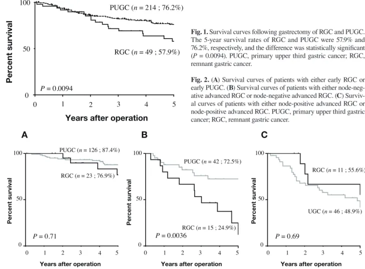
The 5-year survival rates of patients with RGC and PUGC were 57.9% and 76.2%, respectively, and the difference was statistically significant (P = 0.0094, Fig. 1). In detail, no significant difference in prognosis was
T. Matsunaga et al.
Years after operation
0 50 100 P = 0.0094 Percent survival 0 1 2 3 4 5 RGC (n = 49 ; 57.9%) PUGC (n = 214 ; 76.2%)
Years after operation 0
50 100
Percent survival
0 1 2 3 4 5
Years after operation 0
50 100
Percent survival
Years after operation 0 50 100 Percent survival P = 0.71 P = 0.0036 P = 0.69 RGC (n = 11 ; 55.6%) PUGC (n = 42 ; 72.5%) RGC (n = 15 ; 24.9%) UGC (n = 46 ; 48.9%) RGC (n = 23 ; 76.9%) PUGC (n = 126 ; 87.4%)
A
B
C
0 1 2 3 4 5 0 1 2 3 4 5Matsunaga et.al Figure 2
Fig. 1. Survival curves following gastrectomy of RGC and PUGC. The 5-year survival rates of RGC and PUGC were 57.9% and 76.2%, respectively, and the difference was statistically signifi cant (P = 0.0094). PUGC, primary upper third gastric cancer; RGC, remnant gastric cancer.
Fig. 2. (A) Survival curves of patients with either early RGC or early PUGC. (B) Survival curves of patients with either node-neg-ative advanced RGC or node-negnode-neg-ative advanced RGC. (C) Surviv-al curves of patients with either node-positive advanced RGC or node-positive advanced RGC. PUGC, primary upper third gastric cancer; RGC, remnant gastric cancer.
observed between early RGC patients and early PUGC patients (P = 0.71, Fig. 2a). However, the prognosis of patients with nodenegative advanced RGC was signifi -cantly worse than that of patients with node-negative advanced PUGC (P = 0.0036, Fig. 2b). There was no significant difference in prognosis between node-posi-tive RGC patients and node-posinode-posi-tive PUGC patients (P = 0.69, Fig. 2c). Since a signifi cant difference in prognosis was observed between node-negative advanced RGC and node-negative advanced PUGC, the sites of recur-rence of either RGC or PUGC were determined in those patients. Peritoneal, hematogenous, and lymph node recurrence were observed in 3 (20%), 3 (20%), and 2 patients (13.3%) with node-negative advanced RGC and 1 (2.3%), 2 (4.7%), and no patients (0%) in node-negative advanced PUGC. Peritoneal recurrence was observed signifi cantly more in RGC than in PUGC patients (P = 0.049), although there was no significant difference in hematogenous (P = 0.1) and lymph node recurrence (P = 0.064) between RGC and PUGC.
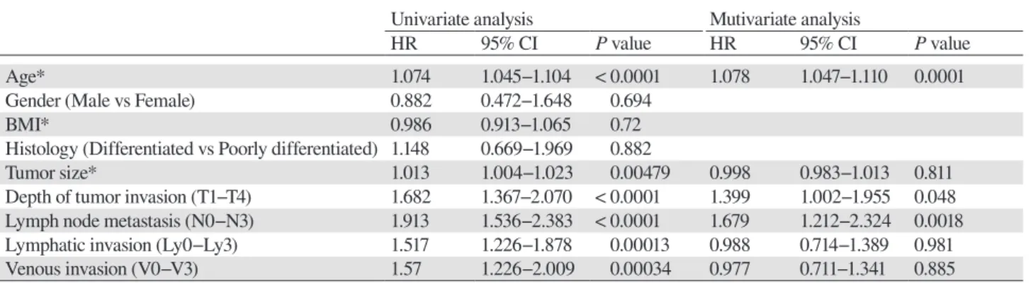
Factors related to prognosis in RGC and PUGC We next examined factors related to prognosis in both RGC and PUGC. Univariate analysis revealed that BMI, tumor size, and depth of tumor invasion were signifi cant prognostic factors in RGC, and that age, tumor size, depth of tumor invasion, lymph node metastasis, lym-phatic invasion, and venous invasion were significant prognostic factors in PUGC (Tables 2 and 3). Further-more, multivariate analysis revealed that BMI and depth of tumor invasion were independent prognostic factors in RGC (Table 2). However, age, depth of tumor invasion, and lymph node metastasis were found to be indepen-dent prognostic factors in PUGC (Table 3).
Because BMI and depth of tumor invasion were in-dependent prognostic indicators, we determined whether an accurate prognosis of RGC could be predicted using BMI and depth of tumor invasion. ROC analysis indicat-ed that an optimal cut-off value for BMI was 20.6 (AUC 0.746, P = 0.0031) (Fig. 3). Based on this value, patients were divided into two groups as follows: BMIHigh (≥
rates of patients with BMIHigh early gastric cancer, BMI-High advanced gastric cancer, BMILow early gastric cancer,
and BMILow advanced gastric cancer were 90%, 83.3%,
64.3% and 33.8%, respectively, and the difference was statistically signifi cant (Fig. 4a). The 5-year disease spe-cifi c survival rates of patients with BMIHigh early gastric
cancer, BMIHigh advanced gastric cancer, BMILow early
gastric cancer, and BMILow advanced gastric cancer were
Matsunaga et.al Figure 3
1-Specificity 0 0 0.5 0.5 1.0 1.0
Fig. 3. ROC analysis indicated that an optimal cut-off value for BMI was 20.6. The BMI had good diagnostic accuracy (AUC 0.746, sensitivity 82.6%, specifi city 61.0%, P = 0.0031). The arrow shows the optimal cutoff point. ROC, receiver operating charac-teristic.
Table 2. Univariate and multivariate analyses of factors prognostic of overall survival in patients with RGC Univariate analysis Mutivariate analysis
HR 95% CI P value HR 95% CI P value
Age* 0.997 0.936−1.063 0.936
Gender (Male vs Female) 0.776 0.180−3.345 0.733
BMI* 0.723 0.579−0.902 0.00417 0.731 0.563−0.949 0.018
Histology (Differentiated vs Undifferentiated)† 0.516 0.216−1.232 0.136
Tumor size* 1.014 1.003−1.025 0.01 0.994 0.973−1.016 0.512
Depth of tumor invasion (T1−T4) ‡ 1.837 1.289−2.616 0.000755 1.989 1.122−3.527 0.004 Lymph node metastasis (N0−N3)§ 1.759 0.735−4.208 0.205
Lymphatic invasion (Ly0−Ly3)|| 1.086 0.698−1.688 0.715
Venous invasion (V0−V3)¶ 1.438 1.038−1.992 0.029 0.994 0.598−1.509 0.6
*Continuous variable
†Histology: Differentiated, papillary or tubular adenocarcinoma; undifferentiated, poorly differentiated or mucinous adenocarcinoma, or signet ring cell carcinoma
‡T1, tumor has invaded lamina propria or submucosa; T2, tumor has invaded the muscularis propria; T3, tumor has invaded the subse-rosa; T4, tumor invasion is contiguous to or exposed beyond the serosa or tumor invades adjacent structures.
§N0, no regional lymph node metastases; N1, metastasis in 1−2 regional lymph nodes; N2, metastasis in 3−6 regional lymph nodes; N3,metastasis in 7 or more regional lymph nodes.
||Lymphatic vessel invasion: ly0-ly3, grade of lymphatic invasion. ¶Blood vessel invasion: v0-v3, grade of venous invasion.
BMI, body mass index; CI, confi dence interval; HR, hazard ratio; RGC, remnant gastric cancer.
Table 3. Univariate and multivariate analyses of factors prognostic of overall survival in patients with PUGC Univariate analysis Mutivariate analysis
HR 95% CI P value HR 95% CI P value
Age* 1.074 1.045−1.104 < 0.0001 1.078 1.047−1.110 0.0001
Gender (Male vs Female) 0.882 0.472−1.648 0.694
BMI* 0.986 0.913−1.065 0.72
Histology (Differentiated vs Poorly differentiated) 1.148 0.669−1.969 0.882
Tumor size* 1.013 1.004−1.023 0.00479 0.998 0.983−1.013 0.811
Depth of tumor invasion (T1−T4) 1.682 1.367−2.070 < 0.0001 1.399 1.002−1.955 0.048 Lymph node metastasis (N0−N3) 1.913 1.536−2.383 < 0.0001 1.679 1.212−2.324 0.0018 Lymphatic invasion (Ly0−Ly3) 1.517 1.226−1.878 0.00013 0.988 0.714−1.389 0.981
Venous invasion (V0−V3) 1.57 1.226−2.009 0.00034 0.977 0.711−1.341 0.885
*Continuous variable
See Table 2 for details of histology, depths of invasion, lymph node metastasis, lymphatic invasion, and venous invasion. BMI, body mass index; CI, confi dence interval; HR, hazard ratio; PUGC, primary upper third gastric cancer.
T. Matsunaga et al.
Years after operation
Percent
survival
BMIlowadvanced RGC (n = 19 ; 42%)
0 1 2 0 50 100 3 4 5 P = 0.0027 (A) (B)
Years after operation
Percent survival 0 1 2 0 50 100 3 4 5
BMIhighearly RGC (n = 13 ; 90%)
BMIhighadvanced RGC (n = 7 ; 83.3%)
BMIlowearly RGC (n = 10 ; 64.3%)
BMIhighadvanced RGC(n = 7 ; 83.3%)
BMIhighearly RGC (n = 13 ; 90%)
BMIlowearly RGC (n = 10 ; 90%)
BMIlowadvanced RGC (n = 19 ; 33.8%)
P = 0.00023
Fig. 4. Survival curves of patients with RGC according to BMI and depth of invasion. (A) The 5-year overall survival rates of patients with RGC. (B) The 5-year disease specific survival rates of patients with RGC. BMI, body mass index; RGC, remnant gastric cancer.
90%, 83.3%, 90% and 42%, respectively, and the differ-ence was statistically significant (Fig. 4b). Furthermore, recurrence was observed in 11 patients (57.9%) out of 19 with BMILow advanced gastric cancer and 3 patients out
of the remaining patients (10%), a difference that was statistically significant (P = 0.0007). Two patients with RGC died of other cancer and 6 patients with RGC died of other disease such as pneumonia or heart failure or myocardial infarction in RGC.
DISCUSSION
We have demonstrated in the current study that the prog-nosis of RGC was significantly worse than that of PUGC. This result is similar to that previous reports.15, 16 The poor
prognosis of RGC may be affected by the stage distri-bution of the patients. In the study by Tokunaga et al., 43.1% of the RGC group comprised tumor penetration of serosa (SE) or tumor invasion of adjacent structure (SI) patients; in contrast, only 18.9% of the primary gastric cancer group included these patients.15 However, this
does not seem to explain the poor prognosis observed in the current study because there was no significant difference in depth of invasion, lymph node metastasis, and stage of disease between RGC and PUGC patients. Another possibility is the molecular changes observed in RGC. The remnant stomach is frequently exposed to bile and pancreatic juice, which can induce molecular chang-es in the gastric mucosa. In a retrospective study with 130 patients, Leivonen et al. analyzed the cell prolifera-tion rate in biopsy specimens from gastric remnants by immunohistochemical staining of Ki-67.17 The Ki-67
labeling index of tumors was significantly higher in the remnant stomach group, and is known to be associated with bile reflux and reconstruction without bile reflux. Baba et al. found that p53 overexpression in tumors was almost twice as common in patients with gastric cancer of the remnant stomach than in patients that had under-gone distal gastrectomy for benign disease.18 It has also
been demonstrated that p53 overexpression and higher Ki-67 labeling index are closely related to poor progno-sis in gastric cancer.19, 20 Therefore, the poor prognosis
observed for RGC patients in the current study might be related to molecular changes. Further investigations are therefore required to determine the correlation between poor prognosis and molecular changes in RGC.
Anatomical alterations and intraabdominal adhe-sions after initial surgery, as well as frequent combined resection of other organs, present difficulties for RGC surgery. Many studies have described an altered lym-phatic pathway for RGC resulting from initial surgery. Furthermore, optimal lymph node dissection remains unclear thus far. It is likely that these surgical limitations are associated with poor prognosis in RGC.
It has recently been reported that preoperative nutri-tional status is a useful predictor of prognosis of gastric cancer patients. Several assessment tools can be applied to nutritional evaluation, such as Subjective Global Assessment (SGA),21 mini-nutritional assessment,22
nu-tritional risk scoring 2002 (NRS2002),23 and Onodera’s
prognostic nutritional index (PNI).24 Nozoe et al. have
recently reported that PNI could predict the prognosis and biological aggressiveness of gastric cancer.25 With
regard to BMI, it has been reported that underweight patients with BMI < 18.5 kg/m2 and low preoperative
albumin levels had a significantly decreased OS after gastrectomy for cancer,26 indicating the close correlation
between low BMI and poor prognosis in gastric cancer patients. It is likely that an initial gastrectomy places patients in a state of malnutrition. In the current study, BMI was significantly lower in patients with RGC than in those with PUGC. Furthermore, multivariate analysis indicated that BMI was an independent prognostic in-dicator, suggesting the usefulness of BMI to predict the prognosis of RGC patients.
Lymph node metastasis is one of the most import-ant prognostic factors for patients who have undergone curative resection for gastric cancer.27, 28 Lymph node
metastasis was shown in the current study to be an in-dependent prognostic indicator for PUGC. However, no significant difference in prognosis was observed between patients with lymph node metastasis and those without lymph node metastasis in RGC. Furthermore, lymph node metastasis was not an independent prognos-tic indicator for RGC in the current study. One possible explanation for this is that most lymph nodes are dis-sected in RGC patients whose initial operation was per-formed for gastric cancer, thus lowering the significance of lymph node metastasis as a prognostic indicator. Another possible explanation is insufficient lymph node dissection as a result of postoperative adhesion induced by an initial operation, which renders subsequent lymph node dissection difficult. Insufficient lymph node dissec-tion is related to stage migradissec-tion and might represent one reason why a significant difference in prognosis between RGC and PUGC was observed in node-negative but not node-positive advanced cancer.
The staging of gastric cancer according to the Japa-nese Classification of Gastric Cancer system is based on the depth of tumor invasion, status of lymph node metas-tasis, and presence of distant metastasis. In general, this staging system is used for RGC, although it is unclear how suitable it is for this disease subset. In this regard, our results indicated that factors reflecting the patient’s nutritional status should be included in the staging sys-tem of RGC.
The Japanese Gastric Cancer Treatment Guide-line recommends adjuvant chemotherapy with S-1 for a year in stage II/III gastric cancer patients who have undergone gastrectomy with D2 lymph node dissection based on the results of the Adjuvant Chemotherapy Trial of TS-1 for Gastric Cancer (ACTS-GC).29 It remains
unclear whether S-1 is also effective in improving the prognosis of RGC. Since the prognosis of RGC is worse than that of PUGC, adjuvant chemotherapy might be
necessary to improve prognosis in RGC patients. Based on our results, adjuvant chemotherapy should be admin-istered to patients with BMILow advanced RGC, given
their extremely poor prognosis. Furthermore, random-ized control trials are required to determine effective adjuvant chemotherapy strategies in patients with RGC.
This retrospective study has several limitations. One major limitation is the small sample size. Furthermore, we included patients with RGC following distal gastrec-tomy for both benign diseases and gastric cancer, which might have influenced the clinical outcome. In this regard, it has been demonstrated that there was no sig-nificant difference in survival between RGC where the initial operation was performed for benign disease and that where the initial operation was performed for gas-tric cancer.15 In addition, the retrospective nature of our
study could be considered another significant limitation, which may have influenced the results. Therefore, our findings should be confirmed by prospective studies.
In conclusion, our retrospective study indicated that the prognosis of RGC was poor compared with that of PUGC, and that BMI is useful to predict the prognosis of RGC. The prognosis of patients with BMILow advanced
RGC was extremely poor, and recurrence rates among this subset of patients was extremely high. Therefore, multidisciplinary therapy, including preoperative chemo-therapy and intensive adjuvant chemochemo-therapy, might be necessary for patients with BMILow advanced RGC.
The authors declare no conflict of interest. REFERENCES
1 Jemal A, Bray F, Center MM, Ferlay J, Ward E, Forman D. Global cancer statistics. CA Cancer J Clin. 2011;61:69-90. PMID: 21296855.
2 Ahn HS, Kim JW, Yoo MW, Park DJ, Lee HJ, Lee KU, et al. Clinicopathological features and surgical outcomes of patients with remnant gastric cancer after a distal gastrectomy. Ann Surg Oncol. 2008;15:1632-9. PMID: 18379851.
3 Nozaki I, Nasu J, Kubo Y, Tanada M, Nishimura R, Kurita A. Risk factors for metachronous gastric cancer in the remnant stomach after early cancer surgery. World J Surg. 2010;34:1548-54. PMID: 20217411.
4 Sinning C, Schaefer N, Standop J, Hirner A, Wolff M. Gas-tric stump carcinoma - epidemiology and current concepts in pathogenesis and treatment. Eur J Surg Oncol. 2007;33:133-9. PMID: 17071041.
5 Svanes C, Salvesen H, Stangeland L, Svanes K, Soreide O. Perforated peptic ulcer over 56 years. Time trends in patients and disease characteristics. Gut. 1993;34:1666-71. PMID: 8282252.
6 Maruyama K, Kaminishi M, Hayashi K, Isobe Y, Honda I, Katai H, et al. Gastric cancer treated in 1991 in Japan: data analysis of nationwide registry. Gastric Cancer. 2006;9:51-66. PMID: 16767357.
Cancer of the gastric stump following distal gastrectomy for cancer. Br J Surg. 2007;94:92-5. PMID: 17054314.
8 Fujita T, Gotohda N, Takahashi S, Nakagohri T, Konishi M, Kinoshita T. Relationship between the histological type of ini-tial lesions and the risk for the development of remnant gastric cancers after gastrectomy for synchronous multiple gastric cancers. World J Surg. 2010;34:296-302. PMID: 20012285. 9 Tanigawa N, Nomura E, Lee SW, Kaminishi M, Sugiyama M,
Aikou T, et al. Current state of gastric stump carcinoma in Ja-pan: based on the results of a nationwide survey. World J Surg. 2010;34:1540-7. PMID: 20182716.
10 Newman E, Brennan MF, Hochwald SN, Harrison LE, Karpeh MS, Jr. Gastric remnant carcinoma: just anoth-er proximal gastric cancanoth-er or a unique entity? Am J Surg. 1997;173:292-7. PMID: 9136783.
11 Sasako M, Maruyama K, Kinoshita T, Okabayashi K. Sur-gical treatment of carcinoma of the gastric stump. Br J Surg. 1991;78:822-4. PMID: 1873711.
12 Bozzetti F, Bonfanti G, Morabito A, Bufalino R, Menotti V, Andreola S, et al. A multifactorial approach for the prognosis of patients with carcinoma of the stomach after curative resec-tion. Surg Gynecol Obstet. 1986;162:229-34. PMID: 3952614. 13 Maruyama K. The most important prognostic factors for
gas-tric cancer patients. Scand J Gastroenterol. 1987;22:63-8. DOI: 10.3109/00365528709091021.
14 Japanese classification of gastric carcinoma: 3rd English edi-tion. Gastric Cancer. 2011;14:101-12. PMID: 21573743. 15 Tokunaga M, Sano T, Ohyama S, Hiki N, Fukunaga T,
Yamada K, et al. Clinicopathological characteristics and sur-vival difference between gastric stump carcinoma and prima-ry upper third gastric cancer. J Gastrointest Surg. 2013;17:313-8. PMID: 23233273.
16 Wang Y, Huang CM, Wang JB, Zheng CH, Li P, Xie JW, et al. Survival and surgical outcomes of cardiac cancer of the remnant stomach in comparison with primary cardiac cancer. World J Surg Oncol. 2014;12:21. PMID: 24468299.
17 Leivonen M, Nordling S, Haglund C. Does Helicobacter py-lori in the gastric stump increase the cancer risk after certain reconstruction types? Anticancer Res. 1997;17:3893-6. PMID: 9427799.
18 Baba M, Konno H, Tanaka T, Kamiya K, Baba S, Sugimura H, et al. Relationship of p53 and Helicobacter pylori to clin-icopathological features of human remnant stomach cancer after gastric surgery for primary gastric cancer. Oncol Rep.
2001;8:831-4. PMID: 11410793.
19 Ikeguchi M, Saito H, Kondo A, Tsujitani S, Maeta M, Kaibara N. Mutated p53 protein expression and proliferative activity in advanced gastric cancer. Hepatogastroenterology. 1999;46:2648-53. PMID: 10522058.
20 Bani-Hani KE, Almasri NM, Khader YS, Sheyab FM, Karam HN. Combined evaluation of expressions of cyclin E and p53 proteins as prognostic factors for patients with gastric cancer. Clin Cancer Res. 2005;11:1447-53. PMID: 15746045.
21 Detsky AS, McLaughlin JR, Baker JP, Johnston N, Whittaker S, Mendelson RA, et al. What is subjective global assessment of nutritional status? JPEN J Parenter Enteral Nutr. 1987;11:8-13. PMID: 3820522.
22 Vellas B, Villars H, Abellan G, Soto ME, Rolland Y, Guigoz Y, et al. Overview of the MNA--Its history and challenges. J Nutr Health Aging. 2006;10:456-63. PMID: 17183418.
23 Kondrup J, Rasmussen HH, Hamberg O, Stanga Z. Nutri-tional risk screening (NRS 2002): a new method based on an analysis of controlled clinical trials. Clin Nutr. 2003;22:321-36. PMID: 12765673.
24 Onodera T, Goseki N, Kosaki G. [Prognostic nutritional index in gastrointestinal surgery of malnourished cancer patients]. Nihon Geka Gakkai Zasshi. 1984;85:1001-5. PMID: 6438478. 25 Nozoe T, Ninomiya M, Maeda T, Matsukuma A, Nakashima
H, Ezaki T. Prognostic nutritional index: a tool to predict the biological aggressiveness of gastric carcinoma. Surg Today. 2010;40:440-3. PMID: 20425547.
26 Ejaz A, Spolverato G, Kim Y, Poultsides GA, Fields RC, Bloomston M, et al. Impact of body mass index on periopera-tive outcomes and survival after resection for gastric cancer. J Surg Res. 2015;195:74-82. PMID: 25619462.
27 Siewert JR, Bottcher K, Stein HJ, Roder JD. Relevant prog-nostic factors in gastric cancer: ten-year results of the German Gastric Cancer Study. Ann Surg. 1998;228:449-61. PMID: 9790335.
28 Kim JP, Kim YW, Yang HK, Noh DY. Significant prognostic factors by multivariate analysis of 3926 gastric cancer patients. World J Surg. 1994;18:872-877; discussion 877-8. PMID: 7846911.
29 Sakuramoto S, Sasako M, Yamaguchi T, Kinoshita T, Fujii M, Nashimoto A, et al. Adjuvant chemotherapy for gastric cancer with S-1, an oral fluoropyrimidine. N Engl J Med. 2007;357:1810-20. PMID: 17978289.