Studies on the Chernical Control of Reproductive Functions in Higher PIan脩
…Physiological and Biochenlicai Characteristics of Rice Sterility induced by Chemical Treatment…
高等植物における生殖機能の化学的制御に関する研究 ―化学的処理により誘導される稲不稔の生理、生化学的性質―
ATSUMISHIMADA
1993
Contents
introduction
Materials and Methods
Assay procedures to rice plants Materials
Assay procedures
PhysiO10gical and biochernical assay procedures to rice steritity Assay for glutarnine synthetase activity in panictes
Assay for ATPase activity in panictes
Extraction and analysis of free anlino acids for sterile anthers Extraction and analysis offree anlino acids forieaves and cul「Ⅵs
Arnrnonia contentin panictes
Totai nitrogen in panictes,leaves and cutrns Protein nitrogen in panicles,leaves and culrns
Free arnino acids contentin panictes,teaves and cutrns Various carbohydrates in panicles,leaves and cul『 ¬s
Deterrnination of chiorophyH content in teaves
Assay for rice root activity by α‐naphthytanline oxidation
isolation,structure analysis,and the physiological and biochernical assay procedures for S35‐
C2
1solation and cuitivation of microorganism Asaay for GS inhibhory act 比y
Procedures for identification of strain S‐ 35 Ferr輛entation,isolation and structure analysis
List of the culture rれ edia for starin S‐35
4 4 4 4 5 5 6 7 7 8 9 9 9 10 13 13 14 14 14 15 20 23
Resutts and Discussion
Effects of twelve chernicals on sterility in rice(in 1990)
Effects of kasugamycin and AOA on sterihy in rice(in 1991) Sex expression of WYamabikoW rice plants treated whh AOA interactive effect between kasugamycin and plant hormones on stenthy
Ettects of inhibitors of protein synthesis on sterility
Effects of inhibitors of nucleic acid synthesis on sterility in rice Effect of bialaphos on steritity in rice(in 1991)
Biochenlical characteristics of rice sterilty treated with bialaphos
Effect of bialaphos on GS activity,'p ylitro
Changes offree arnino acids in anthers
Rice sterility induced by inhibitors of glutarnine synthetase Effects ofinhibhors of GS on sterilhy in rice(in 1992)
Effect of glutarnine on sterility in rice treated with biataphos
Effects of arn!れonia and arnrnoniurn sulfate on sterility in rice
Effect of DCMU on steriltty in ttce treated wtth bialaphos
Physiological and biochernical characteristics of sterility in rice Effects of GS inhiblors on GS activity,in ylitro
GS activity and free arnrnonia content in panicles
Total and protein nitrogen and free arnino acids content in panictes,leaves and cutrns
Changes offree arnino acids in anthers
Changes ofthe component of carbohydrates in panictes, leaves and culrns
28 28 29 34 35 36 38 40 42 42 43 45 45
49
50 51 53 53 54 1 7 6 6 1 7 7 7Changes of chiorophy‖ in leaves ofthe sterile rice plants Effects of GS inhibttors on「oot activtty in rice
Rice sterilhy induced by S35‐
C2
Screening for GS inhiblorfrom actinOmycetes Taxonomy of strain S35
Structure of S35‐
C2
Effect of S35‐C2on steriliv in ttce
Physiological and biochernical characteristics of sterility induced by S35‐(32
Effect of S35‐C2on the growth of rice seedlings
80 84 85 85 85 90 91 91 100 101 107 108 Conctusion Acknowiedgements References
introduction
The irnprOvement of crop plants through the production of hybrid varieties is a ma,Or goal of plant breeding.1)C,「
osses between inbred
plant lines often result in progeny with higher yield, increased
resistance to disease, and enhanced perforrnance in different
environments compared with the parental lines.2)
The production of hybrid seed on a large scale is cha‖enging because
many crops have both male and female reproductive organs(stamen and
piStiりOn the same planti However, the large‐
scale directed crossesbetween inbred lines makes difficuit to accomplish. To guarantee that
outcrossing will occur to produce hybrid seed, breeders have either
manua‖ y or mechanica‖y removed stamens from one parental line, used
natural seif‐incompatibility systems that prevent self中 poWination, or expioited male sterility mutations that disrupt po‖ en development.3,4)
Manual emasculation is iabour intensive and irnpracticai for plants with
smaH bisexual flower(suCh as rice, wheat), many CrOp plants do not
have seif‐incompatib‖
ity or male sterility genes, and use of male
sterility requires a fertility restorer system.2,5)
The chernical induction of male sterility in rice ((9ryza satliya L., Japonica) iS potentia‖ y atractive, because the utilization of heterosis
for rice breeding is timitedi The timiting factor in breeding is the
prolonged process of developing male‐
sterile and restore lines for hybridization. This disadvantage could be overcome if chenlicals wereused to induce male sterility.2,6‐
8)cOnSequentiy, chernicals may be
crop iniury. Further!町
ore, they must be unaffected by environmental
variability such as rain, wind, and temperature during crop growth stage and be suitable for various cuitivars.6‐8)
AIthough 2‐ chioroethylphosphonic acid (ethreり
,9)SOdium l‐
(ρ…chiorophenyり ‐1,2‐dihydro‐4,6‐dirnethy卜2‐oxonicotinate(RH‐ 531)10,11)
and disodiummethanearsonate(DSMA)8)are known to induce male
sterility in rice, these chernicals have several problems associated with their use such as high application dose(ethrei; 55 mM, RH‐ 531, 0.3 mM, and DSMA,l mM),a prOpenshy to cause crop in,ury and a failure to bttng
about complete male sterility (Fig. 1).
RH-531
CI CH2 CH2 lく
:I
EthrelO
CH3‐ Asく。
Na
DSMA
Fig.l The Structures of Male Stettlants k)r Rice PIants
2
For such reasons, they do not have any practical use. in order to
develope effective chenlicals to induce male sterility in rice, it is
necessary to study their mode of action. For example, DSMA reduces the
activities of succinic dehydrogenase and cytochrome oxidase, two
respiratory enzymesi consequently, it suppresses the nor:mal growth ofpO‖en,8)HOwever, littie study has been devoted to the mode of action of
chernicals on male sterility.2,8)
FurtherrⅥore, screening for new rice sterilants among microbial metabolites is the irnportant program because the devetopment of the
chernicals by artificial synthesis is tinlited, and natural products12ぃ 26)
provide with new structural ctasses which can serve as modeis for the
syntheses of the effective rice sterilantsi Frorn this point of view, S35‐
C2, a metabolite frorn actinomyces, was found as an effective rice
sterilant.Moreover, kasugamycin,27,28)aminooxyacetic acid(AOA)29‐
39) and bialaphos40‐
46)were found as the effective rice sterilantst
Bialaphos especia‖y induces complete sterility in rice at a very tow
concentration, and bialaphos is the most effective sterilant of the
known chernicals used k)r inducing sterility.
In this study, the author found the new effective rice sterilants and investigated the physiological and biochenlical characteristics of rice sterility induced by chenlical treatrYlenti
Materials and Methods
Assay
ρ「oce」vres to r,ce prar7tS
Maどer,arst Kasugamycin hydrochio
de, aminoOxyacedc acid(AOA),
gibbereitine (GA3), α‐naphthytacetic acid (NAA), benZOtriazole, D‐
cycloserine, hydroxyproline,
α,α‐dipyridil, benzyladenine (BA),kanamycin suifate, tetracycline hydrochioride, rifampicin, guanidine
thiocyanate, 1‐
(2‐tetrahydrofuryl)‐ 5‐刊uorouracil, L‐giutarnine,arnrnonium suifate, and arnrnonia solution were purchased from Wako
Pure Chemical Co..1ぃ
Aminocyclopropane‐ 1‐carboxylic acid (ACC),
sperrnine tetrahydrochloride, methylglyoxal bis‐
(guanythydrazone)
dihydrochioride (MGBG), L…
aZetidine‐2‐carboxylic acid (ACA), 2‐
chloroethylphosphOnic acid (Ethrel), erythrOmycin, spectinomycin
dihydrochioride, and Lいrnethionine suifoxir¬ine (L‐
MSC))were purchased
from Sigma Chemical Co..Bialaphos and L‐ phOsphinothncin(L‐
PTC)Was
kindly provided by Metti Seika Co..Assay proce」
vres, Rice plants(Oryza sarya L., cv. Yamabiko)were
ger!minated and planted in paddy soil. At the two‐ or three‐leaf stage, three weeks after the gerrnination, the seedlings were transplanted into 2 kg Of paddy soil in 16‐ cm diameter pots, Each pOt held one seedling
and was fertilized with 120 ppm N and 180 ppm P as diammonium
hydrogenphosphate, and with 140 pprn K as pOtassium chioride.The rice plants were grown under natural suntight at a temperature from 20 to35
°C. The developmentai stages in rice were ascertained by measuringlast two leaves in the main ti‖
er.47) The developmental stages are
defined as the panicte forrnation stage when the young panicte reachedO.4 ‐ 0.5 Rlrn in tength, and as the reduction division stage when this distance was ‐4 to +4 cmi
The chernicats to be tested were forrnulated as an aqueous
solution containing O.1 0/O Tween‐ 80 as a wetting agent and 5 0/o acetone to aid solubility. The aqueous solution of each chernical was adiuSted to
pH 7.O with lN NaOH and sprayed on a‖ leaves of the rice plant with an atonlizer at the rate of 10 rni per pOti Three replications were used for each treat!Ⅵ ent.
At ear emergence, a‖
the heads frorn each pot were placed in
giassine bags to prevent cross poHination. After the bagged ears hadbeen harvested at maturity, the seed set, spike length, cutrn tength and
weight of the ripe seeds were recorded and compared with the values
for an untreated control.
PrP/s,OrOgFcar arP」
bす。crPemttcar assa/ proceJtrres
を0
「'ce
ster,「,ど
/
Assay rOr grvta阿加e Sypttetase acry,y tt paЯ ん′
es.Panicles(lg)
were washed and cut into 5 Rlm tength. The sample was homogenized
with a Polytron(Kinematica Co.)at the medium speed setting for 60 secat O
°C. The homoginizing medium was composed of imidazole‐
HCIbuffer(pH7.2;50 mM),EDTA(pH7.2;0.5 mM)and 2‐
mercaptoethanol(5
mM). ARer the extract was filtered through four iayers of cheesecioth, the filtrate was centrifuged at 1 5,000Xg for 15 min and the supernatant
Gtutamine synthetase (GS)actiVity was measured by the formation
of v‐giutamythydroxamate in the sythetase reaction.44‐
54)The reaction
mixtures contained enzyme(100μ
り,SOdium L‐glutamate(50
μM),NH20H
(pH7.2; 100 μM), NaATP(10
μM), MgC12(20
μM), imidazoleⅢ HCi buffer (pH7,2; 50 μM), 2‐rnercaptoethanol(25 μM), in a finat votume of l.O mi.ARer incubation at 37 。C for 15 min, the reaction mixture was treated with l.5 mi of ferric chioride reagent contained ferric chioride (0.37 M),
HCI(0.67M)and TCA(0,20M).The fOrmauon Of Y…
glutamylhydroxamatewas measured at 530 nm (Shimazu UV 2200 spectrophotometer)・ The
standard curve was plotted in the manner described above by utilizing l mi samples of v‐glutamythydroxamate(0.l t0 0,7 μmole).
Assayた
》rAア
Pase actliylity tt parPjcres. Panicies(l g)were collected before 2 days from anthesis, and they were washed and cut into 5 Rl『 ηlength.The sample was homogenized with a Polytron(Kinematica Co.)at
the medium speed setting for 60 sec at O OC. The homoginizing mediumconsisted of sorblol(0,25M),MOPS‐
KOH(pH 7.0;50 mM),EDTA(2 mM)
neutralized with KOH, 2.5 mM pOtassium metabisulfite, and l.5 0/o
soluble PVP (rY101 Wt 24,000)・
ARer the extract was filtered through
four layers of cheesecloth, the filtrate was centrifuged at 15,000xg for 15 nlin and the supernatant was used for the assay as ATPase activity. ATPase activities were deterrnined by measuring Pi hydrolyzed from ATP,55‐
63)Pi was determined by the Du‖
ey modification64)Of the
Fiske and SubbarRowi65)The reaction mixtures contained enzyme (50
μり
,ATP(2Na)(3
μM),MgS04(3
μM),HEPES(pH 6.5,25
μM),KCI(50
μM),T ton X…100(0.03 0/o)and inhibitor, in a final volume of O.25 mi.A■ er
stopped by the addition of 2.5 ml of ice cold stopping reagent contained
ammonium molybdate(o.25 0/o), SOdium dodesitsuifate(1.3 0/o),and
concentrated H2S04(1・4 0/0)and placing the tubes on ice. ARer the
solution was added O.5 ml of the reducing agent contained l‐ amino‐2‐
naphto卜4‐
suronic acid(0,1
°/0), NaHS03(5,70/o),NaS03(0・ 20/o),the testtube was thoroughiy shaken by hand and incubated for 10 nlin at room
temperature ,and measured at 530 nm (Shimazu UV 2200
spectrophotometer)for deterrnining reteased inorganic phosphate i The standard curve was plotted in the manner described above by utilizing
250
μl samples of KH2P04(10t050
μg).fxtractliorP an」 aЯarys,s Or rree aЛttrPo ac′Js ror sどer,r a19fん9「si For
analysis of free arnino acids,45,66)abOut 800 anthers were co‖ ected
before 2 or 3 days from anthesis. The sample was ground in mortar with
70 0/o Et(DH, kept for 2 days at rooRl temperature and then centrifuged at
3,000xg for 10 mini The precipitate was further extracted once with 70
0/o Et(DH. The two supernatants were combined and concentrated at 35∼ 40 。C under reduced pressure, The extract was dissoived in 2 nll of water
and washed twice with the same volume of Et20・ ARer the water
sotution was dried up in vacuunl, the extract was dissoived in waterand the quantity of free anlino acids was deterrnined with an amino acid
analyzer(Shimazu LC‐ 6A, columni ISC‐ 07′
S1504 Na).
fχtractliorP an」 alTarystt Or rree a″,lir70 aCliJs ror reayes arPJ parpデ cres, For analysis of free arnino acids,45,66)ieaVes or panictes(1 9)Were
coHected before 2 days from anthesisi They were dried at 80 °C for 3
dayst The dried sample (1 9)WaS grOund by a coffee mill. The dried
in 15‐
rni test tube and heated at 100
。C for l hr. ARer the sample
solution was cooled in a tapped water bath, the etuate separated by
centrifugation (3000xg, 5 min). The etuate was concentrated at 35∼ 40。
C
under reduce pressure. The extract was dissolved in water and the
quantity of free amino acids was deterrnined with an anlino acid
analyzer(shimazu LC‐ 6A, columni ISC‐ o7/S1504 Na).Am′ηon,a corPtenf′ β par9Fc′es, Panicies were coWected after l, 3 and
5 days from chemical treatment.The sample(lg)waS Cut 5 mm long
and homogenized in lo mi methano卜
chioroforrn‐ water (12:5:3, v′v′v) with a Polytron(Kinematica Co.)at the medium speed setting for 60 sec at O °C. The nonturbid iayers were produced by the addition of 3 mi
water per lo mt homogenate fo‖
owed by centrifugation (3000xg, lo
rnin)・The upper iayer of methano卜 water was contained the polar
solutes and was taken for deter!Ⅵ ining anlrnonia content,67‐
70) A■
er 5ml of the sample solution was added in 30‐ rni tube, the tube was placed
on ice and added 4 nll of water and 2 nli of pheno卜
nitroprusside solution contained phenol(6 0/o), nitrOprusside (0.02 0/o), phOSphate‐chrate buffer(o,l M, pH 12). ARer the solution mixed thoroughly, 3 mi of the alkaline hydrochiorite solution, contained sodiurn hypochiorite
(0.1 0/0)and SOdium hydroxide (l Ml, Was added. The solution mixed
thoroughty and incubated for 45 min at room temperature, and was
measured at 635 nm by Shirnazu UV‐
2200 spectrophotometeri The
standard curve was piotted in the manner described above by utilizing 5
mi samples of NH4CI(10t050
μg).ァοtar plitrogerP tt reayes arPJ cy′阿s, arp」 ρa19んres. Leaves and culrns,
and panicies were co‖
ected before 2 days from anthesis. They were
dried at 80 °C for 3 days. The dried sample (1 9)WaS grOund by a coffee
mi‖.The ground sample(0,1 9), 2 ml of concentrated H2S〈 D4 and a bК of
catalyst(Se‐Cus04‐
K2S04, 1 : 10 :50w/w/w)added in 300‐
rni Kieldahifiask and heated for about 5 hr using the electric‐
heated six‐fiaskdigestion stands,71‐
73)The c001ed digest was diluted to volume in a
20‐mi messttask,l ml of the sotulon was neutralized(methyt red as an
indicator)With l N NaOH.The neutralized solution was diluted to volume
in a 10‐rⅥI messfiask, and o.5 ml of the diluted solution was used for
deter!ηination of anl,Ⅵ
onia content by the indophenol method described
above,
Prote,p rPitrogen lirP reayes arPJ cv′阿,S an」 ρanん′es. The dried sample (01l g)Of leaVes and culrns or panictes and 4 mi of disti‖
ed water added
in 30‐mi test tube and heated at 85
°C for l hri ARer the sample
solution was cooled in a tapped water bath, the residue separated by centrifugation (3000xg, 5 min). The residue was used for determining
protein nitrogen by the method described in the section of totai nitrogen assay.
Free a″,デβo acli」s coЯterTど lirP reayes anJ cv,「ηs, arP」 parP,c′es, The dried
sample (0.1 9)Of leaves and cutrns or panictes and 10 Rl1 0f disti‖ ed
water added in 15‐ rni test tube and heated at 100 °C for l hr. After the sample solution was cooled in a tappe「 water bath, the etuate separated by centrifugation (3000xg, 5 Rlin). The eluate was diluted to volume in a
solution and 2 nll of the ninhydrin reagenti74‐
76)This reagent was
composed of ninhydrin (0.8 g), hydrindantin (0.12 g), methyI Ce‖ osoive
(30 mi)and 4 M acetate buffer(pH 5.5, 10 ml). ARer the reaction was
carried out at 10o oC for 15 rnin in a boiling water bath, the reaction
was stopped in a cooled water bath and added 3 mi of 50 0/o ethanol. ARer the test tube was thoroughty shaken on a mixer and incubated for 10 min at roonl temperature, the absorbance was measured at 570 nm by Shirnazu UV… 2200 spectrophotometer.74ぃ
79)The standard curve was
plotted in the manner described above by utilizing 2 mi samples of L‐ aspartic acid (3 to 30 μg).
yarlioys carborP/Jrares,p reaves an」 cylms, arPJ parMcres. Leaves and
cutrns, and panicles were coWected before 2 days from anthesisi They
were dried at 80 °C for 3 days. The dried sample (1 9)WaS grOund by a
coffee nli‖ . For analysis of the reducing sugar, the total sugar, the
crude starch and the total carbohydrates in ieaves and cuirns, and
panictes, the analyticai methods described below were exanlined.80)
月θJvcling svgaぇ A sample(100 mg)of the ground ieaves and culrns Or
panictes was extracted by hot 80 0/o ehtanol in a boiling water bath. The residue was further extracted twice with 80 0/o Et(9H. The 80 0/o ethanol etuates were co‖
ected and concentrated under reduced pressure. The
residue was dissolved in 5 rnl of hot water, and added O.5 rni of Oi3 N
Ba(OH)2 and O.5 ml of 5 0/o ZnS04・ After the solution was m ed, the
solution was centrifuged at 3000xg fOr 10 rnini The supernatant was
diluted to volume in a 20‐ rni messflask. l ml of the diluted solution (the sugar solution)waS further diluted to volume in a 5‐
mi messlask.
TOtar sygaL After l ml of the sugar solution was hydrolyzed by l mI
of 4 0/o H2S04 in a bOiling water bath for 15 min, the reaction was
neutralized(methyi red as an indicator)With l N NaOH. The neutralizedsolution was diluted to volume in a 20‐ rni messfiask, and l ml of the
dituted solution was further diluted to volume in a 20‐ rvli messfiask.
CrvJe starcrP, The residue of the leave and cutrn or panicte, prepared by the extraction of 80 0/o ethanol, was dried at 80 °C for 5 hr. ARer
dried, the residue added 2 ml of water and heated in a boiling water bath for 10 min. Aner coOled in a water bath, the mixture added 2 ml of 60 0/o
perchioric acid and heated in a boiling water bath for 15 min. The
supernatant of the reaction mixture was separated by centrifugation
(3000 xg, 10 min), After the rection residue added 4 ml of 30 0/o
perchloric acid and heated in a boiling water bath for 15 rnin, the
supernatant was separated by centrifugation (3000x g, 10 nlin). The
reaction residue was further reacted once by the same method described
above, and the supernatant was coHected. The supernatants were
co‖ected and diluted to volume in a 100‐
rni messfiask, 2 rni of thediluted solution was heated in boiling water for 2 hr. A■ er cooled, the
reaction was neutralized ( methyl red as an indicator)with l N NaOHi
The neutralized solution was diluted to volume in a 10‐rni messfiask.
ァOtar carbOr7ydratesi A sample (100 mg)of the ground leaves and
cuims or panictes was hydrolyzed by 3 mi of O.7 N HCt in a boiling water
bath for 3 hr. The reaction rnixture was centrifuged at 3000 g fOr lo
mini The supernatant was neutralized(methyt red as an indicator)wlh
ARer the solution was rnixed, the solution was centrifuged at 3000 g
for 10 min. The supernatant was diluted to volume in a 20‐
『ⅥImesslask, l ml of the diluted solution (the Sugar solution)Was further
dituted to volume in a 20‐ rni messfiask,
Detemttatlioη Or trPe svgar cOntent or tte prepareJ sattpres,l mi of the diluted sample sotution of reducing sugar, total sugar, crude starch, or total carbohydrates and l rni of a copper reagent were pipeted into a
30‐rni test tube and nlixed thoroughiy, The reaction mixture was heated
in a boiling water bath for 15 nlin and cooled in a cooled water bath. 1
ml of the arsenomolybdate reagent was then added and mixed thoroughiy.
A■er 7 ml of water was added and the mixed solution incubated for 20
rnin at roorn temperature, the sugar concentration was measured at 500
nm by Shirnazu UV‐
2200 spectrophotometeri81‐85)The Standard curve
was plotted in the manner described above by utilizing l mi samples of
DngluCose (10 t0 100 μg).
The copper reagent and the arsenomolybdate reagent was prepared by
the methods described below.85)The Copper reagent 24 9 of Na2C03 and
12 g of Roche‖ e salt were dissoived in about 250 ml of water, then the dissoived copper suifate (40 rni of a 10 0/o solution)was intrOduced with stirring, and this was fo‖owed by the addition of 16 9 of NaHC03. 180 g
of Na2S04 (anhydrOus)was diSSOived in about 500 mi of hot water and
boiled to expel air. ARer cooling, the two solution were united and
diluted to volume in a 1000‐ rni messflask. The arsenomolybdate reagent
25 9 of arnrnoniurn molybdate and 21 ml of concentrated H2S04 Were
dissoived in about 500 ml of wateri 3 g of Na2HAs04・
7H20 WaS
dissoived in 25 ml of water. The two solution were united and diluted to
volume in a looo_rni messfiask. The mixed solution placed in an
incubator at 37 °C for 24 hri
Deterrw,patlior9 or cr7′ OrOp力y′′ corPどerPt FrP reayes. Leaves were cut 5 rnrn iong and 1 9 of thern was extracted in 5 ml of 80 0/o ethanol. ChlorophyH cOntent was deterrnined by measuring at 665 nm by Shirnazu
UVⅢ
2200 spectrophotometer.86)Another method for deterrnination of
chlorophyH cOntent was carried out by the chlorophy‖
meter(SPAD‐
502,MINOLTA Co.).
Assay
どo rlice root actliy,どy by
α‐ηaprPtrP/raplirpe oχ′」aJοη. Roots
were washed, cut l to 2 cm iongi 2 9 of thenl, 25 ml of α‐naphthylarnine(40 ppRl)and 25 ml of phosphate buffer(pH 7.0, 0.l M)were added in
100‐rni fiask and incubated at 25 °C for 6 hr in a water bath. 2 ml of the
sample,10 ml of water,l ml of sulfanilic acid(1° /0)in acetic acid(20 0/0),and l ml of NaN02 aqueous solution(100 ppm)were added in 30‐ mi
test tube, and then it was fiHed up to 20 ml with wateri After 30
rninutes, α‐
naphthylamine concentration was measured at 510 nm by
Shirnazu UV‐ 2200 spectrophotometer,87‐
89)The standard curve was
plotted in the manner described above by utilizing 2 mi samples of α…,sOrattOrP′ sど
ryctvre arParys,s, arPJ
どrpe prP/s,0'OS'Car arPJ
bす。
cr7em,car assa/ρ
「。ceJvres ror s35‐
C2
Screenttg rest rO「
GS lirPんlibfror rrOm acrlinOm/cetes′sOrat,OPP arPJ cυ′tiyaど,oη or m√crOOrgarP,s阿
. TeSt strains were
isolated fronl soil by conventional methods. About 600 strains of
actinomycetes were isolatedi These strains were inoculated into testtubes wnh 5 ml of modwied Chuken medium (pH 7.2). A■ er shaking at
30
°C for 5 days, the cuiture broth was centrifuged (3000xg, 5 min)and the supernatant was used for assay of GS activity.Assay rOr es ,19rP,b,ど Ory ac∬y,1/. Foutteen‐day‐ old light‐
grown
shoots(10g)Were Washed and cut into 5 mm iength.The sampte was
ground in a mortar with 1 5 nll of extract solutioni The extract solution was composed of imidazole…
HCi buffer(pH7.2i50 mM),EDTA(pH7.2;0.5
mM)and 2‐
mercaptoethanol(5 mM). ARer the extract was litered
through four iayers of cheesecloth, the filtrate was centrifuged at
15,000 g for 15 min and the supernatant was used k)r the assay of GSactivity.
GS activity was measured by the forrnation of v‐ glutamythydroxamate in the sythetase reaction,46‐
54) The reaction rnixtures contained
enzyme(100μ
り,SOdium L…giutamate(50
μM),NH20H(pH7.2, 100
μM),NaATP(10
μM), MgC12(20
μM), imidazole…HCi buffer(pH7.2; 50 μM), 2‐mercaptoethanoI(25
μMl, and inhibitor, in a final volume of l.O mi. ARer incubation at 37 。C for 15 min, the reaction mixture was treated with l.5 rnl of ferric chloride reagent contained ferric chloride (0,37 M),HCI(0,67M)and TCA(0.20 M).The formation of Y‐
gtutamythyd『oxamate
was measured at 530 nm (Shirnazu UV 2200 spectrophotometer)and
compared with a control in which no inhibitor was added. The standardcurve was plotted in the manner described above by utilizing l mI
samples of v… glutamythydroxamate (0.l t0 0・ 7 μmole).
ProceJvres ro「
crPa「acterlizatliott or stra,p S‐35
Morp力o,og,car c力aracrerlizatliοrP, Morphological characteristics90‐
100) were observed on several media described by Shirling and
Gottlieb.91)incubation was carried out at 30 °C, for 21 daysi
Tettperatvre or grOM的 . Siants of Bennett's agar Or yeast dextrose
agar were inoculated, quickty heated or cooled to the desired
temperature in water bath and then placed in water bath at the same
temperature inside a constant‐ temperature incubatori The water ievel
and Temperature of the water bath were carefu‖ y maintained. The
cuitures were exanlined for growth aner 5 to 7 days at temperatures of35
°C or above, and after 3 weeks at 10 °C.101,102)汀ydrorysる o「 geratlirP.Duplicate plates of the medium were streaked
once across with each cuiture. After 5 daysi incubation at 28 °
C, one
plate was covered with 8 to 10 nll of the fo‖ owing solution i HgC12, 15 gi concentrated HCt, 20 mii disti‖ ed water, loO RlI. Hydrolysis of the
gelatin103)waS measured by a ciear zone underneath and around the
μy」「oryslis Or sど arcヵ
.The medium for determining hydrolysis of
starch103)waS used with the substitution of 10 g of potato starch for getatint Duplicate plates were streaked and stored at 28 °
C, ARer 5
daysi incubation, one plate was flooded with 8 to 10 nli of 95 0/o ethanolOr i2 / KI solutioni the second plate, aner lo daySi A clear zone
surrounding the growLh indicated the extent of hydrolysis.
Mlilた ρθρどoη″aron aβ」 coagvrat,Oη.Skim milk(instant nonfat milk)
10 g and brom cresol purple (BCP)Were diSSolved in loo mi disti‖
edwateri The solution was then autoctaved, inoculated and incubated at 30 °C for 5 to 10 days,97)
MeranOtt pデ gttept pro」vctlioη,P『
oducdon of melanoid pigments was
determined on agar stants of peptOne iron agar and tyrosine agari
Cuitures used as inoculum source should be less than 3 weeks old.
Duplicate stants were streaked and incubated at 30 °C' for 4 to 7 days.
The inoculated test tubes were compared with uninoculated controls.
Cuitures forrⅥing a brown to black diffusible pigment or distinct brown pigment modified by other color sha‖ be recorded as positive. Absence
of brown to black colors, or total absence of diffusible pigment, sha‖
be
recorded as negative for melanoid pigment productioni91)
OxygerP re9v″er77enti Oxgen requirement test104)waS deterrnined on yeast‐rnalt extract agari Duplicate stants were streaked and incubated
at 30° C for 4 to 7 days.
打/Jrogen svrrli」 e prOJvctliorP, Peptone‐ iron agar was employed in tube
siants for determining the hydrogen sulfide reaOtions of the
organisms,105) inOCulum fronl actively growing cuitures was used to
streak the surface of the agar stants. After a short incubation period(15 to 20 hr)at 28
°C, the stants were observed. A pronounced bluish black discoloration of the medium surrounding the colonies indicatedthe production of hydrogen suifidei whereas, those organisRl nOt
producing hydrogen suifide effected no change in the mediunl, or ernitted onty faint tints Of other colorsi
Re」vcrOЛ O′ rP,traどθ, Cuitures in nitrate broth were tested for the
presence of nitrate after 5, 10, 14 days Of incubation at 28 °C, One rnl of broth cuiture was withdrawn aseptica‖ y and rnixed with 3 drops Of each
of the foWowing two solutionsI(1)sulphanilic acid, 8 gi 5 N acetic acid,
1000 rni.(2)dimethy卜a…naphthytanline, 6 mI; 5 N acetic acid, 1000 mI.
The appearence of a red color indicated the presence of nitrate.103)
Satt to′erarPce. Salt tolerance was deter!Ⅵ ined on yeast‐ IⅥalt extract
agar containing NaCi prepared at concentration of O, 3, 7, 10 and 14 0/0, respectively.101)Duplicate stants were streaked and incubated at 30 °
C
for 4 to 7 days.
Svgar ytlillizatliorp. ChenlicaHy pure carbon sources were used for sugar utilization testi Carbon sources for this test are : D‐ giuCose, L‐
arabinose, sucrose, D‐ Xy10se, 卜inositol, D‐ rnannitol, D‐ fruCtose,
appropriate amount of the dry carbon source was weighed and spread as
a sha‖ow iayer in a pre‐ sterilized Ertenmeyer fiask fitted with a loose
Cotton plug. Ethylether was added to cover the carbohydrate. Ether was
a‖owed to evaporate at roorn temperature under a ventilated fume hood
over night, ARer an ether has evaporated, sterile disti‖
ed water was
added asepticalit to make a 10 0/O w/v solution of the carbon sourcetARer autociaving the Pridhanl and Gottlieb90) basal agar mediurn,
cooled it to 60 °C and added sterile carbon source aspectica‖ y to give a
concentration of approxirnately 1 0/o. The medium was agitated and
poured 15 mi per dish into Petri dishes, Duplicate plates were streaked
and incubated at 30 °C. After 20 days incubation, plates were observed
and compared growth on a given carbon source with the two controls i
growth on basai mediurn alone, and growth on basal medium plus
glucose.
Ce〃 va〃 arParyslis. strain S‐
35 was suspended in 5 ml of modified
Chuken mediurn108)and incubated with constant shaking at 30 °C for l
day. The cuiture suspension was added to a 500‐rni ¶ask containing 200
mi rnodified Chuken medium and incubated with constant shaking at 30
°C for 5 days. The ce‖ s were killed with for:η alin (final COncentration, 1 °
/0)fOr 24 hr at room temperature and co‖
ected by centrifugation(6000xg, 15 min). The ceWs were washed once in disti‖
ed water and
once in 95 0/o ethanol and then dried by overnight drying at 45 °
C. The
dried ceWs were analyzed for diaminopirnelic acid (DAP)109‐112)and
carbohydrates109)as described belowiThe procedure was fo‖ owed for the hydrolysis of whole ceWs
preceding DAP analysis.Approximatety 3 mg(dry Weight)of Celis was
placed into a sma‖ lask with l ml of 6 N HCI.The sealed flask was kept
at 100
°C, in oil bath for 18 hr. A■ er cooling, the hydrolysate wasfiltered through What!man no・ l paper. The filtrate was evaporated to
dryness and redissoived in l rnl of distiled water, and taken to dryness again. This residue was dissoived in O.3 nll of disti‖
ed water, and 2
μIwas applied at the base line of the TLC sheet(Advantec No.50 1iter
paper). Ascending TLC was performed with the soivent system
methano卜disti‖ed water…6 N HC卜
pyridine (80:26:4:10, v′ v′ v′v) forapproxirnatety 3.5 hr. After the chromatogranl was air dried, spot were visualized by spraying with O.2 0/o ninhydrin in acetone and heating at
l10
°C for 5 min, As a DAP standard, l
μl of O.01 M DL‐DAP(Sigma
Chemical Co.),whiCh COntains both ttesoⅢ and LいDAP isomers,was usedi
The DAP spots were seen as gray‐
green fading to ye‖ow, with the Lisomer moving ahead of the tteso isomeri
The carbohydrate analysis was carried out the fo‖ owing method,
Approximately 25 mg(dry Weight)of Celis was placed into an sma‖
刊ask wКh l.5 ml of l N H2SOtt The sealed lask was heated at 100°C in oil bath for 2hri A■ er cooling, the hydrolysate was transferred to a 15‐
rni centrifuge tube, and saturated barium hydroxide was added dropwise until the pH was between 5.2 and 5.5 (deterrnined with pH paper). The
precipitate was removed by centrifugation (3000x g, 10 rnin) and
discarded. The supernatant fluid was evaporated under reduced pressure, and the residue was redissoived in O.3 nll of disti‖ ed wateri l μl of this hydrolysate was applied to the base line of the TLC sheet as we‖ as two
standard solution. The first contained galactose, arabinose, and xylose,
perforrned with the soivent systern n‐ butanolぃdisti‖ed water‐pyridine‐
toluene (10:6:6:1, v′ v′
v/v) for apprOxirnately 4 hri Spots were
visualized by spraying the chromatograrn with acid aniline phthalate
(3.25 g Of phthalic acid dissoived in 100 ml of water‐ saturated butanol
plus 2 Rl1 0f aniline)and heating at 100 。
C for 10 mini Hexose spots
ye‖ow a■er heating, and pentose spots were marooni The carbohydrates rnigrated in the foHwing sequence fronl origin i galactose, giucose,
arabinose, rnannose, xylose, ribose, and rhamnose,
FertterPrarliOrP夕 ′soratliOrp arP」 strvoど
ure ar7arys,s Or s35‐
C2
FermerPratliOr9 0r stra,rP s_35i One loopful of strain S‐
35 was
aseptica‖y transferred from an agar siant into 30‐ rni tube containing 5mi of modified Chuken medium and cuitured with shaking for 24 h at 30
°C. The cuiture was transk)rred into 500‐ rni Sakaguchi ftask containing 200 Rl1 0f the same medium and cuitured with shaking for 24 h at 30 °C.
This cuiture was transferred into 500‐ :ηI Sakaguchi fiask containing 600 rnl of the same medium and cultured with shaking for 120 h at 30
°C.
′sOratliOn aЛ」 ρvrliFlica∬orP or s35‐ C2. The filtrate separated by
centrifugation fronl a cuiture broth (30 1iters)waS adiuSted to pH 9
with 5 N NH40H, and activated charcoal(1
°/0, W/v)waS addedi The
activated charcoal fraction was coWected ,and dried at room
temperature. This fraction was eluted with 80 0/o acidic methanol(pH 2).
The eluate was concentrated under reduced pressure and dissoived with water. The solution was partitioned with chroloforrn and the organic
layer was concentrated under reduced pressure. The extract was put on a
silica gei column etuted with chloroforrn‐
methanol. The fraction
containing S35‐ C2 (chiorOfOrrn‐rrlethanol, 99 : l v′
v)was purified by
crysta‖izing from Et(DAc and obtained as coloriess plates in yield of 7.2mg/1ler(Fig.2)。
rPTstrymerPrar aparyses, Instrumental analyses were perforrned using
the fo1lowing instrumentsi mp, Yanaco MP‐ S3 micro meiting point
apparatus, opticat rotation, Horiba SEPA‐ 200 high sensitive
polarimeteri lR, Jasco FT′
IR 7000; UV, Shimazu UV2200 UV‐
VISrecording spectrophotOmeteri l H and 13C NMR spectra, JEOL JNM‐
GX270
Streptomyces sp.No.S‐35 Active charcoal Cuhure broth l Centrifuged(6000 rprn,10『 nin) Filtrate
mOdried chuken medium 30 1iter 30°C,,days,shaking cuiture
adiuSted to pH9 whh 5N‐ NH4(Э
H
added 1 0/o active charcoaletuted with 80 0/o acidic methanol(pH2)
EIuate
l cOncentratedinyacvo Extracts
dissolved into water
partitioned with chlorofor!¬
Chioroform iayer
concentrated,p yacyo
Chloroform extracts(2.6g)
VVakogel C‐200 column chromatography
eluted with chloroforrη …rηethanol
Fraction 2(10/O methanoり
S35‐
C2
recrystallized from EtOAc (215 mg)
Fig.2 1solation Procedure of S35‐
C2
と
'st or trTe cvrtyre meJlia ror stralirP s35
Cv′tvre tteJlivtt for tte proJyc∬orP
Modiled Chuken medium
Meat extract Yeast extract
Peptone
D‐giuCose
(NHか2S04 KCIK2HP04
CaC03
Distilled waterpH
Or S35‐C2
1 2.5 10 20 5 4 0.2 4 1000 7.2 g g g g g g g g mCv′どυre meJlia ror ′JerPrFttca∬orp ο「 Trace salts solution
FeS04・
7Hρ
MnCIを
4H20
ZnS04・
7H20
Distilled water Sucrose‐nitrate agarSucrose
NaN03
KttP04 MgS04・7H20
KCI FeS04・7H20
Agar
Distilled waterpH
sどra,PP S‐35
0.1 0.1 0,1 100 g g g m 30 2 1 0,5 0.5 0,01 15 1000 7.2 g g g g g g g mGlucose‐ asparagine agar D‐giuCose L‐
aSparagine
KttP04Agar
Distilled waterpH
Glycero卜
asparagine agar
Glycerol
L…
aSparaglne
KttP04
Trace salt solution
Agar
Distilled water
pH
Starch…yeast extract agar Yeast extract Soluble starch
Agar
Distilled waterpH
Tyrosine agar Glycerol L‐tyrOsine L…aSparagine
KttP04 Mg(Xら7Hp
NaCI FeS04・7H20
Trase salts solution
Agar
Distilled waterpH
10 g
O.5 g
O.5 g
15 g
1000 rni 7.0 10 1 1 1 15 1000 7.2 2 10 15 1000 7.215 g
O.5 g
1 90.5 g
O.5 g
O.5 g
O.01 g
l mI15 g
1000
■lI 7.2 g g g m g m g g g m 24Nutrient agar
Peptone
Meat extract NaCIAgar
Distilled waterpH
Yeast extract‐ rnalt extract Yeast extract Malt extract D‐
giuCOse
Agar
Distilled waterpH
Oatrneal agarOatmeal
Trace salts solution
Agar
Distilled water
pH
agar
Medium for getatin liquefaction
Peptone
Meat extract GelatinAgar
Distilled waterpH
5 5 5 15 1000 7.3 4 10 4 15 1000 7.3 g g g g m g g g g m g mi g m 20 1 18 1000 7.2 5 3 4 15 1000 7.0 g g g g mMedium for starch hydrolysis
Peptone
Meat extract Potato starchAgar
Distilled waterpH
Mediurn for milk peptonization and
Skim milk
Bromcresol purple Distilled waterpH
5 3 20 15 1000 7.0 coagulation10 g
O.1 9
100 rni 7.2 g g g g mPeptone‐yeast extract iron agar
Peptone
tron arvlrnonium citrate
KttP04 Na2S203 Yeast extract
Agar
Distilled waterpH
for production of15 g
O.5 g
l gO.08 g
l g15 g
1000 mi
7=2 melanoid pigmentsMediurn for hydrogen suifide production
Meat extract Yeast extract
Peptone
D‐giuCose
LactoseSucrose
FeS04・7H20
NaCI Na2S〆兆・5H20
0.2 0/o phenoired solution
Agar
3 g
3 g
20 g 1 910 g
10 g
O.2 g
5 g
O,3 g
12 mi
20 g
26Distilled water
pH
Mediurn for reduction of nitrate
Peptone
Meat extractm03
Distilled water
pH
Medium for sugar utilization Pridham and Gottlieb trace
CuS04 5H20
FeS04・
7Hρ
MnCIか
4H20
ZnS04・
7H20
Distilled water Basal mineral salts (NHぅ2S04 KH2P04 KttP04・3H20
MgS04・7H20
Pdham &Gotuieb
Agar
Distilled water 5 3 11000
7.0 salt O.64 0.11 0,79 0.15 100 agar 2.64 2.38 5.65 1.00 trace salt l 15 1000 1000 rni 7.4 g g g m g g g g m g g g g m g m 7.0Resuits and Discussion
fttects or加絶rye ctte阿んars On sterlil,ty tt r'ce rЯ
799り
113)The tweive chenlicals to be tested were applied at a
concentration of l,ooo mg′ l at meiosis in 1990 (Tablel). Kasugamycin
and A()A induced high ieveis of sterility in rice of 89。4 0/o and 95.3 0/o, respectively. However, the cutrn tength of rice plants treated with A()A
was shorter than that of the control. GA3 fOr maizel14‐
116) and
benzotriazole for wheatl17) aS male sterilants produced no marked
effect on sterility for rice, ACCl18‐122)aS an irnrnediate precursor of
ethytene29)and NAA induced as the same leveis of sterinty as that in
the controli Sperrnine and MGBG,123,124)which are related to the
regulation of ethylene production, induced sterility sirnilar to that in the control. Azetidine‐ 2‐
carboxylic acid ,125)which is an analogue of
azetidine‐3‐carboxytic acid that induced male sterility in wheat,126)
produced no effect on sterility in rice. D‐
Cycloserine,
hydroxyproline,127)and α,αとdipyridil,128)which are inhibitors of proline
metabolisnl, were applied at meiosis, but these did not induce effective sterility in rice. it is we‖ known fronl biochenlical studies that the
proline content of such cytoplasnlic male sterile anthers as
rice,129,130)wheat, maize,131)sOrghunl,132)and suger beet133)is tessthan that of nor!Ⅵal anthers, The inhibitors of proline metabolisrYI Were applied thus at meiosis, but they did not effectively induce sterility in rice.129,130)
Table l. WIcan Nunber oF Sceds, Steriltty, spike of Ripe Sceds Of W Yamabiko.l Rice as Aftected
Length, CulHll Length and Weight by Twelve Chenicals at ヽleiosis
Chemicala Number of seeds (■0.ISpike) Ster』詭y (%) Spike length em) Cuhm Weight of
length raped seeds
(Cm) 僧Xl,000 1) Kasuganycin AOA GA3 NAA Benzotriazole ACC Speruune MGBG ACA D―Cycユoserine Ⅱydroxyproline α,αttDipyridil Control 73 57 52 5S 66 62 69 69 70 71 70 71 71 89.4db 95,3d 26.3b 14.2a 26.3b 9.l a 9.l a 10.Oa 8.9 a 12.la 9.9 a 9。4 a 10,6a 17。2 15.9 21.7 16.3 16.7 17.0 17.5 17.9 17.2 17.8 17.5 18.1 17.0 48.2 28.8 79.5 52.9 46.9 54.1 53.1 54.0 56.6 54.3 53.7 53.S 51.4 25.4 25.4 26.0 24.9 26.0 24.3 26.2 26.9 25。S 26.5 25.5 a Applied at a cOncentration Of l,000 mgll.
b lcans in a coluttn followed by the sane letter are ■ot signincantly difFerent at the % level oF signincance by Duncan's lultiplc Range Test.
frrects Or々asvgattyclirP arP」
Kasugamycin and AOA
sterility in rice in 1990, so plants in 1991 (Fig. 4, 5)
dosage for the induction of
AOA Oη
sterF′,ty,Я r,ce rlin ,997)(Figi 3)were fOund to induce effective
these two chernicals were applied to rice to find the optimum application period andHO
Kasugamycin
NH2
O CH2
COOH
AOA
Fig日
3 The Structures of Kasugamycin and AOA
(Cm) (NO.)
90
80
70
60
50
40
Fig.4 GrowLh Process of Oryza satliya(cv Yamabiko)in 1991
〓 , O C 〇 一 , C o 一住 ∽ ﹂ 0 〓 , 一0 ﹂ 0 0 ″L コ Z
10
30 ― Piant iength ― Number of ullers6,27'61320278/310 172431
Date
_ Ear length
_ Auricte distance
7/23(8/13 57 91113151719 Date
Fig. 5 Cirowth Process of Ear Length and Auricte Distance PF,Panicte Formation stagei MS,Meiosis
in 1991 (Table 2), kasugamycin also induced a high ievel of
sterility in rice at a concentration of l,Ooo mg′ l at meiosis, but its ratio for sterility was sirvlilar to that of the control when applied at the
same concentration at the panicle for:Ⅵ
ation stage. When AOA was
applied at the concentration of loo and l,000 mg/1 at the panicte
fOFiⅥation stage, the ratio of sterility was 81.8 0/o and 10o O/0,
respectively. However, the number of seeds, spike length, cuirn tength and weight of ripe seeds in the rice plants treated with A()A at both
concentrations were inferior to those of the control. When A()A was
applied at the same concentrations as those mentioned above at
meiosis, the ratio of sterility was 35.3 0/o and 97.2 0/o, respectively. The spike length and culi¬
length of rice plants treated with A()A were
shorter than those of the control when applied at a concentration of l,000 mg′I. These resuits show that kasugamycin was effective for
inducing high sterility in rice at meiosis without crop iniury, and that
O O C “ , ∽ 一O ① 一 O E コ く 0 〓 ︺O C 〇 一 ﹂ “ Ш
A()A was effective for inducing complete sterility in rice at the panicte forrnation stage,
Tab■● 2. M● an NuHb● r of
Woigit Of Rip● 3oodB oE AOA at T▼ o crowth atagoa
日ooこar Stori■ ity, apik● L●4gtと, ■Yュnabi■oB Rice as Affocted by
and at TwO Rat● B
Cu■nL● ngth and
КaBugaコ呼cin aad
Ch●niC4■
Nunb●r st●ri■ity
of ao●da
(nO./8,ikO) (4)
g,lk● cu■n w● lght of
■●ngth ■●ng七h rip● D●0こB
(on) (cm) (g X 工,000‐ i》
Panic■● =ornatiOn stago КaDuganvcin ェoo ng/■ i000 ng/■ AOA ■00 Hg/■ ■000 ng/■ ng/■ ng/■ ng/■ ng/■ ■4.3■・ 23.■ a 8■.8 c i00 o 工3.O a Si。3 b 35。3b 97.2 c i3.6 4 工8.8 ■8.エ ■■.S ■0。 ■ ■8。3 ■8。3 工7.3 ■4.8 ■7.4 5■.7 ■9。3 38。S 2■ .エ 50。 S ■6。3 ■2.■ 2■.2 49.0 25。エ 23。■ 20.■ 2■ .6 22。2 22,7 2■.2 22.3 77 8エ 46 2エ 67 68 63 63 6■ M●■oa■B КaBugaコ呼cin ■oo ■000 AOA ■00 ■000 Coこ tro■
a M.ana in a cO■ uコm =o■■owoa by th. 3an● ■●tter ar● not aignificant■ y
difr●r.nt at th● 5 L ■●vo■ of aignificanc● by Duncan'a Xu■ tip■O Rango
T●Bt.
in this investigation, kasugamycin and A()A were found to induce
high ieveis of sterility in riceH Kasugamycin induced high sterility in
rice at a concentration of l,000 mgメ l at meiosis, while A()A induced
complete sterility in rice at a concentration of l,000 mg/1 at the
panicte forrnation stage. These resuits show that AOA was the most
effective for inducing sterility in rice among the chernicals testedi However, rice plants treated with AOA suffered from crop iniury suchas straightheadi This phenomenon suggests that AOA inhibited Plant
senesence, since A()A is an inhibitor of ACC synthase30)which produces ACC which is an immediate precursor of ethylene(Fig.6).29)Methionine
PPi+Pi
S‐adenosylrnethionine
Fig.6 Regulation of Ethylene Biosynthesis
□
ぽ
Kasugamycin had a weaker activity for inducing sterility in
rice than AOA. Rice plants treated with kasugamycin were sirnilar to the control in the number of seeds, spike length, culrn iength and weight of
ripe seeds, while rice plants treated with A()A were inferior to the
control in these respects. These resuits suggest the possibility of
inducing a high ievel of sterility in rice without crop iniury if moreeffective kasugamycin analogues can be developed.
Seχ express,oη ο′W yattab′κo W rlice prapts treateJ vlitrP AOA
Table 3 shows sex reversal from stamens to pistils in a glumous
fiower of " Yamabiko " rice plants when AOA was applied at a
concentration of l.ooo mg/1 at the panicle forrnation stage. AIthoughthe totai number of stamens and pistils per a glumous flower which
were affected by A()A application was the same as those of the control,
the number of pistils affected by AOA was 3.4 times as many as that of
the control, These resuits show that a stamen was changed to a pistil in
a gtumous fiower when AOA was applied at a concentration of l,ooo
mg/1 at the panicle forrnation stage. AOメヽ induced the conversion of sex expression frorn a stamen to a pistil in a glumous flower of the rice
plant. メヽIthough ACC is known to convert sex expression from starninate
flowers to pisti‖ate fiowers in cucumber,134,135)the Conversion of sex
expression in rice plants by AOA is proved by this investigation. It is
suggested that endogeneous ACC or ethylene may play an irnportant role
in sex expression in rice at the initiat reproductive stage (Fig. 6).
Table 3. Sex Expression ofWYamabiko''Rice Plants
as Affected by AOA Apptication
Number of starnensa
Numberof
げ
StiISaAOA(1,000 mg洲
) (:,'(:〕「‖lVOI 3.6± 0.56上
0 3.4上 0.51
上0a Data presented are the rnean of three replications
of seven glumous lowers
′Яterac∬ye erect beAveerP kasvgattycFp arP」 prant ヵOrmOrpes orP srer〃舟y
As kasugamycin induced high sterility in rice without crop iniury,
the interactive effect between kasugamycin and plant horrY10neS on
sterility was tested (Table 4). Treatrnent by kasugamycin with ethrelinduced higher sterility than that with kasugamycin treat『 Ⅵent alone.
Treatment by kasugamycin with NAA or BA, in contrast, decreased the
ratio of sterility when compared with that by kasugamycin treat!¬ entalone, In particular, treatrnent by kasugamycin with NAA caused the
same level of sterility as that in the control. In the interaction between
kasugamycin and the four plant hormones of GA3, NAA, BA, and ethrel,
fertility was restored to rice when NAA was applied sirnuitaneously
with kaSugamycini However, it is not ctearly understood yet what is the mode of action of NAA on the restoration of fertility.
Tab■● ■。 MOan HuHb● r of
Woight Of Rip● 3●●da oE
of rour P■ ant HormOnoB
S●Oda, 日teri■ity′ gpikO L● ngthP
■Yanabiko口 Rioo aB Affooと oこ by
at Moェoロ エ臼
CulH L● ngth and
Кasugaコ呼cin and on●
Chonical
Nuコb●r oE steri■ ity
so●da (a。./api■o) (Ъ ) apiた。 cuin ■ength ■●agth (on) (cm) Woight of rip● B。●da (gX工 「000‐ ■ ) Rasuga面呼cin I NAA + BA + GA3 + ●thr●■ Controユ 工000 ng/■ 300 ng/■ 300 ng/■ 300 ng/■ 300 ng/■ 68 54 SO SS ■9 6■ Si。3 ba 22.7 ■ 30.2 a 62.2 b 87.2 ● ■3.6 れ ■9。3 ■7.7 ■7.3 ■9。 S iS.8 ■7.■ 46.3 SO.■ 45.6 58.エ ■■,■ ■9.0 22.2 22.7 22.8 25.3 24.■ 22.3
a M.ana ■▲ ■ 00■unE Eo■■Ov●d by tho a■H●
■●tt●r ar● 40t aignificant■ y differ● nt at tho s と とOv●■ of aigniFicanc● by DuncanIB Mu■ tip■O Rang0
T● st。
frrects Or′ηヵゎFtors or ρ「 οどθ加 射胴的esた on srerlility
Since kasugamycin is we‖ known as an inhibitor of protein
synthesis,28) erythromycin,136,137) kanamycin,138‐
141) tetracyctine,142)and spectinomycin143‐ 147) which are other inhibitOrs of prOtein synthesis, were tested for their effect on sterility in rice (Table 5). HoweVer, none of these four chenlicals induced high sterility in rice when applied at a concentration of l,ooo mg′l at meiosis.Tab■● 5。 Moュュ Nunbor
Woight OE Rip● g。●dB
at Moとoa■a
of gooこB▼ Steri■ ity,
of ttY―abiko・ Rico as
Spiko とongth′
Affoct●ユ by
Cu■H Lo■gth and PrOt014 1nhibitora
Chenica■ a
Hunb●r of ateri■ ity
s●oda
(n。./sPiたo) (Ъ)
Cu■H Woight of
■ongth rip● s●oda (CH) (釘 X ■ 「000‐・ ) Sp■ko ■●nght (on) KaDuganvo■ n ■rythroコ vcin Кananycin TotracyO■ ino Sp●ctinoコ呼Oia Si。3 bb ■2.S a ■6.8 a 27.S a ■6.O a 68 66 60 S■ 68 ■e。3 ■8。 S i7.9 ■7.7 ■9。3 46.3 50。 7 5工.9 48.7 ■9。S 2■.6 23.8 22.2 23.7 23.■
a App■i●d at a conc● ntritioと
b Moans in a co■ 4- fo■ ■owod
diEE●r●nt at th● 5 Ъ ■●v●■ ToB七。
of 工′000 ng/■ 。
by th● BaH● ■●七七or ar● 4oこ βignificant■ y
of aigni=icanc● by Dunoan'B Mu■ tip■● Rang0
Kasugamycin as an anti‐
rice blast antibiotic was isolated fromStrepどo阿
yces kasvgaens,s in 1965,27)subSequently, it has been shown
that kasugamycin inhibited protein synthesis of P/r′ cv′
arFa oryzae and
f.co′
, (Fig. 7).28) BaSed on this action, erythromycin, kanamycin,
tetracycline, and spectinomycin, which are inhibitors of protein
synthesis, were apptied at meiosis, but these had littie effect to induce sterility in rice. These results suggest that kasugamycin as an inhibitor of protein synthesis is a specific inducer of sterility in rice.
DNA
― nascent mRNA
Kasugarnycin
IF‐1,IF‐2
Fig.7 Active Site of Kasugamycin in Protein Synthesis 30S,50S,70S:ribosomei lF‐1,‐ 2,‐3:initiation factors EF‐ G,‐T:elongation factors
frrects Or,η ヵrib′どors or nycleん acli」 syptrpesね On sterFr,ty tt r,ce
Rifampicin,148‐ 150) guanidine thiocyanate,151‐ 153) and l‐ (2‐ tetrahydrofuryI)‐ 5‐fiuorouracil which are the inhibitors of nucteic acid
synthesis, were tested for their effect on sterility in rice (Table 6). However, none of these three chenlicals induced high sterility in rice when applied at a concentration of l,000 mgメ l at meiosis. By the way, it
is known that seif‐ incompatibility in A′′cotFatta arata is related to ribonuciease activity of styte S‐ glycoproteinsi154‐
162) BaSed on this
action, rifampicin as an RNA polymerase inhibitor, guanidine
thiocyanate as an RNase inhibitor, and l‐ (2‐tetrahydrofuryI)‐ 5‐ftuorouracil as an DNA pOlymerase inhibitor were applied at meiosis, but these had little effect to induce sterility in rice. These resuits
suggest that the applicatiOns of inhibitors of nucleic acid have littte effect against sterility in rice,
Tab■● 6。 M●an Hunb●r of ao●dBr Steri■ ity, spiko Longth aュ d cu■H L●ngth of ttY―abiたo・ Rico AB AffoOt● d by lnhibitora of nuc■ eic acid aynthoaia at
H●とosiβ Ch●nica■ Hunbor of ao●da (a。./Splたo) St● ri■ity (名) ap■k。 ■●ag th (cm) Cu■H ■ongth (om)
Ri=4コぃiCin ■ooo コ呵/■
Cuanidin● thiocyanat● ■‐(2‐tetrahvdrofury■ )‐ 5‐f■uorouraci■ Control 6■ S2 52 64 工8.3 ■7.0 ■7.■ ■7.4 5工 。O ■7.8 ■8。2 ■9。 O 23.エ 23.エ 工8.7 a ■3.6 a aa a a M●n4S in れ 00■uコa difforent at tho S T●at. tho Bano ■●七七or significanca by
aro not Bignifio■ nt■y Duno■ュ'B Mu■tip■● Rang●
fo■■owod ヽ ■●v●■ y f る 0
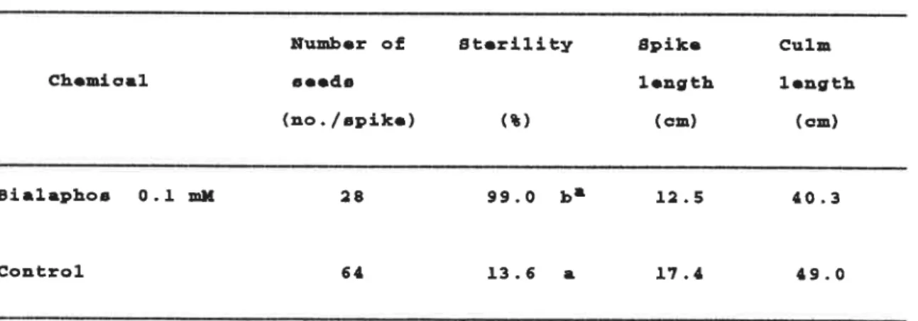
frrect Or D′araρんOs OrP ster〃1/ 1irP rFce rliP7 7997)163)
When bialaphos was applied at meiosis (Table 7), 99.0 0/o Sterility was induced by O.l RlM bialaphosi Furtherrnore, po‖ en grains were littie
stained with acetocarrnine solution (Fig. 8), when ObServed by
rnicroscopy. The resuit of acetocar!Ⅵ
ine staining suggested that
sterility induced by bialaphos treatrnent was caused by inhibition ofpo‖
en growth.(9n the other hand, rice plants treated with bialaphos
were inferior to the control plants with regard to number of seeds,
spike length and cuirn iength. As high sterility in rice is induced at a
low concentration (o=l RlM)Of bialaphos, it may be regarded as an
effective sterilant if it does not iniure the crop.
Tab■o 7. Moan Nunb● r Of ao●ds, st●ri■ity, spl■● L●ngth ana Cuin L● ngth of ttYanabikoB Ric● AB Affocted by Bia■ aphoB at Moioa■ a
Hunb●r oE St●ri■ity Spi■ ● cu■ n
Ch●mica■ ao● da ■●ngth ■●ngth
(■。./spiko) (L) (cn) (cm)
Bia■apとo8 0.工 HM 28 99,O ba ■2.5 ■0.3
Contro■ 64 ■3.6 a ■7.■ ■9.0
a MOanD in a 00■ unn fo■■owoa by tho aan● ■●tt●r ar● 4o七 日ignificant■ y
difFer●at at th● 5 名 ■●v●■ of aigttificanc● Ьy Duncanea Mu■ tip■● Rango T●at.
A
B
Fig.8 Rice Pollen Grains Stained with Acetocarmine Solution(X200) A:Bialaphos Treatment(0。 lmM)BI ContrOl
β 'oc力
em,car crParacrettst,cs or rlice sterlir,ど
/ treareJ vlitrP
bFaraρ ttOs
f旅増ど or bねraprIOs Oη
GS ac∬
yliり,加
ylirroThe assay was carried out on GS extracted from shoots of the rice
plantsi GS activity was deterrnined by the v‐
glutamythydroxamate
procedure.48)At the concentrations tested in this experiment(0‐ 3 nlMl (Fig, 9), bialaphos did not inhibit GS lin ylitro. However, L‐ PTC inhibited GS by 81 0/o at a concentration of O,l mM. Furthermore, LⅢ PTC inhibited completely GS at a concentration of l rnMi Since it is known in Japanese barnyard Rli‖et (fcrP,P90crP,oa vど ,′
,s OHwI)45) that bialaphos is
metabolized into L‐ PTC, it is suggested that Lい PTC is the real inhibitor of GS frorn rice plants and the real inducer of sterility in rice.
(°/0)
100
40
20
0 0 8 6 E O 中や いね 一〓 E 一o.1 0.3 1
Concentration
3(mM)
Fig.9 Effects of Bialaphos and L‐ PTC on Glutamine Synthetase frorn Shoots of Rice Piants,lirt yftro
42 ―
BialaphOS
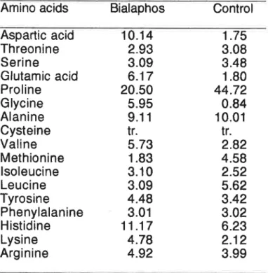
Cんattges or rree am∫ βO acliJs ,n arpどんers
The total contents of free anlino acids in untreated and bialaphos‐
treated anthers were 15,41 nM and l.36 nM per anther, respectively, The rate of proline was about a half of free anlino acids in control anthers.
Proline is utilized in new protein synthesis164)and Can be used as an
energy source,165) hOwever, the role of proline in connection with
po‖en fertility is not ctearly understood.131,166)
The untreated anthers had about twice as much proline as the anthers
treated with bialaphos, while the treated anthers had about 4″ 6 times
more aspartic acid and giutanlic acid than the untreated anthers (Table 8). The decrease of proline in bialaphos treated anthers was sirnilar to
that found for cytoplasnlic male sterile anthers as of rice,1291130)
wheat, maize,131)sOrghum132)and Sugar beet,133)The Characteristic
changes in the free amino acids in anthers treated with bialaphos arethe increases of giutamic acid and aspartic acid. Bialaphos, an inhibitor
of GS, inhibits the conversion of glutarnic acid to giutanline.45)As a
result of glutanline deficiency, the conversion of aspartic acid to
asparagine is inhibited because giutanline plays an irnportant role as the
arnide nitrogen donor in asparagine synthesis in rice plants.167,168)
This in turn means that proline synthesis is also inhibited, since
asparagine is a precoursor of prolinei169,170)Therefore, the decrease of proline and the increase of glutarnic acid and aspartic acid which occur in sterile anthers treated with biataphos is easily explained (Figi lo).
Table 8.Free Amino acids Composition(0/0)Of Anther Treated whh Bialaphos
Amino acids Bialaphos Control Aspattic acid Threonine Serine Glutamic acid Proline Glycine Alanine Cysteine Valine Methionine isoleucine Leucine Tyrosine Phenylalanine Histidine Lysine Arginine 10.14 2.93 3.09 6.17 20.50 5,95 9.11 tr. 5.73 1.83 3.10 3.09 4.48 3.01 11.17 4.78 4.92 1.75 3.08 3.48 1.80 44.72 0.84 10.01 tr. 2.82 4.58 2.52 5.62 3.42 3.02 6.23 2.12 3.99 tr.,trace Aspartic acЮ ― Asparagne Gtttarntt add+
〔
BialaphOSゝ引
GlutamineFig lo lhe Possible Pathway of Abnormal Amino acid Metabolism in the Stente Anthers Treated wtth Bialaphos
Eコ :Accumulauoni( 〕:Decrease
月
lice sterlilliり li「PJvceJ by lirP力 libFtors or g,vね阿
lirPe s"廿rPetasefttecrs or ′ηPP′b'どors οr g′vtam′rpe syr7:力eS's orP ster〃,ty FrP r,ce rlirP
T992)
Bialaphos was fOund to induce effective sterility in rice in 1991.
Three inhibitors of glutarnine synthesis, bialaphos,41‐ 46) L‐ phosphinothricin (L…
PTC)40)and L‐
methionine sulfoximine (L‐MSO)
,69,70,171‐173)were applied to rice plants in 1992 to find the optimum
application dosage for the induction of sterility (Fig. 11),
Bialaphos
O
ギ
鳴
7H3 7H3
H C―
CONH―
―C―
COOH
CH3 甲
C眺
C鳴
C―
CONH―
山
h
山
OH
L‐PTC(L‐phOsphinothricin)°
平
鳴
││CH3 甲
―C馳
CH2 C COOH
占
OH
L‐
MSO(L‐
methionine suroximine)ギ
鳴
O
llCH3 S C鳴
―CH2 C COOH
‖
占
NH
,L‐PTC,and L‐MSO
In 1992 (Table 9), bialaphos induced a high level of sterility in rice at a concentration of o.l mM at rmeiosisi L‐
PTC was also induced a high
ievel of sterility in rice at a concentration of O.l RlM at meiosisi The number of seeds, spike length and cuirn tength in the rice plants treated with bialaphos and L‐
PTC at a concentration of O.l mM were stighJy
inferior to those of the control. Bialaphos induced complete sterility in rice at a concentration of o=3 RlM at meiosis, but rice plants withered before anthesis by treatrnent with LttPTC at a concentration of O.3 rniⅥ .
These results indicate that the effective dosage for the induction of
sterility is in the narrow range of the concentrations from o.l RlM t0 0,3
mM.
Tab■● 9. 盟oan N4Hb● r of s●●dBr St●ri■ity and ap■kO L●ngth of ttYanabiko・ R10● aD Affoct● d by Thr●o Ch● Hica■a nt Hoと0日ia
Chomica■ Hunb●DO●da 驚 of tt t● ri■ity splk●■●ngth iongth rip● cu■H woight oE 3●●こa (n。・/Bplk● ) (鼈) (oH) (om) (g, ユ000い 工) B■ ■laph03 L‐PTC LいMB0 Co4tro■ 0。 0■ mM O.03 0。と 0.3 0。0工 HH O.03 0。エ 0.3 0.Oi nH O.03 0。i O.3 5。3 3.6 9■.8 工00 5。4 6。O 9S.4 ■.` S。1 3.2 2.B 4.■ 63 66 ■8 ■8 67 59 ■3 68 7■ 62 Se Se an a b b a a b n a ■ a a 工6。 9 57.8 ■7.3 57.0 iS。 2 S2.■ ■0.8 46。 O ■7.2 60.8 ■6。 S SB。 2 ■■.7 ■■.7 ■7.2 59。 9 工7.7 50.■ i6.9 S9。 エ ユ7.i SeoS 工6.e SS.S 26.8 25.7 25.7 26.0 25.2 2SoS 2S.0 24.9 25.3
a Moana in a cO■ unn fo■■ov●a by tho sano ■●tter ar● ■ot a10ni=■oAnt■y diffor●nt at tho s も ■●v●ユ oE Bignificano● by DuncanIB Muitip■ ● Rango Toat。
When the stamens and the pistils in the gtumous flowers treated
with O.1 ■
lM of bialaphos were observed by RliCrOscopy and compared
with the untreated control before anthesis (Fig. 12), the Shape of the
pistil treated with bialaphos was the same as that of control. However,
a‖ stamens treated with bialaphos became albinos, and po‖en grains in
the anthers were littie stained with acetocar!η ine solution and iodine‐ potassiurvl iodide (lKり SOlution, respectively (Fig. 8). The results of IKI
staining suggested that po‖
en sterility induced by bialaphos was
related to the absence or the decrease of pomen carbOhydrate content. The resuits from the observation of the giumous flowers suggest thatthe growth of the pistils treated with bialaphos is nor:mal and the
stamens alone are inhibited the norrnal growth by treatrnent with
bialaphos,
Fig。 12 Stamen and Pistilin the Glumous Flower Treated with Bialaphos
Figure 13 shows the panicles ater maturationi
panicles, which was sterilized by treatrnent with O.1 and L‐
PTC, is pale green. The resuits suggest that
occurs by the disaccumulation of nutrients in panictes.
The mM the color of the of bialaphos
phenomenOn
Fig.13 The Panicies Treated Bialaphos,L‐ PTC,and L‐
MSO
IAter Maturation)
fttecど or g′ vtam′ne orp ster,′ Fty 加「,ce treateJ v,ど rP bliaraprPOs
Bialaphos inhibits glutanline synthetase activity and is a potent herbicide for a wide range of weedsi41‐
46)Glutamine has been reported
to antagonize bialaphos through its action mechanism in inhibiting
growth of poHen tubes of Came〃
Fa ブaρOβ′Ca L..174)This suggest thataction mechanisrn of bialaphos invoives the GS inhibition and the
shortage of glutarnine. However, an exogenous application of giutamine does not antagonize bialaphos in Japanese barnyard mi‖ et(fCrP,ηOcrP′οavtlillis OHWり・45)in order to exarnine the interaction between bialaphos
and glutarnine on sterility in rice, glutanline applicated at the
concentrations Of l to 100 nlM to rice plants sirnuitaneousty treated with bialaphos(Table 10).Tab■o ■0。 Moan Nunber of g。 。ds, Steri■ ity and Sp■ kO Longth o至 田Yanabiko田
Rico ■B AEEocted by Bia■ aphOs and e■ utanino at Moiosia
Chonica■
Nunbor of Steri■ ity seeds
gpike Cu■ H Woigh七 oE
■ength ■●4gth ripe aooda
(n。./Spiko) (4) (cm) (Om) (gr■ 000‐・ ) Bia■aphoB + G■utanino Contro■ 0。■ mH ■ ■O ■00 48 ■7 ■2 59 6B 9■.8 93.8 95。 6 6,S 4.■ ■5.2 ■5。2 ■5.0 ■7.0 ■6.8 52.4 S■.■ 5■.7 57,0 55。 S 25。 3 25。 3 ド b b a a
a Moans in a co■ uHH differont at the S
ToB七.
fo■■owod by tho 8■ mO ■Otter 亀 ■ova■ of significanca by
aro not aignificant■ y Duncan's Mu■ tip■● Rango