ORIGINAL
Electroneurography cannot predict when facial synkinesis
develops in patients with facial palsy
Takahiro Azuma1, Katsuhiko Nakamura1, Mika Takahashi1, Hitomi Miyoshi1, Naoki Toda2, Hidetaka Iwasaki3, Teruhiko Fuchigami1, Go Sato1, Yoshiaki Kitamura1, Koji Abe1, and Noriaki Takeda1
1Department of Otolaryngology, University of Tokushima School of Medicine, Tokushima, Japan, 2Department of Otolaryngology, Anan Med-ical Center, Tokushima, Japan, 3Department of Otolaryngology, Yoshinogawa Medical Center, Tokushima, Japan
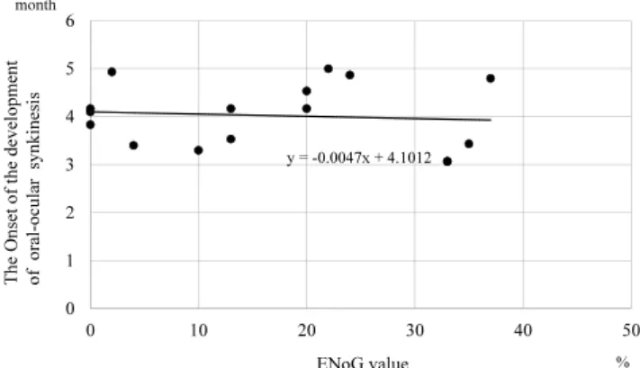
Abstract : The objective of this study is to clarify when facial palsy patients with lower value of Electroneurogra-phy (ENoG) should begin the rehabilitation to prevent the development of facial synkinesis. For this purpose, we examined the relationship between the value of ENoG measured 10-14 days after facial palsy onset and the onset day of the development of oral-ocular synkinesis. Sixteen patients with facial palsy including 11 with Bell’s palsy and 5 with Ramsay Hunt syndrome (7 men and 9 women ; 15-73 years old ; mean age, 41.6 years) were enrolled in this study. There was no correlation between ENoG value and the onset day of the development of oral-ocular synkinesis (ρ = .09, p = .73). Oral-ocular synkinesis began to develop in 4.0 ± 0.7 months (mean ± SD ; range : 3.1-5.0 months) after facial palsy onset regardless of ENoG value. In conclusion, ENoG value cannot predict when facial synkinesis develops in patients with facial palsy. We recommend that facial palsy patients with a high risk for the development of synkinesis begin the biofeedback rehabilitation with mirror to prevent the development of facial synkinesis 3 months after facial palsy onset. J. Med. Invest. 67 : 87-89, February, 2020
Keywords : facial palsy, facial synkinesis, electroneurography, onset, predictive factor
INTRODUCTION
The most unpleasant sequela of peripheral facial palsy is facial synkinesis, which develops after facial nerve aberrantly regenerates in affected facial muscles. Because facial nerve func-tion hardly recovers completely once facial synkinesis is estab-lished (1), preventing synkinesis with biofeedback rehabilitation is effective for its better outcomes. Indeed, in our previous study, facial biofeedback rehabilitation using a mirror that began when the first signs of facial synkinesis developed resulted in a de-crease of facial synkinesis (2). We also showed that facial palsy patients with lower value of electroneurography (ENoG) have a high risk for the development of facial synkinesis (3). These findings suggested that facial palsy patients with low value of ENoG should receive the biofeedback rehabilitation with mirror to prevent the development of facial synkinesis.
In the present study, an attempt was made to clarify when facial palsy patients with lower value of ENoG should begin the rehabilitation, because it was controversially reported that facial synkinesis began to develop in 3-14 months (1), 4-8 months (4), or 4-7 months (5) after the onset of facial palsy. For this purpose, we examined the relationship between the value of ENoG mea-sured 10-14 days after facial palsy onset and the onset day of the development of oral-ocular synkinesis in patients with facial palsy to use ENoG as a predictive indicator of facial synkinesis development.
MATERIALS AND METHODS
This study was approved by the Committee for Medical Ethics of Tokushima University Hospital. This study was conducted in accordance with the principles of the Declaration of Helsinki.
Patients
Sixteen patients with facial palsy were included in this study. In 16 patients (7 men and 9 women ; 15-73 years old ; mean age, 41.6 years), of them 11 had with Bell palsy and 5 with Ramsay Hunt syndrome. Our previous study showed that 46.5% of ENoG was the cut off value as a predictor for the development of oral-oc-ular synkinesis (3). Based on this result, facial palsy patients with less than 46.5% of ENoG value were enrolled in this study. Corticosteroid was administered to those with Bell’s palsy, while corticosteroid with ant-viral agent was administered to with Ramsay Hunt syndrome within 7 days after the onset of facial palsy. Each patient then visited our clinic once a month to be examined for the onset day of the development of oral-ocular synkinesis. No patients received any facial rehabilitation. No decompression surgery was performed in all patients.
Development of oral-ocular synkinesis
In the present study, we defined the first sign of development of oral-ocular synkinesis by visual assessment as any involuntary contraction of the orbicularis oculi muscle in the affected side during three designated mouth movements, lip pursing [u], teeth baring [i], and cheek puffing [pu] (2,3).
Electroneurography
Electroneurography was performed 10-14 days after the onset of facial palsy. A rectangular 0.2 msec impulses of 1000 Hz with stepwise increase of the stimulating current from 35 mA to 50 mA was given by a bipolar stimulator placed on the skin over the stylomastoid foramen, and the maximal compound action potential were recorded through the surface electrodes placed on the skin in the nasolabial fold (6, 7). The percentage ratio of
The Journal of Medical Investigation Vol. 67 2020
87
Received for publication July 1, 2019 ; accepted December 11, 2019. Address correspondence and reprint requests to Takahiro Azuma, M.D., Ph.D., Department of Otolaryngology, University of Tokushima School of Medicine, 3-18-15, Kuramoto, Tokushima 770-8503, Japan and Fax : +81-88-633-7170.
88
T. Azuma, et al. When does facial synkinesis develop?the peak to peak amplitude of the maximal compound action po-tential of the affected side relative to that of the normal side was calculated as value of ENoG. This ENoG value indicates the per-centage of fibers without degeneration in facial nerve axons (7).
Statistical Analysis
Spearman’s rank correlation coefficient was used for statis-tical analysis (SPSS 22.0 for Windows ; SPSS Inc, Chicago, Illinois). p < 0.05 was considered significant.
RESULTS
There was no correlation between ENoG value measured 10-14 days after facial palsy onset and the onset day of the de-velopment of oral-ocular synkinesis in patients with facial palsy (ρ = .09, p = .73) [Fig 1]. Oral-ocular synkinesis began to develop in 4.0 ± 0.7 months (mean ± SD ; range : 3.1-5.0 months) after facial palsy onset regardless of ENoG value.
DISCUSSION
In the present study, we demonstrated no correlation between ENoG value and the onset day of the development of oral-ocular synkinesis in patients with facial palsy. Because ENoG value in-dicates the degree of facial nerve injury, this result suggests that the onset of the development of facial synkinesis after facial paly is unaffected by the degree of facial nerve injury. Therefore, it is concluded that ENoG value cannot predict when facial synkine-sis develops in patients with facial palsy. We also demonstrated that oral-ocular synkinesis began to develop 3.1-5.0 months after the onset of facial palsy regardless of the degree of the facial nerve injury. Because facial palsy patients with low value of ENoG have a high risk of developing of facial synkinesis, they are recommended to begin the biofeedback rehabilitation with mirror to prevent the development of facial synkinesis 3 months after the facial palsy onset.
Facial nerve runs in the fallopian canal of the temporal bone, which is a narrow bony canal from the fundus of the internal au-ditory canal to the stylomastoid foramen. Then, facial nerve ex-tratemporally passes through the parotid gland and innervates the mimic muscles of facial expression. After herpes simplex virus in Bell’s palsy or varicella zoster virus in Ramsay Hunt syndrome reactivate at geniculate ganglion of the temporal bone in patients (8-11), the viral inflammation spreads along the facial
nerve in the fallopian canal both rostrally and caudally, and the inflamed swollen facial nerve is compressed and injured (12,13). But, it was reported that within one week following injury, injured facial nerve gave rise to axonal sprouts (14). According to the anatomical reports on facial nerve (15), the length of the fallopian canal of the temporal bone was 30 mm and that of the extratemporal segment of facial neve was 110 mm (16,17). Based on these reports, it is assumed that the distance of facial nerve from the fundus of the internal auditory canal to mimic muscles is 140 mm and that the distance of facial nerve from the stylomastoid foramen to mimic muscles is 110 mm. Because the injured motor nerve axon regenerates at the rate of 1 mm/day (18), it is calculated that facial nerve that was injured in the fallo-pian canal regenerates to innervate mimic muscles in the period from 3.7 months (110 mm) to 4.7 months (140 mm). During the regeneration process, aberrant axonal sprouting of facial nerve re-innervates inappropriate facial muscle and misdirection of the facial re-innervating leads to the development of synkinesis (19). Therefore, the present finding that oral-ocular synkinesis began to develop in 3.1 to 5.0 months is well in accordance with the above-mentioned assumption of 3.7 to 4.7 months.
In conclusion, ENoG value cannot predict when facial synki-nesis develops in patients with facial palsy. Since facial synkine-sis began to develop 3.1-5.0 months after the onset of facial palsy regardless of the value of ENoG, facial palsy patients with a high risk for the development of synkinesis should begin the biofeed-back rehabilitation with mirror to prevent the development of facial synkinesis 3 months after facial palsy onset.
CONFLICT OF INTEREST
The authors declare not having any financial support or rela-tionship that may pose a conflict of interest.
ACKNOWLEDGEMENTS
We thank Dr. Kalubi Bukasa for his critical reading of the manuscript.
REFERENCES
1. Yamamoto E, Nishimura H, Hirono Y : Occurrence of se-quelae in Bell’s palsy. Acta Otolaryngol Suppl 446 : 93-6, 1988
2. Nakamura K, Toda N, Sakamaki K, Kashima K, Takeda N : Biofeedback rehabilitation for prevention of synkinesis after facial palsy. Otolaryngol Head Neck Surg. 128 : 539-43, 2003
3. Azuma T, Nakamura K, Takahashi M, Miyoshi H, Toda N, Iwasaki H, Takeda N : Electroneurography in the acute stage of facial palsy as a predictive factor for the develop-ment of facial synkinesis sequela. Auris Nasus Larynx 45 : 728-31, 2018
4. Celik M, Forta H, Vural C : The development of synkinesis after facial nerve paralysis. Eur Neurol 43 : 147-51, 2000 5. Kanaya K, Ushio M, Kondo K, Hagisawa M, Suzukawa K,
Yamaguchi T, Tojima H, Suzuki M, Yamasoba T : Recovery of facial movement and facial synkinesis in Bell’s palsy pa-tients. Otol Neurotol 30 : 640-4, 2009
6. Fisch U : Maximal Nerve Excitability Testing vs Electro-neuronography Arch Otolaryngol 106 : 352-7, 1980 7. Gantz BJ, Gmuer AA, Holliday M, Fisch U :
Electroneuro-graphic evaluation of the facial nerve. Method and technical Figure 1. Relationship between ENoG value 10-14 days after the
onset of facial palsy and months at the onset of the development of oral-ocular synkinesis after facial palsy. n = 16, ρ = .09, p = .73.
89
The Journal of Medical Investigation Vol. 67 February 2020
problems. Ann Otol Rhinol Laryngol 93 : 394-8, 1984 8. Furuta Y, Takasu T, Sato KC, Fukuda S, Inuyama Y,
Nagashima K : Latent herpes simplex virus type 1 in human geniculate ganglia. Acta Neuropathol 84 : 39-44, 1992
9. Furuta Y, Takasu T, Fukuda S, Sato-Matsumura KC, Inuyama Y, Hondo R, Nagashima K : Detection of varicel-la-zoster virus DNA in human geniculate ganglia by poly-merase chain reaction. J Infect Dis 166 : 1157-9, 1992 10. Murakami S, Mizobuchi M, Nakashiro Y, Doi T, Hato N,
Yanagihara N : Bell palsy and herpes simplex virus : identi-fication of viral DNA in endoneurial fluid and muscle. Ann Intern Med 124 : 27-30,1996
11. Murakami S : Bell palsy and Ramsay Hunt Syndrome. In : William H. S, Azizzadeh B, editors. The Facial Nerve. Thieme, New York, 2014, pp70-80
12. Liston SL1, Kleid MS : Histopathology of Bell’s palsy. La-ryngoscope 99 : 23-6, 1989
13. Fisch U, Esslen E : Total intratemporal exposure of the facial nerve. Pathologic findings in Bell’s palsy. Arch Otolar-yngol 95 : 335-41, 1972
14. Georg K : Neurobiology of regeneration and degeneration. In : May M and Schaitkin B, editors. The Facial Nerve. Thieme, New York, 2000, pp67-79
15. Kudo H, Nori S : Topography of the facial nerve in the human temporal bone. Acta Anat (Basel) 90 : 467-80, 1974 16. May M : Anatomy for the clinician. In : May M and
Schaitkin B, editors. The Facial Nerve. Thieme, New York, 2000, pp19-56
17. Tzafetta K, Terzis JK : Essays on the facial nerve : Part I. Microanatomy. Plast Reconstr Surg 125 : 879-89, 2010 18. P Weiss, HB Hiscoe : Experiments on the mechanism of
nerve growth. J Exp Zool 107 : 315-395, 1948
19. Crumley RL : Mechanisms of synkinesis. Laryngoscope. 89 : 1847-54, 1979