INTRODUCTION
Pituitary adenomas are generally benign tumor, and the majority of pituitary adenomas do not metastasize to distance sites, but about 30% of them invade to the local tissues, such as cavernous sinus, sphenoid sinus, orbit and clivus (1-4). The radical surgical treat-ment of the invasive pituitary adenoma is more difficult than the noninvasive tumors. Therefore, the tumor
re-currence may be seen in a short period after operation. Tumor invasion into surrounding tissues is a com-plex phenomenon and characteristic of more aggres-sive and often malignant tumor behavior. The invaaggres-sive factors involve the heparinase, serine proteinases, cathepsins, and matrix metalloproteinases (5). MMPs are proteolytic enzymes that have the capability of breaking down basement membrane and connective tissue, and are important for tissue breakdown in the process of tumor invasion (6, 7). Among these invasive factors, MMP-2 and MMP-9 have been widely studied in lung cancers, breast cancers and other malignant neoplasms (8). Several studies examined the expres-sion of MMPs in malignant gliomas (9-12). However, the expression of MMP-2 and MMP-9 in the invasive
ORIGINAL
Matrix metalloproteinase 2 and 9 expression correlated
with cavernous sinus invasion of pituitary adenomas
Weiping Liu, Yoshihito Matsumoto, Masaki Okada, Keisuke Miyake, Katsuzo Kunishio,
Nobuyuki Kawai, Takashi Tamiya, and Seigo Nagao
Department of Neurological Surgery, Faculty of Medicine, Kagawa University, Kagawa, Japan Abstract : Object : Matrix metalloproteinase (MMP) 2 and 9 are important for tissue breakdown in the process of tumor invasion. The aim of this study is to evaluate the relationship between the expression of MMP-2, MMP-9, MIB-1 LI and cavernous sinus invasion in pituitary adenomas.
Methods : Tissue samples from 54 patients with pituitary adenomas were studied. Expression
of MMP-2, MMP-9, and MIB-1 labeling index (LI) were evaluated by immunohistochemical method. In sixteen cases, the expression of MMP-2 and MMP-9 mRNA was also examined by RT-PCR assay.
Results : Thirty-four patients were women and 20 were men, with a mean age of 49.9 years old
(range 18-76 years). There were 12 cases with cavernous sinus invasion, and 42 were noninvasive cases. MMP-2 and MMP-9 score of invasive case (3.9±0.5, 4.1±0.4) were significantly higher
than those (2.3±0.2 ; p<0.01 ; 2.6±0.2 ; p<0.01) without invasion. The MIB-1 LI of this study presented no significantly difference between the invasive and noninvasive pituitary adenomas. The percentage of MMP-2 mRNA/β-actin mRNA and MMP-9 mRNA/β-actin mRNA were also observed significantly higher in invasive pituitary adenomas (68.2±15.3 % ; 59.7±12.5 %)
than noninvasive pituitary adenomas (21.8±8.2 % , p<0.05 ; 33.3±5.4 % , p<0.05).
Conclusions : Our study suggests that the expression of MMP-2 and MMP-9 may have a value
to assess the invasive pituitary adenomas, and proliferation and invasion of pituitary adenomas may present a different mechanism. J. Med. Invest. 52 : 151-158, August, 2005
Keywords : MMP-2 ; MMP-9 ; Ki-67 ; pituitary adenomas ; tumor invasion
Received for publication December 8, 2004 ; accepted March 15, 2005.
(the order of the first two authors is to be considered arbitrary) Address correspondence and reprint requests to Yoshihito Matsumoto, Department of Neurological Surgery, Kagawa Medical University, Miki-cho, Kita-gun, Kagawa 761-0793, Japan and Fax : +81-87-891-2208.
The Journal of Medical Investigation Vol. 52 2005
pituitary adenomas showed different results in the previous studies. This may be related to different meth-ods, or different value of assessing the data.
The aim of this study was to determine whether expression of MMP-2 and MMP-9 might play a role in cavernous sinus invasion of pituitary adenomas. The possible mechanisms was investigated in cavern-ous sinus invasion of pituitary adenomas.
MATERIALS AND METHODS
Samples of pituitary adenomas were obtained from 54 patients who had undergone surgery from Oc-tober 1984 to September 2001 at the Kagawa Medical University Hospital. Thirty-four patients were women and 20 were men, with a mean age of 49.9 years (range 18-76 years). There were 12 cases with cavernous in-vasion (Fig. 1), and 42 cases without inin-vasion. There were 12 growth hormone (GH)-producing adenomas, 11 prolactin (PRL)-producing adenomas, 4 adrenocor-ticotropic hormone (ACTH)-producing adenomas, one thyroid stimulating hormone (TSH)-producing adenomas, one follicle-stimulating hormone (FSH) -producing adenomas, and 25 nonfunctioning (NF) pituitary adenomas. Invasion to cavernous sinus space of pituitary adenomas was defined on the basis of the
classification by Knops. Invasive and non-invasive ade-noma are distinguished from each other by a medial target, the intercarotid line, through the cross-sectional centers (Fig.1) (13).
IMMUNOHISTOCHEMISTRY
MMP-2 and MMP-9 expression and proliferative potential were examined immunohistochemically on formalin-fixed, paraffin-embedded sections using mon-oclonal antibodies (MAbs) against MMP-2 (Ab -2 ; Fuji-chemistry, Japan), MMP-9 (Ab-2 ; Fuji-chemistry, Japan) and MIB-1 MAb (Immunotech, Marseille, France). Six-micron sections were deparaffinized in xylene, rehydrated through graded alcohols, and immersed for 15 min in phosphate-buffered saline (PBS). For an-tigen retrieval, the sections were microwaved in 0.01 M citrite buffer (pH 6.0) for 20 min. After microwave pretreatment, the endogenous peroxidase activity was blocked with 3% hydrogen peroxidase in methanol for 10 min, and nonspecific staining was then blocked by 20-min incubation with normal horse serum. The sections were then incubated overnight at 4℃ with primary antibodies (MMP-2,×100 ; MMP-9,×100 ;
MIB-1,×50) in a humidity chamber. The sections were
treated for 30 min with biotinylated horse secondary
Figure 1. MRI imaging of invasive pituitary adenomas. Non-invasive pituitary adenomas are disguished from invasive adenomas by a medial tangent, the intercarotid line-through the cross-sectional centers-and a lateral tangent on the intra and supracavernous internal carotid arteries. Case A ; The pituitary adenoma is huge but is classified in non-invasive type. Case B and C ; The adenomas are not large but are considered as non-invasive type.
W. Liu, et al. MMP-2 and MMP-9 expression in pituitary adenomas
antibody against mouse immunoglobulins (ABC Elite, Vector Lab., Burlingame, CA, USA) and for 30 min with avidin-biotin complex (ABC Elite, Vector Lab.), followed by 0.06 % diaminobenzidine (Sigma, MO, USA) with 0.01% hydrogen peroxidase for 5 min. The slides were lightly counterstained with hematoxylin. Controls were performed by omitting the primary antibody.
A semi-quantitative assessment of the immuno-histochemistry of MMP-2 and MMP-9 was employed by grading the staining as (0) negative, (1)<30%, (2) 30 - 60%, (3) 60 -100% of tumor cells showing positive staining. The intensity of the immunostaining was also graded as (0) negative, (1) weak, (2) moderate, and (3) strong. The total immunohistochemical score was determined as the sum of the frequency and intensity scores for tumor cells (14). Twenty representative fields were counted, and the immunohistochemistry scores were determined by concordance among the scores of two independent reviewers. The score was divided into 0 - 6 for each sample.
The MIB-1 LI was calculated as the percentage of immunostained nuclei, excluding nuclei of vascular components and hematogenous cells, by counting approximately 200 tumor nuclei.
Reverse Transcription-Polymerase Chain Reaction (RT-PCR) Analysis
RNA was extracted from the frozen tissue sample by using ISOGEN regent (Nippon Gene, Toyama, Japan). Briefly, 100 mg of pituitary adenomas tissue was homogenized in 1 ml of ISOGEN. Subsequently, 0.2 ml of chloroform was added and the mix was cen-trifuged. This separated the solution into an aqueous phase containing RNA, an interphase containing DNA, and an organic phase containing protein. The aqueous layer was aspirated and added to 0.5 ml of isopropanol for RNA precipitation. Following this, the solution was centrifuged and the pellet was washed with 70% ethanol and centrifuged. After that, RNA was collected into 50µl of diethylpyrocarbonate (DEPC)-treated water. RT-PCR was performed using a First-Strand cDNA Synthesis Kit (Amersham Pharmacia Biotech UK Lim-ited). Two microlitre of total RNA (2µg) was added to 5.5µl of the RT mixture. After mixing, the samples were incubated at 37℃ for 45 min, 95℃ for 5 min, and 4℃ for at least 5 min. 17.5 microlitre of PCR-mixture containing 12.5µl primers and Taq DNA polymerase (Amersham Pharmacia Biotech UK Limited) was added to the RT products. Initial denaturation for 2 min at 94℃ was followed by 30 cycles of 1 min at 94℃, 1 min at 55℃, and 2 min at 72℃, and a final extension
for 6 min at 72℃. The PCR products were separated on 2% agarose gels, and ethidium bromide-stained bands were recorded by Mupid-2R (Cosmo-Bio, Tokyo, Japan). The primer sequences were as follows :
MMP-2 : forward primer : 5’primer : 5’-GCTGAT ACTGACACTGGTACTG-3’, reverse primer : 5’-CAA TCTTTTCTGGGAGCTC-3’.
MMP-9 : forward primer : 5’primer : 5’-AAGGAT GGTCTACTGGCAC-3’, reverse primer : 5’-AGAGA TTCTCACTGGGGC-3’(15).
β-actin : forward primer : 5’primer : 5’-ATCACCA TTGGCAATGAGGG-3’, reverse primer : 5’-TTGAA GGTAGTTTCGTGGAT-3’.
The results of percentage of MMP-2 mRNA/β-actin mRNA and MMP-9 mRNA/β-actin mRNA were as-sessed by NIH software using computer.
Statistical analysis
Student’s t-text was used to evaluate the relation-ships among various parameters. The effect of various parameters, i.e., tumor size, MMP-2 score, MMP-9 score, MIB-1 LI and percentage of MMP-2 mRNA/ β-actin mRNA and MMP-9 mRNA/β-actin mRNA was assessed, using the STAT VIEW for windows software performed statistical analysis.
RESULTS
Immunohistochemistry findings
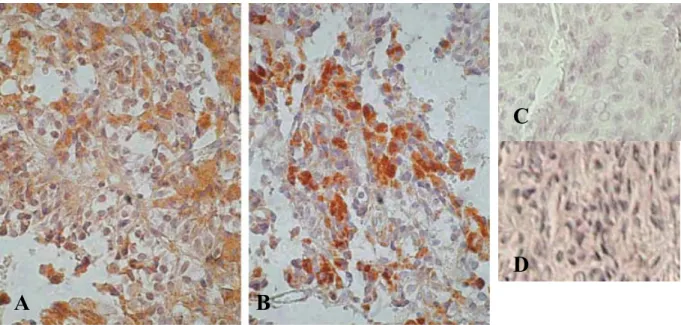
The cytoplasm and/or nuclear of tumor cells were stained as brown color in pituitary adenomas (Fig. 2). In this study, Forty-six cases (82 %) of 54 pituitary ade-nomas were observed positive immunoreactivity, whereas, staining intensity was variable. MMP-2 and MMP-9 positive staining could be seen in all 12 cases of invasive pituitary adenomas. The MMP-2 score of invasive pituitary adenoma (3.9±0.5) (mean±SE) were significantly higher than those without invasion (2.3± 0.2) (p<0.01). The MMP-9 score of invasive pituitary adenoma (4.1±0.4) were significantly higher than those without invasion (2.6±0.2 ; p<0.01) (Table 1). We found that invasive pituitary adenomas were sig-nificantly more likely to express MMP-2 and MMP-9 compared with noninvasive pituitary adenomas. The MIB-1 LI tended to be higher in invasive pituitary ade-nomas (1.2±0.4 %) than that (0.6±0.2 %) in noninva-sive tumors, however, there was no significant differ-ence (p=0.1528). There was no correlation between the MMP-2 and MIB -1 LI (r= -0.05, p=0.72), MMP-9 and MIB-1 LI (r=0.004, p=0.98). According to tumor size, we divided the 54 pituitary adenomas into 4 groups The Journal of Medical Investigation Vol. 52 August 2005 153
(≦10 mm ,>10 mm to≦20 mm,>20 mm to≦40 mm, and>40 mm), and compared the expression of MMP-2, MMP-9, and MIB-1 LI in the different groups. There were no significant differences among the 4 groups (Table 2). There are also no correlation in MMP-2, and MMP-9 expression among ACTH, PRL, or GH producing adenomas and NF pituitary adeno-mas (Table 3).
mRNA Expression of MMPs
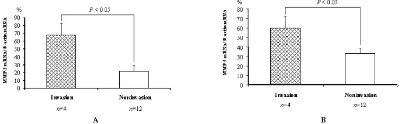
We examined MMP-2 and MMP-9 mRNA by RT-PCR from 16 cases including 4 invasive and 12 non-invasive pituitary adenomas. The expression of MMP-2 and MMP-9 mRNA was also observed in the invasive pituitary adenomas and noninvasive tumor (Fig. 3). The percentage of MMP-2 mRNA/β-actin mRNA was observed significantly higher in invasive pituitary ade-nomas (68.2±15.3 %) than those without invasion (21.8±8.2 % ; p<0.05). The percentage of MMP-9 mRNA/β-actin mRNA was observed significantly higher in invasive pituitary adenomas (59.7±12.5 %) than those of noninvasive adenomas (33.3±5.4 % ; p<0.05) (Fig. 4). There are significant correlation between mRNA and immunohistochemistry score in MMP-2 and MMP-9 (r=0.582, p<0.05 ; r=0.669, p<0.01) (Fig. 5).
Table 1. The score of MMP-2 and MMP-9 expression in invasive and noninvasive pituitary adenomas
Group No. of patients Age Score of MMP-2 Score of MMP-9 MIB-1 LI Invasion 12 52.7±3.6 3.9±0.5** 4.1±0.4** 1.2±0.4 Noninvasion 42 48.9±2.6 2.3±0.2 2.6±0.2 0.6±0.2 Patients age, score of MMP-2, MMP-9 expression and MIB-1 LI are expressed as means±SE.
**
: P<0.01 vs noninvasion group LI : labeling index.
Table 2. Correlation with the expression levels of MMP-2 and MMP-9 and the tumor size
Group Tumor size(mm) No. of patients Age Score of MMP-2 Score of MMP-9 P value 1 ≦10 10 31.6±4.5 2.1±0.5 2.1±0.5 NS 2 >10 to≦20 20 52.2±3.2 3.0±0.4 3.0±0.3 NS 3 >20 to≦40 21 56.3±2.9 2.7±0.4 3.2±0.4 NS 4 >40 3 48.7±6.7 2.3±0.3 3.0±0.6 NS Patients age, and score of MMP-2, MMP-9 expression are expressed as means±SE.
NS : not significant
Table 3. Correlation with the expression of MMP-2 and MMP-9 and the hormone secreting pituitary adenomas
Group No. of patients Score of MMP-2 Score of MMP-9 P value ACTH 4 2.0±0.7 2.5±0.9 NS GH 12 2.8±0.5 3.1±0.4 NS PRL 11 2.7±0.5 2.9±0.6 NS NF 25 2.7±0.3 3.0±0.3 NS
The score of MMP-2, MMP-9 expression are expressed as means±SE.
NS : not significant. NF : nonfunction
Figure 2. Immunohistochemical staining of MMP-2 (A) and MMP-9 (B) in invasive pituitary adenomas. Photograph showing cytoplasmic and/or nuclear immunoreactivity of the tumor cells. Negative control for MMP-2 (C) and MMP-9 (D) (magnification ×400)
W. Liu, et al. MMP-2 and MMP-9 expression in pituitary adenomas
Figure 4. The graph showed the significant differences of MMPs mRNA expression between in the invasive cases and in the noninvasive cases (*p<0.05). (A) MMP-2. (B) MMP-9.
Figure 5. The graph showed that there are significant correlation between mRNA and immunohistochemistry score in MMP-2 and MMP-9. (A) MMP-2 and MMP-2 mRNA (r=0.582, p<0.05) ; (B) MMP-9 and MMP-9 mRNA (r=0.669, p<0.01) Figure 3. Expression of MMP-2 (A), MMP-9 (B) mRNA by RT-PCR withβ-actin serving as a control for equal loading in pituitary adenomas. (A) Positive bands are oberserved in the invasive cases (case 3, 8, 12, and 14). (B) Positive bands are found in the invasive cases (case 3, 8, 12, and 14). The levels of MMP-2 were quantitated by using NIH image (version 1.61).
DISCUSSION
MMPs are a family of zinc-dependent endopeptidases that regarded as promoting of invasion and metastasis (16). The family includes five subclasses : collagenases, gelatinases, stromelysins, metalloelastase and membrane-type metalloproteinases. MMPs have been implicated in various physiological and tissue morphogenesis as well as tumor cell invasion and metastasis (17). The MMP-2 and MMP-9 are the common studied gela-tinases and can degrade the basement-membrane type Ⅳ collagen. Tumor cell invasion utilizes numerous proteases in vitro, of which the predominant ones may be gelatinase-A and -B (MMP-2 and MMP-9 re-spectively) (18). Nakada, et al. (19) demonstrated that the production levels of MMP-2 are significant higher in glioblastomas multiforme than in other grades of astrocytic tumors. Several studies indicated that there presented significant correlation between the MMP-2 and MMP-9 expression and invasion in the malignant tumors (20 -25).
The majority of pituitary adenomas are benign and do not metastasize. However, it always doubted the neurosurgeons that parts of pituitary adenomas are locally invasive and more than 20% cases recur (26 -28). The invasive behavior of pituitary adenomas is poorly understood. In present study, we evaluated the expression of MMP-2 and MMP-9 by immunohis-tochemistry and MMP-2 and MMP-9 mRNA level by RT-PCR assay in pituitary adenomas. We found that invasive pituitary adenomas have a significantly higher expression of MMP-2 and MMP-9 protein, and mRNA than noninvasive tumors. Turnere, et al. (9) found that invasive macroprolactinomas were signifi-cantly more like to express MMP-9 than noninvasive tumor. Kawamoto et al. described that the incidence of tumor cells secreting MMP-9 was significantly higher in invasive pituitary adenomas than in nonin-vasive ones (11). Pereta, et al. found the high expres-sion of MMPs and low levels of TIMP-1 in pituitary adenomas, and the active form of the MMP-2 was found in some pituitary tumors (29). They also showed that high level of MMP activity could be to contribute the regulation of proliferation and hormone secretion in pituitary adenomas (28). We failed to find the sig-nificant difference of MMPs expression among the hormone (GH, PRL, and ACTH) secreting adenoma and NF pituitary adenomas. Our data suggest that proteases, MMP-2/-9, may play a role in tumor inva-siveness of pituitary adenomas and did not correlate with hormone secreting activity.
A number of studies have been widely considered
that Ki-67 LI is the best means to evaluate cell prolif-eration in clinical practice. It is also considered a useful tool for assessing the recurrence of the tumor (30, 31). We have previously shown that no correlation between MMP-2/-9 expression and the Ki67 LI expression in gliomas (32). Mastronardi’s study showed that the Ki-67 LI was a useful marker in determining the invasive behaviour of pituitary adenomas, and his results also seem to exclude significant correlations between MIB 1 LI and tumor size of pituitary adenomas (33, 34). For the first time, we have demonstrated that there was no difference between MIB1 LI of invasive pitui-tary adenomas and that of noninvasive adenomas, and no correlations were found between the expression of MMP-2/-9 and MIB-1 LI, MMP-2/-9 expression and tumor size. The present data suggested that the expression of MMP-2 and MMP-9 were the index markers of tumor invasion, whereas the expression of MMP-2 and MMP-9 had no correlation with the proliferation of the pituitary adenomas. Some re-searcher (6-9) found that the expression of MMP-9 was related with the invasiveness in pituitary adenomas, however few authors examined the MMP-2 expression in pituitary adenomas. Our study demonstrated the correlation between the expression of MMP-2, as well as MMP-9, and cavernous invasion in pituitary ade-nomas. We also found that some of the tumors, in spite of their small size, invade the cavernous sinus or the surrounding area, whereas some huge pituitary ade-nomas did not invade the surrounding structures. Our data may explain this phenomenon. Turner’s study the adenomas removed when they recurred were more likely to express MMPs (9). On the other hand, Knappe et al. found no differences in expression of MMP9 in recurrent adenomas (35). Sampling of tumor tissue is done to the opening in the intrasellar parts of the tumor during transsphenoidal surgery and the areas of lateral invasion are cleaned by the suction rather than excision. Therefore, we could not investigate differences of expression of MMP-2/-9 in distinct areas of possible dural invasiveness, nor compare them with central intrasellar areas of the tumors.
Although there may be many biochemical mecha-nisms about the invasion of tumors, the present data can partially explain the invasive behavior of pituitary adenomas. Our study indicated that the expression of MMP-2 and MMP-9 had a value to assess the invasion in pituitary adenomas, while the MIB-1 LI was not the index of tumor invasion.
W. Liu, et al. MMP-2 and MMP-9 expression in pituitary adenomas
REFERENCES
1. Acquati S, Pizzocaro A, Tomei G, Giovanelli M : A comparative evaluation of effectiveness of medi-cal and surgimedi-cal therapy in patients with macro-prolactinoma. J Neurosurg Sci 45 : 65-69, 2001 2. Meij BP, Lopes MB, Ellegala DB : The long-term significance of microscopic dural invasion in 354 patients with pituitary adenomas treated with transsphenoidal surgery. J Neurosurg 96 : 195-208, 2002
3. Nakase H, Ohnishi H, Matsuyama T, Morimoto T, Sakaki T : Two-stage skull base surgery for tu-mours extending to the sub- and epidural spaces. Acta Neurochir (Wien) 140 : 891-898, 1998 4. Lucas T, Astorga R, Catala M : Preoperative
lanreotide treatment for GH-secreting pituitary adenomas : effect on tumour volume and pre-dictive factors of significant tumour shrinkage. Clin Endocrinol (Oxf) 58 : 471- 481, 2003 5. Liotta LA, Steeg PS, Stetler-Stevenson WG :
Cancer metastasis and angiogenesis : an imbal-ance of positive and negative regulation. Cell 64 : 327-336, 1991
6. Di Nezza LA, Misajon A, Zhang J, Jobling T, Quinn MA, Ostor AG : Presence of active gela-tinases in endometrial carcinoma and correlation of matrix metalloproteinase expression with increasing tumor grade and invasion. Cancer 94 : 1466-1475, 2002
7. Hirano H, Tsuji M, Kizaki T, Sashikata T, Yoshi Y, Okada Y: Expression of matrix metalloproteinases, tissue inhibitors of metalloproteinase, collagens, and Ki67 antigen in pleural malignant mesothe-lioma : an immunohistochemical and electron microscopic study. Med Electron Microsc 35 : 16-23, 2002
8. Hrabec E, Strek M, Nowak D, Greger J, Suwalski M, Hrabec Z: Activity of type IV collagenases (MMP-2 and MMP-9) in primary pulmonary carcinomas : a quantitative analysis. J Cancer Res Clin Oncol 128 : 197-204, 2002
9. Turner HE, Nagy Z, Esiri MM, Harris AL, Wass JA : Role of matrix metalloproteinase-9 in pitui-tary tumor behavior. J Clin Endocrinol Metab 85 : 2931-2935, 2000
10. Nakagawa T, Kubota T, Kabuto M, Sato K, Kawano H, Hayakawa T : Production of matrix metalloproteinases and tissue inhibitor of metal-loproteinases-1 by human brain tumors. J Neu-rosurg 81 : 69 -77, 1994
11. Kawamoto H, Kawamoto K, Mizoue T, Uozumi T,
Arita K, Kurisu K : Matrix metalloproteinase-9 secretion by human pituitary adenomas detected by cell immunoblot analysis. Acta Neurochir (Wien) 138 : 1442-1448, 1996
12. Beaulieu E, Kachra Z, Mousseau N, Delbecchi L, Hardy J : Matrix metalloproteinases and their inhibitors in human pituitary tumors. Neurosur-gery 45 : 1432-1441, 1999
13. Knosp E, Steiner E, Kitz K, Matula C : Pituitary adenomas with invasion of the cavernous sinus space. Neurosurgery 33 : 610-618, 1993 14. Strojnik T., Zidanik B, Kos J, Lah TT : Cathepsin
B and L are markers for clinically invasive types of minigiomas. Neurosurgery 48 :598-605, 2001 15. Knittel T, Mehde M, Kobold D, Saile B, Dinter C, Ramadori G : Expression patterns of matrix met-alloproteinases and their inhibitors in parenchy-mal and non-parenchyparenchy-mal cells of rat liver:regu-lation by TNF-alpha and TGF-beta 1. J Hepatol 30 : 48 - 60, 1999
16. Foda HD, Zucker S : Matrix metalloproteinases in cancer invasion, metastasis and angiogenesis. Drug Discovery Today 6 : 478 -482, 2001 17. Woessner JF : The family of matrix
metallopro-teinases. Ann N Y Acad Sci 732 : 11-21, 1994 18. Rooprai HK, Rucklidge GJ, Panou C, Pilkington GJ :
The effects of exogenous growth factors on matrix metalloproteinase secretion by human brain tumour cells. Br J Cancer 82 : 52-55, 2000 19. Nakada M, Kita D, Futami K, Yamashita J, Fujimoto N, Sato H : Roles of membrane type 1 matrix metalloproteinase and tissue inhibitor of metalloproteinases 2 in invasion and dissemi-nation of human malignant glioma. J Neurosurg 94 : 464 -473, 2001
20. Nordqvist AC, Smurawa H, Mathiesen T: Expres-sion of matrix metalloproteinases 2 and 9 in meningiomas associated with different degrees of brain invasiveness and edema. J Neurosurg 95 : 839-844, 2001
21. Wild-Bode C, Weller M, Wick W : Molecular determinants of glioma cell migration and in-vasion. J Neurosurg 94 : 978 -984, 2001
22. Bello L, Lucini V, Carrabba G, Giussani C, Machluf M, Pluderi M:Simultaneous inhibition of glioma angiogenesis, cell proliferation, and invasion by a naturally occurring fragment of human metalloproteinase-2. Cancer Res 61 : 8730 -8736, 2001
23. Yao J, Xiong S, Klos K, Nguyen N, Grijalva R, Li P : Multiple signaling pathways involved in activation of matrix metalloproteinase-9 (MMP-9) The Journal of Medical Investigation Vol. 52 August 2005 157
by heregulin-beta1 in human breast cancer cells. Oncogene 20 : 8066-8074, 2001
24. Park DW, Ryu HS, Choi DS, Park YH, Chang KH, Min CK : Localization of matrix metalloprote-inases on endometrial cancer cell invasion in vitro. Gynecol Oncol 82 : 442-449, 2001
25. Westermarck J, Kahari V : Regulation of matrix metalloproteinase expression in tumor invasion FASEB J 13 : 781-792, 1999
26. Barbetta L, Dall’Asta C, Tomei G, Locatelli M, Giovanelli M, Ambrosi B : Assessment of cure and recurrence after pituitary surgery for Cush-ing’s disease. Acta Neurochir (Wien) 143 : 477-481, 2001
27. Zhang X, Gu J, Fei Z : Transsphenoidal micro-surgical removal of suprasellar extension pitui-tary adenomas. Zhonghua Yi Xue Za Zhi 79 : 819-821, 1999
28. Kreutzer J, Vance ML, Lopes MB, Laws ER : Surgical management of GH-secreting pituitary adenomas : an outcome study using modern remission criteria. J Clin Endocrinol Metab
86 : 4072 - 4077, 2001
29. Pereda PM, Ledda MF, Goldberg V, Chervin A, Carrizo G, Molina H : High levels of matrix metalloproteinases regulate proliferation and hormone secretion in pituitary cells. J Clin Endocrinol Metab 85 : 263 -269, 2000
30. Hui AM, Shi YZ, Li X, Sun L, Guido T, Takayama T : Proliferative marker Ki-67 in gallbladder carci-nomas : high expreesion level predicts early recurrence after surgical resection. Cancer Letters 176 : 191-198, 2002
31. Espay AJ, Azzarelli B, Williams LS, Bodensteiner JB : Recurrence in pituitary adenomas in childhood and adolescence. J Child Neurol 16 : 364-367, 2001
32. Kunishio K, Okada M, Matsumoto Y, Nagao S : Matrx metalloproteimase-2 and -9 expression in astrocytic tumors. Brain Tumor Pathol 20 : 39-45, 2003
33. Mastronardi L, Guiducci A, Spera C, Puzzilli F, Liberati F, Maira G : Ki-67 labelling index and invasiveness among anterior pituitary adenomas : analysis of 103 cases using the MIB-1 monoclonal antibody. J Clin Pathol 52 : 107-111, 1999 34. Mastronardi L, Guiducci A, Puzzilli F : Lack of
correlation between Ki-67 labelling index and tumor size of anterior pituitary adenomas. BMC Cancer 1 : 12-20, 2001
35. Knappe UJ, Hagel C, Lisboa WL, Waldemar W, Ludecke DK, Saeger W : Expression of serine proteases and metalloproteinases in human pi-tuitary adenomas and anterior pipi-tuitary lobe tissue. Acta Neuropathol 106 : 471-478, 2003
W. Liu, et al. MMP-2 and MMP-9 expression in pituitary adenomas