Studies on antimicrobial susceptibilities of porcine
mycoplasmas in Thailand, rapid detection and
genetic diversity of Mycoplasma hyosynoviae
2013
i
Studies on antimicrobial susceptibilities of porcine
mycoplasmas in Thailand, rapid detection and
genetic diversity of Mycoplasma hyosynoviae
A Thesis submitted to the Tokyo University of Agriculture
for the Degree of Doctor of Agricultural Science
Pacharee Thongkamkoon
Laboratory of Animal Hygiene,
Department of Animal Science, Graduate School of
Agriculture, Tokyo University of Agriculture
ii
Supervisor : Professor Dr. Koshi YAMAMOTO
Advisor :
Dr. Masato KISHIMA
iii
Declaration
I hereby certify that this thesis, submitted to the Tokyo University of Agriculture for the degree of Doctor of Agricultural Science upon Pacharee Thongkamkoon, has not been submitted as an exercise for a degree at any other University. I also certify that the work described in this thesis is entirely my own.
………. Pacharee Thongkamkoon
iv
Table of contents
Title page………..…… i Declaration………... iii Table of contents………..… iv List of tables……….……… viList of figures……….…….. vii
General introduction 1 Background on livestock production system in Thailand………..……... 2
2 Pig production in Thailand……… 3
3 Diseases of pig in Thailand………...…… 5
Chapter 1 Antimicrobial susceptibilities of Mycoplasma hyopneumoniae field isolates and occurrence of enrofloxacin, macrolides and lincomycin resistance 1.1 Introduction……… 9
1.2 Materials and Methods……….……….. 9
1.3 Results……… 12
1.4 Discussion……….………. 14
1.5 Abstract……….………. 18
Chapter 2 Antimicrobial susceptibilities of Mycoplasma hyosynoviae field isolates in Thailand during 2008-2011 and in vitro development of resistance to tylosin and lincomycin in type strain S16 of M. hyosynoviae 3.1 Introduction……… 20
3.2 Materials and Methods……….…….. 20
3.3 Results……… 23
3.4 Discussion……….………. 25
3.5 Abstract……….. 32
Chapter 3 Development of semi-nested PCR for detection of 16S rRNA gene of Mycoplasma hyosynoviae 3.1 Introduction……… 34
3.2 Materials and Methods………... 35
3.3 Results……… 39
v
3.5 Abstract……….…. 44
Chapter 4 Genetic diversity of Mycoplasma hyosynoviae field isolates in Thailand 4.1 Introduction……… 47
4.2 Materials and Methods……….….. 48
4.3 Results……… 50
4.4 Discussion……….………. 52
4.5 Abstract……….…. 56
General discussion 1 Antimicrobial susceptibility of porcine mycoplasmas in Thailand…………..………. 58
2 Use of semi-nested PCR in detecting M. hyosynoviae in Thailand……...………... 60
3 Diversity of Mycoplasma hyosynoviae in Thailand…..…… 61
Abstract... 64
Japanese abstract……… 71
References……... 78
vi
List of tables
Table Page
1 Number of pigs by region in Thailand in 2011……….….. 4 2 Frequency distribution of minimal inhibitory concentrations (MICs)
of 12 antimicrobials for 159 Thai isolates of M. hyopneumoniae
isolated during 2006-2011………... 13 3 Comparison on the MIC for M. hyopneumoniae between this study
and the previous one………..………...……...……….. 15
4 Frequency distribution of minimal inhibitory concentrations (MICs) of 10 antimicrobials for 41 Thai M. hyosynoviae field isolates
during 2008-2011……….... 24
5 Minimal inhibitory concentrations (MICs) of lincomycin and macrolides for 13 Thai M. hyosynoviae isolates obtained G745A
transition in domain II of 23S rRNA……….………. 26 6 In vitro development of tylosin and lincomycin resistance in
M. hyosynoviae strain S16 and MIC level of the mutants... 28
7 Microorganisms used in the development of semi-nested PCR for
Mycoplasma hyosynoviae………... 36
8 Comparison between semi-nested PCR and cultivation for the
vii
List of figures
Figure Page
1 In vitro development of tylosin and lincomycin resistance in
M. hyosynoviae strain S16……….. 27
2 Oligonucleotide primers specifically amplified 16S rRNA gene of M. hyosynoviae and showed the PCR products at the predicted
sizes………... 39
3 Detection limit of the semi-nested PCR procedure in detecting
extracted genomic DNA of M. hyosynoviae………. 40
4 Detection limit of the semi-nested PCR procedure in detecting extracted genomic DNA of M. hyosynoviae from simulated
lung……….…… 41
5 PCR products from homogenate tonsils for detection of
M. hyosynoviae in clinical specimens………. 42
6 Dendrogram of PFGE fragments of 37 M. hyosynoviae Thai
isolates and the type strain S16………. 54
7 Dendrogram of RAPD profiles of 41 M. hyosynoviae Thai
1
2
1. Background on Livestock production system in Thailand
At present, Asian countries comprise more than 4.2 billion people or 60 percent of the world’s population (ESCAP, 2012). Economic development in Asia has generated growing trends of livestock production and increased demand for livestock products. This has led to the implementation of intensive livestock production systems (Cameron, 2000). Although growing rapidly, average meat consumption in ASEAN countries is still low compared to industrialized countries. Nevertheless Thailand has good potential to produce livestock and livestock products. With regards to livestock production, Thailand production system is categorized as:
1. Production for own or domestic consumption: The number of animal raised is minimal but enough for family consumption and some animal can be sold. 2. Commercial production: A cluster of farmers who gain high experience extend
their production for sale in the localities. This production system is more developed with systematic and standard animal housing, husbandry and management. However, a higher level of biosecurity and good farming practices are required. These farmer clusters could be strengthened through the formation of farmer cooperatives.
3. Industrialized or intensive production: This category produces large volume of livestock products with sophisticated technology in production practices. Farmers under this category are mostly contract farmers under production scheme of major enterprisers (Limlamthong, 2012).
The scaling up of livestock production became apparent in developing countries after the introduction of modern livestock 30 years ago. In Thailand, it was introduced in the period of 1975 to 1978. As a result, farm size has increased dramatically in all sectors of livestock industry. For example, in a present average broiler farm is now 10,000 birds are reared in one poultry house and a total number
3
of the birds is between 20,000 and 100,000. An average contract swine farm keeps 100-300 sows since early 2000. The major livestock species produced in Thailand in 2012 were broiler chicken (247.5 million birds), native chicken (82.3 million birds), layer chicken (54.3 million birds), pig (11 million heads), duck (10.4 million birds) and beef cattle (6.4 million heads) (DLD, 2012).
2. Pig production in Thailand
The Department of Livestock Development (DLD) has started the development of pig production in Thailand by importation of exotic pig breeds including Large White, Tamworth, Berkshire, Landrace and Duroc pigs from the United Kingdom and The United States since 1960s. Thus, indigenous native pigs have been increasingly mated with imported breeds to improve their performance in economically important traits. Native pigs have gradually become crossbreeds and are replaced by commercial breeds in the pig and pork industry (Charoensook et al., 2013). Pig breeding has steadily been industrialized in Thailand since 1981. Therefore, pig production increased by 5% annually, the standing stock of sows was 0.8 million in 2011 with production of fattening pigs reaching 12.13 million pigs per year in the last few years.
At present, pigs have been produced by large enterprisers, such as Charoen Pokphand (CP), Betagro, Laem Thong and Mittraparp, which account for more than 20% of the pig industry in Thailand. However, the swine sector is still dominated by the independent producers who access to modern technology and own many large modern farms. In 2011, there were 9.7 million head of pigs throughout the country with 0.71 million native pigs, almost 1 million breeding sows and 6.1 million fattening pigs. The native pigs and breeding pigs were mostly found in the Northern and the lower part of Northern Thailand. The fattening pigs were mostly raised in the Western, Eastern and Central regions of Thailand (Table 1). The Southern part
4
has the lowest number of pigs due to the fact that a population of Muslim that does not consume pork meat in the region is relatively high.
Thailand ranks the eleventh on pork production (946.25 thousand tons) in the world in 2011. Most of the pork produced in Thailand was consumed domestically (930.25 thousand tons), whereas 16 thousand tons were exported to Hong Kong and Malaysia as fresh product and to Japan, Hong Kong, Singapore and Vietnam as processed pork product.
Table 1. Number of pigs by region in Thailand in 2011
Region Native pig Breeding boar Breeding sow Breeding piglet Fattening pig Total Central 29,854 8,280 95,822 189,881 774,866 1,098,703 Eastern 17,813 9,747 159,108 312,489 1,322,146 1,812,213 Northeastern (upper) 143,104 29,076 117,559 211,036 583,728 1,084,503 Northeastern (lower) 91,037 23,034 55,585 143,118 271,180 583,957 Northern (upper) 204,221 19,354 115,274 207,184 521,474 1,067,507 Northern (lower) 88,331 33,285 76,284 120,678 348,190 666,768 Western 51,942 12,801 223,059 385,084 1,580,650 2,253,536 Southern (upper) 57,201 9,667 46,520 99,691 328,442 541,521 Southern (lower) 28,440 8,477 53,465 112,121 361,563 564,066 Total 711,943 153,724 942,586 1,781,282 6,092,239 9,681,774
5
3. Diseases of pigs in Thailand
The incidence of pig diseases is high in areas of intensive farming, whereas in the isolated areas the infection rate was low to moderate. The Eastern, Western and Central regions have lots of disease problems since they are the areas of most intensive farming. The exotic diseases entered Thailand through imported breeding pigs from European and North American during the period for genetic improvement of breeding stock in that country. For instance, the first outbreak of Aujeszky’s disease (AD) in Thailand was found in 1977 (Sunyasootcharee et al., 1978) and that of swine fever (SF) in 1980 (Kongsamak, 1980). The Thai AD virus showed a high similarity to the virus in central Europe (Nishimori et al., 1987). SF vaccine (Chinese strain) developed by DLD has been used to prevent the disease since 1976, but severe SF outbreaks occurred during 1986-1988. So lots of commercial vaccines of SF and AD were imported to Thailand and intensive vaccination programs were implemented to control these diseases. At present clinical AD is rarely found due to effective vaccination program but SF still causes problems in Thai pig farms. In recent years emergence of two new viruses, porcine reproductive and respiratory syndrome (PRRS) virus and porcine circovirus type 2 (PCV-2), causes many disease problems all over the world. The US genotype of PRRS virus was introduced to Thailand in 1996 (Damrongwatanapokin et al., 1996) and PCV-2 in 1999 (Tantilertcharoen et al., 1999). Bacterial diarrhea has been a major problem of piglets in almost all of the pig farms in Thailand and Escherichia coli is the most common pathogen of the disease. Clostridium perfringens is also found in diarrhea affecting neonatal piglets. Brachyspira hyodysenteriae and B. pilosicoli are rarely found in weaning to grower pigs. Many bacterial pathogens of respiratory organs have been found in weaning and fattening pigs. Mycoplasmal pneumonia has been found in slaughter pigs since late 1970s though the isolation of the causative agent,
Mycoplasma hyopneumoniae, could not be made until 1988 (Saitanu et al., 1988).
6
pleuropneumoniae (App) were made during 1983–1989 (Suksaithaichana, 1983;
Patanasophon et al., 1985; Neramitmansook et al., 1989a; Neramitmansook et al., 1989b) in Thailand.
Before the emergences of PRRS virus and PCV-2 the etiology of respiratory diseases of pigs was not complicated. For example, M. hyopneumoniae caused mycoplasmal pneumonia and App caused pleuropneumonia though concomitant infections were sometimes observed. But in recent years the word PRDC (porcine respiratory disease complex) has been used, because many organisms including those which were not pathogenic to the respiratory organs of pigs before began to associate with the respiratory diseases. The economic loss due to PRDC is the greatest among disease-related decrease of production in every swine herds. Though the reason why many organisms have to come to associate the recent respiratory diseases has not been fully clarified as yet it has been assumed that PRRS virus enhances the multiplication of these organisms in the lungs by damaging alveolar macrophages (Thanawongnuwech et al., 2004). The PRRS virus infection by itself does not last for a long period, however, if M. hyopneumoniae infects the animal simultaneously persistent infection of the virus may occur resulting the severe aggravation of the disease (Thacker et al., 1999). Thus PRRS virus and M.
hyopneumoniae play an essential role in the etiology of PRDC.
Mycoplasma hyosynoviae causes nonpurulent polyarthritis in breeding and
fattening pigs. Though the morbidity of the disease is not as high as PRDC, the disease is prevailed worldwide including Thailand leading to decreased production in swine herds. The disease can be a recurring problem in certain farms when new gilts or boars are brought in.
As mentioned above mycoplasmal diseases are very important in swine production, however, little is known on porcine mycoplasmas in Thailand because the organisms are too fastidious to be handled in ordinary laboratories and their
7
isolation and cultivation are very laborious. The present investigations were carried out to get information which will improve the diagnostic techniques of porcine mycoplasmosis and contribute to the control of mycoplasmal diseases in that country.
8
Chapter I
Antimicrobial susceptibilities of
Mycoplasma hyopneumoniae field isolates
and occurrence of enrofloxacin, macrolides
and lincomycin resistance
9
1.1 Introduction
Mycoplasma hyopneumoniae is recognized as one of the most important
pathogens in pigs. Management practices, medication and vaccination are control measures of the disease (Maes et al., 2008). In Thailand, antimicrobials are generally given to piglets to control diarrhea and respiratory problems during weaning to fattening as well as to gilts and sows during acclimatization and lactation (Prapasaranukul et al., 2010). Excessive medication may cause a decrease of susceptibility of mycoplasmas against antimicrobial agents (Le Carrou et al., 2006; Vicca et al., 2004; Yamamoto et al., 1986). To date, antimicrobial resistance of porcine mycoplasmas has been reported to tetracyclines, macrolides, lincomycin and flumequine, the first generation fluoroquinolone in some countries (Bousquet et al., 1997; Hannan et al., 1997b; Stakenborg et al., 2005a; Vicca et al., 2004; Vicca et al., 2007). In Thailand susceptibility of M. hyopneumoniae to antimicrobial agents was investigated for the isolates collected in 1997-1998 and no resistant isolates were found in that period (Narongsak and Thongkamkoon, 2002). In this study, susceptibilities of recent field isolates of M. hyopneumoniae collected during 2006-2011 were examined. M. hyopneumoniae field isolates showing resistance to macrolides and lincomycin were examined for 23S rRNA transition as an evidence of in vivo acquired resistance of M. hyopnuemoniae to macrolides and lincomycin in Thailand.
1.2 Materials and Methods
Mycoplasma strains: One hundred and fifty nine Thai isolates of M. hyopneumoniae and the type strain J obtained from National Institute of Animal Health (NIAH), Japan were used. Of the Thai isolates, 7 were isolated from pneumonic lungs of pigs from 5 farms in 2006, and 20, 39, 76, 14 and 3 isolates were isolated from 10, 11, 13 ,6 and 1 farms in 2007, 2008, 2009, 2010 and 2011, respectively.
10
Culture media: BHL broth and BHL agar medium described previously (Yamamoto et al., 1986) were used throughout the study. Briefly, the basal medium was prepared by dissolving the following constituents [Brucella broth (BD), 5.8 g; Lactalbumin hydrolysate (Difco), 2.0 g; NaCl, 4.0 g; KCl, 0.2 g; Na2HPO4 12H2O, 0.06 g; KH2PO4, 0.03 g; glucose, 1.0 g; 0.4% phenol red solution, 7.5 ml] in 750 ml of distilled water and autoclaved at 115°C for 15 minutes. For agar medium 10 grams of DifcoTM Agar Noble was added before autoclaving. The basal medium was supplemented with 100 ml each of porcine and horse serum, 50 ml of fresh extract of baker’s yeast (Fermipan®) and 10 ml of methicillin solution (10mg/ml). The final pH was adjusted to 7.8 with 5% Na2CO3 solution.
Isolation procedures: The lung samples collected from pneumonic lesions of pigs, either by the farmers whose pigs were clinically affected with respiratory problems or by the veterinarians at the slaughter house to monitor the respiratory diseases in the farms, were submitted to NIAH, Thailand for identification of the causative agents. Focusing on M. hyopneumoniae infection, the primary isolation was carried out in BHL broth and BHL agar medium. Cultures identified as M. hyopneumoniae by colony characterization and by specific PCR (Mattsson et al., 1995) were stocked at -80°C until use.
Antimicrobial agents: The following 12 antimicrobial agents generally applied in pig industry in Thailand were used; Chlortetracycline (CTC) [Sigma, St. Louis, MO, U.S.A.], Oxyteracycline (OTC) [Sigma], Doxycycline (DOXY) [Sigma], Lincomycin (LCM) [Sigma], Josamycin (JM) [Sigma], Kitasamycin (KT) [Bayer, Leverkusen Germany], Spiramycin (SPM) [Sigma], Tylosin (TS) [Sigma], Tiamulin (TM) [Sandoz, Kundl, Austria], Erythromycin (EM) [Sigma], Florfenicol (FFC) [Zhangjiagang Hengsheng Pharmaceutical Co. Ltd., Jiangsu, China] and Enrofloxacin (ERFX) [Bayer].
11
Determination of minimal inhibitory concentration (MIC): Tests were carried out by broth microdilution method with some modification (Hannan, 2000; Senterfit et al., 1983) using flat bottomed microplates with 96 wells. Serial two-fold dilutions of a drug were made from column 1 to 10. Test organisms were diluted to 10-3 to contain 104-105 color changing unit (ccu)/ml and dispensed into two rows of column 1 to 11 per each strain. The column 11 served as organism control and column 12 as medium control. To obtain a constant growth of test organisms, they were passaged in BHL broth until they showed good growth within 5 to 7 days before use. Tests were read after incubation of 7 days at 37°C and the lowest concentration of a drug inhibiting any detectable color change of the medium was regarded as MIC of the drug.
PCR amplification and sequencing of domain V of 23S rRNA of M. hyopneumoniae: PCR amplification and sequencing of domain V of 23S rRNA of
M. hyopneumoniae were performed as follows, One ml of well grown M. hyopneumoniae isolates showing high MICs level of macrolides and lincomycin was
centrifuged at 12,000 x g for 10 min (model 1920, Kubota, Tokyo, Japan). After washing the pellet with 1 ml PBS, the pellet was mixed with Instagene matrix (Biorad, Hercules, CA, U.S.A.) to prepare the DNA template for PCR according to the manufacturer’s instructions. The specific primer sequences for amplification of domain V of 23S rRNA of M. hyopneumoniae were as follows: Mhp Dom5F 5´- AAG CGG AGA TGT TAG CCT A -3´ and Mhp DomR 5´- TCT CCC ACC TAT CCT ACA CA -3´, amplification size of 277 bp (Kobayashi et al., 2008). Amplification was performed in a total volume of 50 µl containing Go taq green (Promega, Medison, WI, U.S.A.), 25 µl; 0.8 µM of each primer; 4 µl of DNA template and DW up to 50 µl. The PCR assay was carried out in a Thermal cycler (Hybaid, Thermo electron, Needham, MA, U.S.A.), comprising pre-incubation at 94°C for 2 min, followed by 30 cycles of 30 sec at 94°C, 30 sec at 60°C, and 30 sec at 68°C. Final extension was performed for 5 min at 68°C. The amplicons, purified
12
by 2% agarose gel electrophoresis, were sequenced with ABI 3130 Genetic Analyzer (Applied Biosystems, Foster City, CA, U.S.A.), according to the manufacturer’s instructions.
1.3 Results
The distribution of the MICs obtained for 159 Thai isolates tested is shown in Table 2. MIC for type strain J as well as the MIC breakpoint values of the 12 tested antimicrobials is also presented in Table 2. Tiamulin (TM) showed the lowest MICs of 0.013 or lower to 0.2 µg/ml against 158 of 159 isolates (MIC90=0.1 µg/ml). Next to TM, tylosin (TS) and lincomycin (LCM) showed high activity with MIC90 of 0.39 µg/ml, however two isolates were resistant to these drugs showing a MIC value of higher than 12.5 µg/ml. Of the two isolates, one was isolated from an affected pig in a farm in 2007, and the other was isolated from a healthy pig in another farm in 2010. However, another two isolates obtained from different pigs in the latter farm were sensitive to lincomycin and macrolides. Three macrolides, spiramycin (SPM), josamycin (JM) and kitasamycin (KT) also showed high potency with MIC90 of 0.78, 0.78 and 1.56 µg/ml, respectively. Of the five macrolides tested erythromycin (EM) showed exceptionally low activity with MIC90 of 200 µg/ml. Florfenicol (FFC) showed moderately high activity with MIC range from 0.2 to 6.25 µg/ml and MIC90 of 1.56 µg/ml. Oxytetracycline (OTC) and doxycycline (DOXY) showed similar activity with FFC and their MIC range distributed from 0.39 to 12.5 µg/ml with MIC90 of 6.25 µg/ml. On the other hand activity of chlortetracycline (CTC) to Thai isolates of M. hyopneumoniae was low with MIC range from 3.12 to 100 µg/ml and MIC90 of 50 µg/ml. MIC values of enrofloxacin (ERFX) distributed in a broad range from 0.1 to 25 µg/ml and MICs for 76 of 159 (47.7 %) Thai isolates, including 13 from affected pigs, exceeded 2 µg/ml which is the breakpoint of this drug.
13
Table 2. Frequency distribution of minimal inhibitory concentrations (MICs) of 12 antimicrobials for 159 Thai isolates of
M. hyopneumoniae isolated during 2006-2011
Antimicrobial
Number of strains with MIC(µg/ml) of MIC(µg/ml)
400 200 100 50 25 12.5 6.25 3.12 1.56 0.78 0.39 0.2 0.1 0.05 0.025 0.013 Strain Break J pointc) Tiamulin 1 4 54 60 22 18(≤) 0.05 ≥16 Lincomycin 2(>) 10 32 80 32 2 1(≤) 0.05 NA Tylosin 2(>) 1 10 15 37 60 21 13(≤) 0.05 ≥4 Spiramycin 2 1 1 4 25 61 51 14 0.39 NA Josamycin 2(>),1 3 5 39 53 35 21 0.2 NA Kitasamycin 1(>),1 2 3 35 77 33 6 1 0.39 NA Erythromycin 2(>)a),11 23 14 40 55 10 4 25 ≥4 Florfenicol 1 2 100 50 3 3 0.39 ≥8d) Doxycycline 24 79 46 9 1 0.39 NA Oxytetracycline 8 82 52 13 4 0.78 ≥16 Chlortetracycline 8 44b) 60 39 4 4 3.12 NA Enrofloxacin 1 1 24 50 17 18 27 4 17 0.2 ≥2
a) : > : equal or higher than MIC indicated ≤ : equal or lower than MIC indicated
b) : The underline indicates that the group includes macrolides and lincomycin resistant strain c) : data from Hannan (2000)
d) : data from CLSI (2010) NA: not available
14
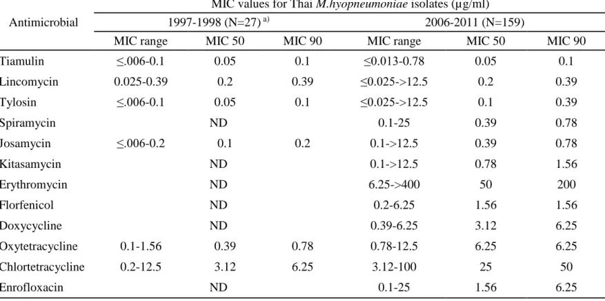
Table 3. shows the comparison of susceptibilities of the present 159 isolates with those isolated from 1997 to 1998 in Thailand (Narongsak and Thongkamkoon, 2002). In the previous data CTC, OTC, JM, TS, LCM and TM were tested against 27 Thai isolates of M. hyopneumoniae. Decreases of susceptibilities of the present isolates to some of the drugs were noted. The MIC90 values of CTC, OTC, JM and TS for previous isolates were 6.25, 0.78, 0.2 and 0.1 and those for present isolates were 50, 6.25, 0.78 and 0.39 µg/ml, respectively. No change was observed in MIC90 of LCM and TM between the present isolates and the previous ones.
The sequences of domain V of 23 rRNA of the two macrolides and lincomycin resistant isolates were compared with the sequences of M.
hyopneumoniae J strain and ATCC 27719 strain (Genbank accession number
AE017243 and X68421, respectively). The result revealed a point mutation at A2058G (E.coli coordinates) in both isolates. The MIC values of the antimicrobials for these isolates were shown (underlined) in Table 2.
1.4 Discussion
It was noteworthy that MICs of ERFX distributed in a broad range from 0.1 to 25 µg/ml and MICs for 76 of 159 (47.7%) Thai isolates exceeded 2 µg/ml which is considered the breakpoint of this drug (CLSI, 2010). The ERFX resistant animal mycoplasmas have rarely been reported. Hannan et al. (1997a) reported that some isolates of animal pathogenic mycoplasmas including M. hyosynoviae and M.
hyorhinis were resistant to flumequine (breakpoint: >16 µg/ml), the first-generation
fluoroquinolone which has been removed from clinical use and is no longer being marketed, while all of these flumequine resistant isolates were sensitive to ERFX. Vicca et al. (2004) described that five of 21 field isolates of M. hyopneumoniae were resistant to flumequine (MIC: >16 µg/ml) and had lower susceptibility to ERFX (MIC: 4 isolates; 0.5 µg/ml, one isolate; >1 µg/ml) than the other isolates (MIC: = 0.06 µg/ml). In all five isolates with lower susceptibility to ERFX one point
15
Table 3. Comparison on the MIC for M. hyopneumoniae between this study and the previous one
Antimicrobial
MIC values for Thai M.hyopneumoniae isolates(µg/ml)
1997-1998 (N=27) a) 2006-2011 (N=159)
MIC range MIC 50 MIC 90 MIC range MIC 50 MIC 90
Tiamulin ≤.006-0.1 0.05 0.1 ≤0.013-0.78 0.05 0.1 Lincomycin 0.025-0.39 0.2 0.39 ≤0.025->12.5 0.2 0.39 Tylosin ≤.006-0.1 0.05 0.1 ≤0.025->12.5 0.1 0.39 Spiramycin ND 0.1-25 0.39 0.78 Josamycin ≤.006-0.2 0.1 0.2 0.1->12.5 0.39 0.78 Kitasamycin ND 0.1->12.5 0.78 1.56 Erythromycin ND 6.25->400 50 200 Florfenicol ND 0.2-6.25 1.56 1.56 Doxycycline ND 0.39-6.25 3.12 6.25 Oxytetracycline 0.1-1.56 0.39 0.78 0.78-12.5 6.25 6.25 Chlortetracycline 0.2-12.5 3.12 6.25 3.12-100 25 50 Enrofloxacin ND 0.1-25 1.56 6.25
a) : data from the previous study (Narongsak and Thongkamkoon, 2002) ND : not determined;
16
mutation in quinolone resistant-determining regions of parC of topoisomerase IV was found and in one isolate with MIC value of >1 µg/ml an extra mutation of gyrA was also found. Thai isolates tested here showed higher resistance to ERFX with MIC values of 3.2–25 µg/ml and in addition, the rate of ERFX resistant isolates was also higher than those reported in the previous papers. This may reflect the overuse of ERFX in pig farming in Thailand especially for treatment of diarrhea of lactating piglets and at nursery up to 9 weeks (Prapasaranukul et al., 2010). Since the mechanisms of resistance to ERFX of Thai isolates were not examined in the present study, further study is needed to elucidate whether the resistance to ERFX of Thai isolates comes from alteration of the genes coding for DNA gyrase or reduction of quinolone accumulation by active efflux (Hooper, 1999).
The MICs of chlortetracycline (CTC) for isolates derived from 2006 to 2011 were higher than those for isolates collected during 1997 to 1998 (MIC90: 50 vs 6.25 µg/ml). Similar results were obtained by previous workers (Yamamoto et al., 1986). Most of the Japanese isolates isolated in 1970 were susceptible to CTC at 5 µg/ml or less, while isolates isolated in 1979 to 1981 were clearly separated into two categories (bimodal frequency distribution), one of which was susceptible at 5 µg/ml or less and the other 20 µg/ml or more (Yamamoto et al., 1986). In Thailand tiamulin (TM), tylosin (TS), CTC and amoxicillin have been widely used in fattening pigs, pregnant and lactating sows and replacement gilts during
acclimatization. In this respect, increase of CTC resistant isolates of
M. hyopneumoniae is considered reasonable, however these CTC resistant isolates
were still susceptible to oxytetracycline (OTC) and doxycycline (DOXY) though MICs were higher for isolates isolated from 2006 to 2011 than for those from 1997 to 1998 (Narongsak and Thongkamkoon, 2002).
Lincomycin and all macrolide drugs except erythromycin (EM) showed a bimodal distribution of MICs. MICs of these drugs were clearly higher for two isolates, indicating acquired resistance. In one farm from which one of the two
17
macrolide resistant strains were derived, additional two strains were isolated and examined for their susceptibility. As a result, these two strains were susceptible to all macrolide drugs tested except erythromycin indicating that M. hyopneumoniae strains with different antimicrobial susceptibility coexist among pigs within a same farm. The resistance of two strains to macrolides and lincomycin were shown to be attributable to a transition of A to G at 2058 (E. coli coordinates) in the central loop of domain V of the 23S rRNA. It was in agreement to the occurrence in Japan and Belgium (Kobayashi et al., 2008; Stakenborg et al., 2005). Erythromycin is highly active to M. pneumoniae (Kenny and Cartwright, 1991) and M. gallisepticum (Gautier-Bouchardon et al., 2002), however M. hyopneumoniae has natural resistance to this drug as indicated not only by the present data but also by the previous reports (Takahashi et al., 1978; Tanner et al., 1993; Ter Laak et al., 1991; Yamammoto et al., 1986). This natural resistance of M. hyopneumoniae to EM may be due to a G to A transition at 2057 (E. coli coordinates) in domain V of the 23S rRNA as suggested by Furneri et al (2000).
Tiamulin (TM) had the highest activity among 12 antimicrobials tested with the MIC90 of 0.1 µg/ml and MICs for 158 of 159 strains were lower or equal to 0.2 µg/ml. The MIC for the remaining isolate was 0.78 µg/ml. Although this value was far lower than the breakpoint of this drug, the strain might be the intermediate stage to resistance. Hence, monitoring of antimicrobial susceptibility at regular interval is inevitable.
In conclusion, this study provided the susceptibilities of Thai isolates of
M. hyopneumoniae to 12 antimicrobials widely used in Thailand. Resistances
against CTC, LCM, ERFX and macrolides were detected. In addition, the proportion of ERFX resistant Thai isolates of M. hyopneumoniae was remarkably high. Hence, mechanisms of resistance should be elucidated hereafter. Acquired resistance to various antimicrobials might pose a serious problem for the treatment of
18
mycoplasma pneumonia of swine. Prudent use of antimicrobials is an urgent necessity in pig industry in Thailand.
1.5 Abstract
A total of 159 Thai isolates of Mycoplasma hyopneumoniae isolated from pneumonic lungs of pigs during 2006-2011 were investigated for their in vitro susceptibility to 12 antimicrobial agents. Low activity of chlortetracycline was indicated by the MIC range from 3.12-100 µg/ml and MIC90 of 50 µg/ml. Seventy six isolates showed resistance to enrofloxacin, whereas 2 isolates showed resistance to macrolides and lincomycin. A point mutation at A2058G which conferred the macrolides and lincomycin resistances was revealed by sequence analysis of 23S ribosomal RNA in both isolates. The present results confirmed the rapid increase of resistant M. hyopneumoniae isolates against chlortetracycline, enrofloxacin, macrolides and lincomycin in Thailand. Selection of drugs to control swine diseases in Thailand must be done more prudently in consideration of reducing the antimicrobial resistance.
19
Chapter II
Antimicrobial susceptibilities of
Mycoplasma hyosynoviae field isolates in
Thailand during 2008-2011 and in vitro
development of resistance to tylosin and
lincomycin in type strain S16 of
20
2.1 Introduction
Mycoplasma hyosynoviae is one of the porcine pathogenic mycoplasmas and
distributes world-wide. Adult pigs served as an important reservoir of infection to piglets by direct contact. Pigs are occasionally infected and developed pneumonic lesion, non-suppurative arthritis and lameness (Hagedorn-Olsen et al., 1999). Nowadays, arthritis caused by M. hyosynoviae becomes increasing problem in many countries (Assuncao et al., 2005; Dahlia et al., 2009; Nielsen et al., 2001). Control measures depend on farm management and medication. Primary isolation from tissues of pigs is difficult due to the presence and overgrowth of other mycoplasmas as well as other bacteria (Friis et al., 1991). Hence, reports on the incidence and susceptibility of M. hyosynoviae to antimicrobial agents were limited (Aarestrup and Friis, 1998; Friis and Szancer, 1994; Hannan et al., 1997a; Kobayashi et al., 1996c). In addition, the medication programs in each country might vary according to the differences of susceptibilities of the organisms. Therefore, susceptibility testing of the field isolates in each country is essential. Recently, we reported the occurrence of M. hyosynoviae infection identified by isolation technique and PCR (Makhanon et al., 2012).
In the present study, the susceptibility of M. hyosynoviae field isolates collected from 2008-2011 were determined against 10 antimicrobial agents used in the swine production. All M. hyosynoviae isolates were examined for 23S rRNA transition at domain II and V and compared with the MIC levels of macrolides and lincomycin. In addition, macrolides and lincomycin resistant M. hyosynoviae S16 mutant was also developed to analyze the mechanisms of the resistance to these antimicrobials.
2.2 Materials and methods
Mycoplasma isolates and type strain: Forty one Thai isolates of M. hyosynoviae collected during 2008-2011 and kept at -80°C were used. The type strain S16
21
obtained from National Institute of Animal Health, Japan was also included. Of the Thai isolates, 18 were isolated from tonsils, synovial fluid and lung of pigs from 5 farms in 2008, 13 were isolated from tonsils of pigs from 2 farms in 2009 and 10 were isolated from lungs and tonsil of pigs from 3 farms in 2011.
Culture medium and cultivation technique: Mycoplasma cultivation was primarily carried out both from the lung and tonsil samples collected from healthy pigs at slaughterhouse and synovial fluid samples collected from the swollen joint of pigsin the farm using Hayflick’s medium supplemented with arginine and mucin (HAM) as described previously (Friis et al., 1991). After the mycoplasma was cloned three times, M. hyosynoviae was identified by colony characterization, film and spot production and a specific PCR (Kobayashi et al., 1996a). Finally, the culture was kept at -80°C as our culture collection. For determination of the susceptibility, all isolates from the collection were sub-cultured for 1 to 2 passages to ensure that each isolate was able to grow well within 2-3 days. Then, the colony forming units per milliliter of the culture were enumerated and aliquots of each culture were kept at -80°C until used.
Antimicrobial agents: The following 10 antimicrobial agents generally applied in pig industry in Thailand were used; Oxyteracycline (OTC) [Sigma], Doxycycline (DOXY) [Sigma], Lincomycin (LCM) [Sigma], Josamycin (JM) [Sigma], Spiramycin (SPM) [Sigma], Tylosin (TS) [Sigma], Kitasamycin (KT) [Bayer], Enrofloxacin (ERFX) [Bayer], Tiamulin (TM) [Sandoz] and Florfenicol (FFC) [Zhangjiagang Hengsheng Pharmaceutical Co. Ltd.]. The antimicrobial stock solution was prepared and serially diluted as described in CLSI (2010) at ten times of MIC test concentrations. The test concentrations ranged from 0.008-8 µg/ml for SPM, KT, JM, LCM and TM; 0.016-16 µg/ml for ERFX, FFC and TS and 0.125-128 µg/ml for OTC and DOXY.
22
MIC determination: The determination of MICs was carried out using agar dilution method (Hannan, 2000). HAM agar dilution plates containing antimicrobial agents were prepared by mixing 2 ml of each dilution of antimicrobial agent with 18 ml of molten HAM agar and pouring into a Petri dish. Agar plates without antimicrobial agents for growth control were prepared in the same manner. When the agar was solidified and dried completely, the plates were stored in 2-8°C for a night. MIC assay was performed in the following day. M. hyosynoviae was diluted to 108 CFU/ml in 0.5 ml HAM broth and transferred into the well of the replicator seed block. One µl each of M. hyosynoviae isolate was inoculated on the agar plate using the replicator so that there would be 105 CFU of M. hyosynoviae per spot. The inoculated plates were incubated at 37°C in a humidified incubator with 5% CO2 for 5 days. The lowest concentration of antimicrobial agent that caused ≥50% inhibition of growth compared with that on the growth control plate was interpreted as the MIC value at the fifth day post incubation.
PCR amplification and sequencing of domain II and V of 23S rRNA of M. hyosynoviae: One ml of each M. hyosynoviae culture was harvested at 12,000 x g for 10 min (model 1920, Kubota). Then, the pellet was washed once with 1 ml PBS and mix with Instagene matrix (Biorad, U.S.A.) to prepare the DNA template for PCR according to the manufacturer’s instruction. Six specific primers designed based on 23S rRNA partial sequence of M. hyosynoviae S16 (GenBank accession number JQ670924) for sequencing of domain II and V of 23S rRNA were as follows: Mhs D2F 5´-ACC CAT GAG CAG GTT GAA GC-3´, Mhs D2R 5´-CCA TCC CAT ATT CAG TGC TC-3´, Mhs D5-1F 5´-CAC GAA AGG AGT AAT TAT CTC-3´, Mhs D5-1R AAC TAC CCA CCA CAC ACT GTC-3´, Mhs D5-2F 5´-CTC ATC GCA TCC TGG AGC TGG-3´ and Mhs D5-2R 5´-CCC GCT TAG ATG CCT TCA GCG-3´. Amplification was performed for each pair of the primers in a total volume of 50 µl containing Go taq green (Promega, U.S.A.), 25 µl; 0.8 µM of each primer; 4 µl of DNA template and DW up to 50 µl. The PCR assay was carried
23
out in a Thermal cycler (Hybaid, Thermo electron, U.S.A.), comprising pre-incubation at 94°C for 2 min, followed by 30 cycles of 1 min at 94°C, 1 min at 55°C, and I min at 72°C. Final extension was performed for 5 min at 72°C. The amplicons, separated by 2% agarose gel electrophoresis, were purified by gel extraction kit (Qiagen, Germany) and sequenced with ABI 3130 Genetic Analyzer (Applied Biosystems) according to the manufacturer’s instruction.
In vitro development of tylosin and/or lincomycin resistance in type strain S16 of M. hyosynoviae: In vitro development of tylosin or lincomycin resistance was carried out in the same procedure as previous study (Hannan et al., 1997b) by serial passage in HAM broth containing tylosin or lincomycin at concentrations covering the MIC determination. After four days incubation, the culture showing growth which contained the highest concentration of the drug was inoculated to a fresh series of drug dilutions in HAM broth. The passages were repeated at four days interval for six passages. The S16 mutants from each passage were examined for susceptibility to antimicrobial agents and mutation in 23S rRNA.
2.3 Results
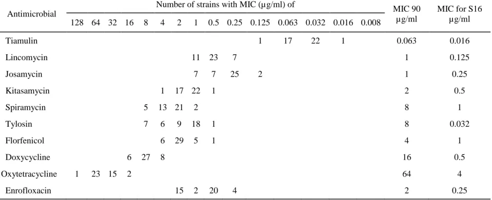
The distribution of the MICs obtained from the 41 isolates tested and MIC for type strain S16 were presented in Table 4. Of 10 antimicrobials tested, tiamulin (TM) showed the lowest MICs of 0.016-0.125 µg/ml against all M. hyosynoviae field isolates. Next to TM, lincomycin and josamycin showed high activity with MIC90 of 1 µg/ml. Kitasamycin also showed high activity with MIC90 of 2 µg/ml, whereas spiramycin and tylosin (TS) showed the lower activity with MIC90 of 8 µg/ml and 13 isolates were resistant to TS according to the MIC breakpoint (Hannan, 2000). Florfenicol showed moderately high activity with MIC range from 0.5 to 4 µg/ml and MIC90 of 4 µg/ml. On the other hand, activity of doxycycline to Thai isolates of M. hyosynoviae was low with MIC range from 4 to16 mcg/ml and MIC90 of 16 µg/ml. Oxytetracycline (OTC) showed the lowest activity with MIC
24
Table 4. Frequency distribution of Minimal inhibitory concentrations (MICs) of 10 antimicrobials for 41 Thai
M. hyosynoviae field isolates during 2008-2011
Antimicrobial
Number of strains with MIC (µg/ml) of
MIC 90 µg/ml MIC for S16 µg/ml 128 64 32 16 8 4 2 1 0.5 0.25 0.125 0.063 0.032 0.016 0.008 Tiamulin 1 17 22 1 0.063 0.016 Lincomycin 11 23 7 1 0.125 Josamycin 7 7 25 2 1 0.25 Kitasamycin 1 17 22 1 2 0.5 Spiramycin 5 13 21 2 8 1 Tylosin 7 6 9 18 1 8 0.032 Florfenicol 6 29 5 1 4 1 Doxycycline 6 27 8 16 0.5 Oxytetracycline 1 23 15 2 64 4 Enrofloxacin 15 2 20 4 2 0.25
25
range from 16 to 128 µg/ml, suggesting all Thai isolates of M. hyosynoviae were resistant to OTC according to the MIC breakpoint (11). MIC values of enrofloxacin ranged from 0.25 to2 µg/ml and MICs for 15 of 41 (36.6%) Thai isolates reached 2 µg/ml which is the breakpoint of this drug (Hannan, 2000).
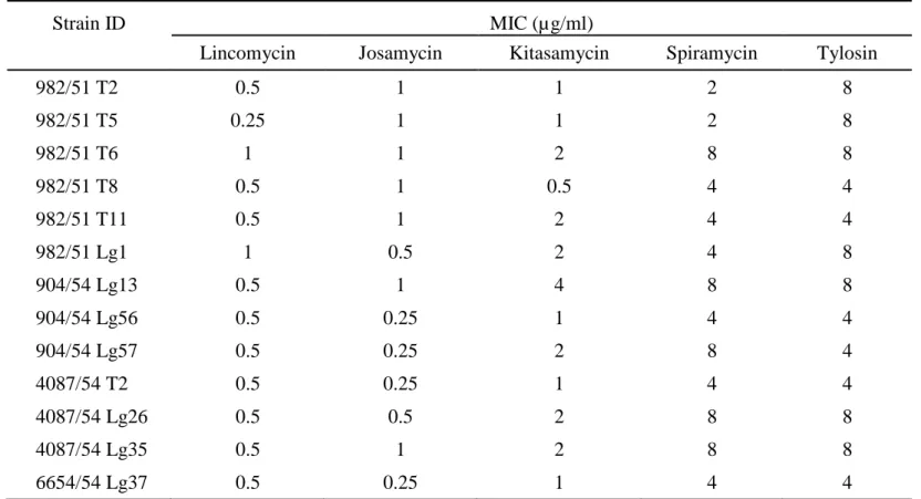
Results from sequencing of the 23S rRNA demonstrated the intrinsic resistance of all M. hyosynoviae isolates to 14-membered macrolide since the nucleotide at 2057 (E. coli numbering) was adenine (A2057). Neither first nor second part of domain V showed other acquired transition. On the other hand, an acquired G745A transition in domain II was found in thirteen isolates of our Thai M.
hyosynoviae. This G745A transition conferred the moderate resistance to tylosin at
4-8 µg/ml but not to lincomycin and the other macrolides tested. The MIC levels of macrolides and lincomycin for those isolates were shown in Table 5.
Before exposure to the antimicrobial agent M. hyosynoviae S16 was highly susceptible to both tylosin and lincomycin, with the MIC of 0.032 and 0.125 µg/ml, respectively. In the first and second passage, the highest concentration of tylosin and lincomycin that S16 survived were the same as that for a wild type. From the third passage, the S16 mutant rapidly obtained resistance to TS or LCM showing four to thirty two fold higher MICs in each passage. At the sixth passage MICs of the drugs reached 256 µg/ml or higher which was 4,000 fold for tylosin and 1,000 fold increase for lincomycin (Fig 1). In lincomycin selection, the A2058C/G or A2059C transitions were obtained in S16 mutants resulting in lincomycin resistance and macrolides cross resistance. While, the A2062G transition was obtained in tylosin selection which conferred only macrolides resistance. Sequence analysis and MIC results for those S16 mutants were shown in Table 6.
2.4 Discussion
The occurrence of tylosin resistant M. hyosynoviae among our Thai isolates was found in this study. In Denmark, M. hyosynoviae strains showed less
26
Table 5. Minimal inhibitory concentrations (MICs) of lincomycin and macrolides for 13 Thai M. hyosynoviae isolates obtained G745A transition in domain II of 23S rRNA
Strain ID MIC (µg/ml)
Lincomycin Josamycin Kitasamycin Spiramycin Tylosin
982/51 T2 0.5 1 1 2 8 982/51 T5 0.25 1 1 2 8 982/51 T6 1 1 2 8 8 982/51 T8 0.5 1 0.5 4 4 982/51 T11 0.5 1 2 4 4 982/51 Lg1 1 0.5 2 4 8 904/54 Lg13 0.5 1 4 8 8 904/54 Lg56 0.5 0.25 1 4 4 904/54 Lg57 0.5 0.25 2 8 4 4087/54 T2 0.5 0.25 1 4 4 4087/54 Lg26 0.5 0.5 2 8 8 4087/54 Lg35 0.5 1 2 8 8 6654/54 Lg37 0.5 0.25 1 4 4
27
Figure 1. In vitro development of tylosin and lincomycin resistance in
28
Table 6. In vitro development of tylosin and lincomycin resistance in M. hyosynoviae strain S16 and MIC level of the mutants
Passage ID Mutation
pattern
MIC range (µg/ml)
Lincomycin Tylosin Spiramycin Josamycin Kitasamycin
P1a)Tb)0.015c), P2T0.03d), P3T0.25, P4T0.5 normal 0.5-2 0.25-2 2-8 0.25-4 0.5-8 P3T1, P4T4, P5T4, P5T16, P5T32, P6T64, P6T128 A2062G 1 16-256 256->256 >256 256->256 P1L0.063, P2L0.125, P3L0.5, P4L0.125, P5L2 normal 0.5-2 0.25-2 1-4 0.25-8 0.5-8 P3L1, P3L2, P4L2 A2058C 64->256 32->256 256->256 >256 256->256 P4L0.5, P4L1 A2059C 64-256 32-256 256 >256 256->256 P5L16, P5L32, P6L128 A2058G >256 64->256 64>256 >256 256->256 a) Number of passage
b) Abbreviation of antimicrobial selected (T: tylosin; L: lincomycin) c) Concentration of antimicrobial in the M. hyosynoviae S16 culture broth
29
sensitivity to tylosin but the MIC levels did not reach the breakpoint (Aarestrup and Friis, 1998; Friis and Szancer, 1994), whereas the macrolides and lincomycin resistant M. hyosynoviae strains were found in Japan (Kobayashi et al., 1996c). In the United States, tylosin tended to have less activity to M. hyosynoviae field strains since 1970, however the recent US M. hyosynoviae strains did not show the resistance against tylosin (Schultz et al., 2012). In addition, macrolide resistant M.
hyorhinis could revert to sensitive by subculturing in macrolide free broth
(Kobayashi et al., 1996b). The above results suggested the occurrence of macrolide resistant strains was induced by the presence of antimicrobials in its environment. Hence, the MIC data may reflect the use of antimicrobial agents in the certain period of time.
As it is known, ribosomal peptidyl transferase center (PTC) is located at the interface side of the 50S subunit of bacterial ribosome and is responsible for protein synthesis. The peptidyl transferase cavity forms by the nucleotides of central loop of domain V in 23S rRNA, and A2058, A2059 implicate as an entrance of the tunnel. Loop of hairpin 35 of domain II in 23S rRNA also forms the wall of the exit tunnel opposite to A2058 (Polacek and Mankin, 2005). Actions of macrolides and lincosamides antimicrobials are binding to the PTC and inhibiting protein synthesis. Therefore, one of the defense mechanisms of bacteria is an alteration of specific nucleotides at the target site in 23S rRNA. As the evidences, various mutations at domain V of 23S rRNA conferred macrolides or lincomycin resistance and cross resistance in the number of bacteria were reported (Vester and Douthwaite, 2001). The mutation of 23S rRNA conferred macrolides and lincomycin cross resistance in
M. hyopnemoniae and M. hyorhinis were also reported (Kobayashi et al., 2005;
Stekenborg et al., 2005). In this study, we confirmed the occurrence of tylosin resistance in M. hyosynoviae Thai strains which had a point mutation at G745A. Tylosin contains a mycinose sugar at position 14 of the lactone ring. Therefore, tylosin not only binds at the upper position of the entrance of the tunnel and reaches
30
to the PTC cavity, but also reaches across a tunnel to interact with loop of hairpin 35
(Pelacek and Mankin, 2005). This might be the reason why the G745A in our
M. hyosynoviae strains conferred tylosin resistance. However, the level of tylosin
resistance of this mutant was not high. This result was in agreement to the previous study (Kobayashi et al., 2005).
To our knowledge, in vitro resistance mechanism of M. hyosynoviae strain against tylosin or lincomycin has never been examined. In this study, M.
hyosynoviae S16 mutants developed quickly within 3 passages for both tylosin and
lincomycin. The result was similar to the studies for M. hyopneumoniae by Hannan and for M. hyorhinis by Kobayashi (Hannan et al., 1997b; Kobayashi et al., 2005). The interaction of lincomycin with E.coli 23S rRNA revealed that lincomycin strongly interact with A2058 but did not interact with A2059 and A2062 (Douthwaite, 1992). However, the mutation at A2059 of 23S rRNA conferred macrolides and lincomycin resistance in M. hyorhinis and Helicobactor pylori (Kobayashi et al., 2005; Wang and Taylor, 1998). In our study, the S16 mutated at A2058C/G or A2059C in lincomycin selection also conferred the lincomycin resistance and macrolides cross resistance. Whereas the S16 mutated at A2062G in tylosin selection conferred only macrolides resistance. This result was similar to the study on josamycin selected M. hominis which mutated at A2062 and showed resistance to macrolides but was susceptible to lincosamides (Funeri et al., 2001). According to the results, we believed that lincomycin might have some part reach to nucleotide A2059 but not to A2062 of central loop of domain V in 23S rRNA of M.
hyosynoviae since A2059G S16 mutants could be obtained and predominated in
lincomycin selection resulting in lincomycin resistance and macrolides cross resistance, but A2062G mutants obtained from the tylosin selection showed the resistance only to tylosin but not to lincomycin.
31
It was noted that 15 of 41 (36.6%) Thai isolates of M. hyosynoviae were resistant to enrofloxacin. The result was different from the previous reports (Aarestrup and Friis, 1998; Hannan et al., 1997a; Kobayashi et al., 1996c; Schultz et al., 2012). On the other hand, enrofloxacin resistant M. hyopneumoniae was found in Belgium and the resistance mechanism has been studied (Vicca et al., 2007). It should be carried out whether the same mechanism would be detected in our M.
hyosynoviae strains. Tiamulin, among antimicrobials tested, showed the highest
activity against M. hyosynoviae strains which is in agreement with the previous reports (Aarestrup and Friis, 1998; Friis and Szancer, 1994; Hannan et al., 1997a; Kobayashi et al 1996c).
Interestingly, all M. hyosynoviae strains were highly resistant to oxytetracycline. In Japan, also the activity of oxytetracycline to M. hyosynoviae has decreased, but MIC level did not reach the break point (Kobayashi et al 1996c). The resistance to oxytetracycline of M. hominis and Ureaplasma parvum was associated with acquisition of tet(M) determinant (Mardassi et al., 2012). The 16S rRNA mutation also conferred tetracycline resistance in Helicobacter pylori, M. hominis and M. pneumonia (Degrange et al., 2008; Wu et al., 2005). The resistance mechanisms of M. hyosynoviae against tetracyclines including oxytetracycline and doxycycline should be elucidated.
In conclusion, this study provided the susceptibilities of Thai isolates of
M. hyosynoviae to 10 antimicrobials used in Thailand. Resistances against
oxytetracycline, enrofloxacin and tylosin were detected. The resistance mechanism of M. hyosynoviae field isolates against tylosin was associated with G745A transition in domain II of 23S rRNA. The veterinarians should carefully choose appropriate drug for the treatment and control of M. hyosynoviae infection to minimize the increase of resistant strains in Thailand.
32
2.5 Abstract
In vitro susceptibilities of 41 Thai isolates of Mycoplasma hyosynoviae
isolated from lung and tonsil of pigs to 10 antimicrobial agents were investigated. All isolates showed resistance to oxytetracycline, while 15 isolates (36.6%) showed resistance to enrofloxacin. The examination for the mutation in domain II and domain V of 23S rRNA revealed 13 Thai isolates of M. hyosynoviae obtained G745A transition (E. coli numbering). The result was correlated to the resistance to tylosin with MIC ranged 4-8 µg/ml. The development of M. hyosynoviae S16 resistance to tylosin and lincomycin showed the A2058C, A2059C or A2058G transitions in lincomycin selection which conferred marked resistance to lincomycin and all macrolides tested. While, A2062G transition was obtained from tylosin selection and this point mutation conferred resistance to macrolides but not to lincomycin.
33
Chapter III
Development of semi-nested PCR for
detection of 16S rRNA gene of
34
3.1 Introduction
Mycoplasma hyosynoviae (M. hyosynoviae), a non-cell wall prokaryote,
belongs to the class Mollicutes, Family Mycoplasmataceae is one of the porcine mycoplasma pathogens and appears world-wide (Kobisch and Friis, 1996). At present, arthritis caused by M. hyosynoviae becomes increasing problem in many countries (Neilsen et al., 2001; Assuncao et al., 2005; Schultz et al., 2012). M.
hyosynoviae is the host specific bacterium in pigs and commonly inhabits upper
respiratory tract such as nasal cavity, pharynx and tonsil of convalescent and adult animals. Adult pigs are an important reservoir of infection to piglets by direct contact. Pigs may occasionally develop pneumonic lesion and arthritis (Hagedorn-Olsen et al., 1999). M. hyosynoviae is an arginine metabolizing mycoplasma, which can be cultivated in Hayflick’s medium supplemented with mucin and arginine. Primary isolation from tissues of pigs is often complicated because of overgrowth of
M. hyorhinis as well as other bacteria (Friis et al, 1991) making it difficult to detect M. hyosynoviae by cultivation.
Although certain breeders have experienced osteoarthritis lesions similar to
M. hyosynoviae arthritis in breeding pigs in Thailand, there has been no report
confirming the detection of this bacterium. However, M. hyosynoviae has been a common pathogen causing acute and severe lameness in grower-finisher pigs in Denmark. Time consumed for the surveillance and treatment of the disease was 30-90 minutes per 1000 pigs daily (Nielsen et al., 2001). Among pathogenic porcine mycoplasmas, M. hyopneumoniae is known as the most important mycoplasma that causes enzootic pneumonia and economic losses in pig industry. Therefore, there have been several techniques developed for specific and rapid detection and identification including PCR based method with high sensitivity such as nested PCR and real time PCR (Stark, et al., 1998; Calsamiglia et al., 1999; Kurth et al., 2002; Dubosson et al., 2004). Although the 16S-23S intergenic spacer PCR was established to differentiate the porcine mycoplasmas in the culture medium, it was
35
not evaluated for use in clinical samples (Nathues et al., 2011). A few PCR protocols for identification of M. hyosynoviae in clinical samples have been developed, however, the assays had limit of detection about 104 CFU of the organism per gram of lung tissue (Ahrens et al., 1996; Kobayashi et al., 1996a).
In this study we developed a semi-nested PCR assay with improving limit of detection targeted to 16S rRNA gene for detection of M. hyosynoviae from tissues of pigs.
3.2 Materials and Methods
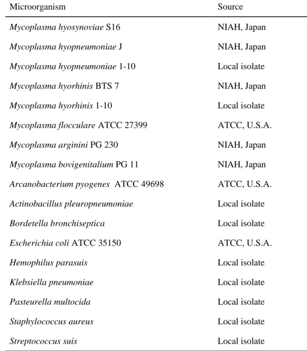
Bacterial strains and growth conditions: The mycoplasmas and other bacteria used in this study are listed in Table 7. Mycoplasma type strains including M.
hyopneumoniae strain J, M. hyorhinis BTS7 and M. hyosynoviae S16 were obtained
from National Institute of Animal Health (NIAH), Japan. M. flocculare,
Arcanobacterium pyogenes and Escherichia coli were purchased from American
Type Culture Collection. The other bacteria were the local isolates derived from naturally infected pigs and collected in our culture collection.
M. hyopneumoniae was cultured in BHL broth, M. hyorhinis, M. arginini and M. bovigenitalium in Hayflick’s broth, and M. hyosynoviae in Hayflick’s broth
supplemented with mucin and arginine. Actinobacillus pleuropneumoniae and
Haemophilus parasuis were incubated on chocolate blood agar and the other
bacteria on 5% sheep blood agar. The incubation times were 3-5 days for mycoplasmas and 18-24 hours for bacteria. Mycoplasma cells were collected in 1.5 ml microtube by centrifugation at 13,000 rpm for 10 minutes. The pellets were washed one time with phosphate buffered saline and followed by DNA preparation step.
36
Table 7. Microorganisms used in the development of semi-nested PCR for Mycoplasma hyosynoviae
Microorganism Source
Mycoplasma hyosynoviae S16 NIAH, Japan
Mycoplasma hyopneumoniae J NIAH, Japan
Mycoplasma hyopneumoniae 1-10 Local isolate
Mycoplasma hyorhinis BTS 7 NIAH, Japan
Mycoplasma hyorhinis 1-10 Local isolate
Mycoplasma flocculare ATCC 27399 ATCC, U.S.A.
Mycoplasma arginini PG 230 NIAH, Japan
Mycoplasma bovigenitalium PG 11 NIAH, Japan
Arcanobacterium pyogenes ATCC 49698 ATCC, U.S.A.
Actinobacillus pleuropneumoniae Local isolate
Bordetella bronchiseptica Local isolate
Escherichia coli ATCC 35150 ATCC, U.S.A.
Hemophilus parasuis Local isolate
Klebsiella pneumoniae Local isolate
Pasteurella multocida Local isolate
Staphylococcus aureus Local isolate
37
DNA preparation: All mycoplasmal and bacterial DNA were prepared using Instagene (Bio-Rad, U.S.A.) following the manufacturer’s protocol. After the cell lysate was centrifuged at the end of the process, the supernatant was collected and kept as DNA template at -20°C until use. For M. hyosynoviae S16, the concentrations of DNA were determined by spectrophotometer (Nanodrop® ND-1000, Nanodrop Technology, U.S.A.). The DNA was then diluted to 1 ng/µl and serial ten-fold dilution were made to 10-6 and used to test the detection limit of this semi-nested PCR assay.
Selection of primers and PCR reactions: The specific forward primer was selected based on the alignments of 16S rRNA gene of M. hyosynoviae (Genbank accession number U26730) with other porcine mycoplasmas including M. hyopneumoniae (E02783), M. flocculare (X63377), M. hyorhinis (M24658), M. arginini (NR041743) and M. hyopharyngis (U58997). The outer specific reverse primer was designed by Kobayashi et al. (1996a). The inner specific reverse primer was also selected from the multi-alignments of 16S rRNA genes as well. Nucleotide sequences of the primers were MHS_F (A): 5' GAA GCG TTT GCT TCA CTA AGA GAT 3' (nt 196-219), MHS_R (B1): 5' TTA GCT GCG TCA GTG ATT GG 3' (nt 825-844) and MHS_R (B2): 5' GCT TTC TAA CAA GGT ACC GTC AGT 3' (nt 468-491).
The PCR was carried out in 0.2 ml tube in a reaction volume of 20 µl. All PCR mixture contained 1x PCR buffer (10 mM Tris-HCl pH 8.8, 50 mM KCl and 0.1% TritonX-100), 1.5 mM MgCl2, 200 µM each of dATP, dTTP, dCTP and dGTP; 0.5 U Taq polymerase (HotstarTaq, Qiagen, Germany), and 0.5 µM of each forward and reverse primers. Then DNase/RNase-free water was added up to 18 µl. For the first round of the semi-nested PCR, primer A and B1 were used and 2 µl of DNA sample was added in each reaction. The amplification performed in thermal cycler (Hybaid, Thermo electron, U.S.A.) with an initial denaturation at 95°C 15 min, followed by 35 cycles of 94°C for 1 min, 59°C for 1 min and 72°C for 1 min,
38
then a final extension at 72°C for 5 min. For the second round of the semi-nested PCR, primer A and B2 were used. The 0.5 µl of PCR product from the first round of the semi-nested PCR used as a DNA template and 1.5 µl DNase/RNase-free water were added in each reaction. The amplification performed in the same condition as the first PCR, but the amplification ended after cycle 25. The PCR products from both amplifications were analyzed by electrophoresis through 1.5% agarose gels containing 0.1 µg/ml ethidium bromide. The gels were run at 100 volts for 30 min and visualized by ultraviolet light in gel documentation (GelDoc. It. UVP, U.S.A.). Specificity: The specificity of the primers was examined by amplification of these primers with the other porcine mycoplasmal and bacterial species (Table 7) which associate with pneumonia and arthritis in pigs.
Limit of detection:
Purified M. hyosynoviae DNA
The serial ten-fold dilutions of M. hyosynoviae S16 DNA started from 1 ng/µl were tested by semi-nested PCR. The minimum concentrations showing positive result in the semi-nested PCR were noted.
Simulated lung samples
A piece of lung collected from a mycoplasma free pig was homogenized and put into a bag approximately 1 gram per bag. Five ml of phosphate buffered saline was added to make a lung suspension and serial 10-fold dilutions of M. hyosynoviae S16 from 107 CFU to 10 CFU were added into each bag. Afterwards, the DNA of simulated lung samples was extracted as described by Kobayashi et al. (1996a). Then semi-nested PCR was performed to determine the minimum number of mycoplasma cells in simulated lung sample that showed positive result.
Detection of M. hyosynoviae from slaughtered pigs: Thirty tonsil samples per farm were collected from slaughtered pigs in 10 farms and tested for M. hyosynoviae by semi-nested PCR as described above as well as by cultivation following to the
39
modified method from Friis et al. (1991). The rate of detection of M. hyosynoviae by each method was compared.
3.3 Results
The expected sizes of the PCR products from the first and the second round of the semi-nested PCR were 649 bp and 295 bp, respectively, because the primers were designed based on 16S rRNA gene of M. hyosynoviae accession number U26730 from the following positions. The position of a forward primer was at 196nt to 219nt whereas the positions of an outer reverse primer and an inner reverse primer were at 825nt to 844nt and 468nt to 491nt, respectively. Using the adequate concentration of the M. hyosynoviae DNA, the first round of semi-nested PCR could generate an amplified fragment about 649 bp followed by an amplified fragment about 295 bp for the second round of semi-nested PCR as shown in Fig 2.
Figure 2. Oligonucleotide primers specifically amplified 16S rRNA gene of M.
hyosynoviae and showed the PCR products at the predicted sizes of 649 bp (A)
and 295 bp (B) with the primers A&B1 and A&B2, respectively. Lane M, 100 bp
DNA marker; Lane 1-4, DNA of M. hyosynoviae strain S16
bp 500 400 300 200 M 1 2 3 4 M 1 2 3 4 bp 500 400 300 200 649 295 bp
40
The first and the second round of semi-nested PCR were examined for their specificity with porcine mycoplasmas and other bacterial species commonly associated with pneumonia and/or arthritis in pig as listed in Table 7. None of the primer pairs yielded PCR products or non specific bands with DNA from the other mycoplasmas and bacterial species. The semi-nested PCR detected as little as 10 fg of purified M. hyosynoviae DNA in a reaction (Fig 3) and showed positive result with lung samples containing 107-103 CFU/g of M. hyosynoviae (Fig 4).
Figure 3. Detection limit of the semi-nested PCR procedure in detecting 10 fold dilution of 1 ng of extracted genomic DNA of M. hyosynoviae (M. hs) from 1 ng to 0.1 fg, Lane M, 100 bp DNA marker; Lane 1 through 8, 1 ng of M. hs DNA to 0.1
fg of M. hs DNA, respectively; Lane 9, DW (negative control); Lane 10, M. hs DNA (positive control) M 1 2 3 4 5 6 7 8 9 10 295 bp bp 500 400 300 200 DNA (g) 10-9 10-10 10-11 10-12 10-13 10-14 10-15 10-16
41
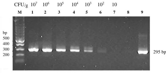
Figure 4. Detection limit of the semi-nested PCR procedure in detecting extracted genomic DNA of M. hyosynoviae (M. hs) from simulated lung with
107-10 CFU/g, Lane M, 100 bp DNA marker; Lane 1 through 7, DNA from
simulated lung with M. hs 107 CFU to 10 CFU, respectively; Lane 8, DW (negative control); Lane 9, M. hs DNA (positive control)
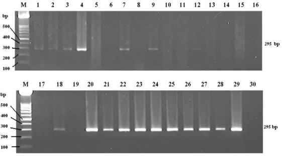
To evaluate the applicability of this method a total of 300 tonsillar samples were collected at a slaughterhouse and compared the detection rate with that by cultivation. As shown in Table 8, 45 of 300 (15%) samples were positive by semi-nested PCR while 17 of 300 (5.7%) were positive by cultivation. As for farm, 5 of ten farms were positive by semi-nested PCR (Fig 5) and 2 of 10 by culture. In cultivation positive two farms, positive rates by cultivation were 7/30 (23.3%) and 10/30 (33.3%) while those by semi-nested PCR were 14/30 (47%) and 21/30 (70%) respectively. Thus the semi-nested PCR was shown to be a useful tool for rapid and sensitive detection of M. hyosynoviae in clinical materials in pig herds.
bp 500 400 300 200 295 bp M 1 2 3 4 5 6 7 8 9 10 CFU/g 107 106 105 104 103 102 10