Japan Advanced Institute of Science and Technology
JAIST Repository
https://dspace.jaist.ac.jp/
Title
Protein Carbonylation Detected with Light and
Heavy Isotope-Labeled 2,4-Dinitrophenylhydrazine
by Matrix-Assisted Laser Desorption/Ionization
Time-of-Flight Mass Spectrometry
Author(s)
KINUMI, Tomoya; OSAKA, Issey; HAYASHI, Akio;
KAWAI, Takaaki; MATSUMOTO, Hiroyuki; TSUJIMOTO,
Kazuo
Citation
Journal of the Mass Spectrometry Society of
Japan, 57(6): 371-377
Issue Date
2009
Type
Journal Article
Text version
publisher
URL
http://hdl.handle.net/10119/8498
Rights
Tomoya KINUMI, Issey OSAKA, Akio HAYASHI, Takaaki
KAWAI, Hiroyuki MATSUMOTO, and Kazuo TSUJIMOTO
(2009) Protein Carbonylation Detected with Light
and Heavy Isotope-Labeled
2,4-Dinitrophenylhydrazine by Matrix-Assisted Laser
Desorption/Ionization Time-of-Flight Mass
Spectrometry, Journal of the Mass Spectrometry
Society of Japan, 57(6), 371-377. ここに掲載した
著作物の利用に関する注意:本著作物の著作権は日本質
量分析学会に帰属します。本著作物は著作権者である
日本質量分析学会の許可のもとに掲載するものです。
Description
Protein Carbonylation Detected with Light and Heavy
Isotope-Labeled 2,4-Dinitrophenylhydrazine by Matrix-Assisted Laser
Desorption/Ionization Time-of-Flight Mass Spectrometry
Tomoya K>CJB>,
1῎ Issey OH6@6,
2Akio H6N6H=>,
3Takaaki K6L6>,
4Hiroyuki M6IHJBDID,
5and Kazuo THJ?>BDID
4῍1
Bio-medical Standard Section, National Metrology Institute of Japan, National Institute of Advanced Industrial Science and Technology (AIST), Tsukuba, IBARAKI, JAPAN
2
Center for Nano Materials and Technology, Japan Advanced Institute of Science and Technology (JAIST), Nomi, ISHIKAWA, JAPAN
3
Agilent Technologies, Hachioji, TOKYO, JAPAN
4
School of Material Sciences, Japan Advanced Institute of Science and Technology, Nomi, ISHIKAWA, JAPAN
5
Department of Biochemistry and Molecular Biology, University of Oklahoma Health Sciences Center, Oklahoma City, Oklahoma, USA
Oxidation of proteins leads to carbonylationῌthe formation of aldehydes or ketonesῌat the amino acid side chain and/or the terminal amino groups. Carbonylated proteins have been conventionally detected by UV absorption spectrometry of the stable adduct with 2,4-dinitrophenylhydrazine (DNPH). However, this routine method is limited to detection of the total carbonyl content and does not provide structural information. We developed an isotope-dilution method for the specific detection of carbonylated proteins using12C
6-DNPH and 13C
6-DNPH. This method has the following steps: the oxidized protein or peptide is divided into two parts, and
these parts are independently labeled with 12C
6-DNPH and 13C6-DNPH; the mixtures of these two labeled
solutions are subsequently measured with matrix-assisted laser desorption/ionization time-of-flight (MALDI-TOF) mass spectrometry. The carbonylated peptide was found by searching for a doublet peak having a mass di#erence of 6 Da. We examined oxidized angiotensin I and oxidized lysozyme prepared by treatment with NaOCl. The oxidized angiotensin I showed four pairs of doublet peaks in the MALDI-TOF mass spectrum. The structure was determined by tandem mass spectrometry. In the case of tryptic digest of the oxidized lysozyme, two carbonylated products could be easily identified even in a complex mixture. The use of13C
6-DNPH provides
rapid and accurate detection of carbonylated peptides even in complex mixtures.
(Received July 6, 2009; Accepted August 25, 2009)
1. Introduction
Protein oxidation is known to occur in a variety of diseases, and is thought to contribute to their patholo-gy. Amino acid residues and the terminal groups in protein are susceptible to oxidation by reactive oxygen species (ROS), including superoxide anion (O2῎ῌ),
peroxy radical (RO2῎), alkoxy radical (RO῎), hydroxyl
radical (HO῎), hydrogen peroxide (H2O2), hypochlorous
acid (HOCl), and singlet oxygen (1
O2).1) For example,
the oxidized proteins cause an etiology or progression of a number of human diseases, e.g. Alzheimer disease, Parkinson disease, arteriosclerosis.2)῍4) In a general
mechanism, the oxidation reactions of proteins with ROS are classified into several reactions including
direct oxidation, secondary reaction via lipid peroxida-tion product, and that via glycaperoxida-tion end product, which result in oxidation of amino acid side chains, terminal groups and/or cleavage of peptide chains.5), 6) These
reactions generate carbonyl groups with aldehydes or ketones at the amino acid residues and finally induce a deterioration of protein.7)῍9)
Detection and quantifica-tion of protein carbonylaquantifica-tion have been studied ex-tensively as a marker for oxidative stress.10)
The carbonyl group reacts readily with phenylhydrazine (DNPH) to provide a stable 2,4-dinitro-phenyl (DNP) hydrazone product. Quantification of the carbonylated protein has been usually performed by a spectrophotometric assay that measures the UV ab-sorbance of DNP-derivatized protein.11), 12) This is the
most widely utilized method of detecting protein carbonylation. An anti-DNP antibody is a highly sensi-tive tool for detection of a DNP-derivatized protein.13)
These methods are powerful techniques for quantify-ing the total carbonyl content of a protein or mixture of carbonylated proteins. Detection of specifically carbonylated proteins in a protein mixture was
῎ Correspondence to: Tomoya K>CJB>, Bio-medical Stan-dard Section, National Metrology Institute of Japan, National Institute of Advanced Industrial Science and Technology, C3῍9 1῍1῍1 Umezono, Tsukuba, Ibaraki 305῍ 8563, JAPAN, e-mail: [email protected]
performed by immunoblot analysis of the proteins with two-dimensional gel electrophoresis using an anti-DNP antibody. However, this method depends on the conditions of electrophoresis-based separation, and the results are complicated by DNP-derivatized com-pounds without structural information. Mirzaei and Regnier reported that heavy and light isotope labeled Girard’s P reagent (1-(2-hydrazino-2-oxoethyl)pyri-dinium chloride), a selective modifier for the carbonyl group, was used with mass spectrometry to identify carbonylated proteins in complex mixtures.14)
A mass spectrometry-based method has been developed to clarify the detailed structure of the carbonylated amino acid residues in the oxidized protein.
In this study, we developed an application to detect and characterize the structure of carbonylated peptides or proteins using unlabeled (12C
6-DNPH) and 13C6
-labeled DNPH by matrix-assisted laser desorption/ion-ization time-of-flight (MALDI-TOF) mass spectrometry. This method was applied to oxidized angiotensin I and oxidized lysozyme after reaction with NaOCl, which is known to be generated by phagocyte enzyme myeloperoxidase under physiological conditions.15)
Here we show that the carbonylated peptide can be easily found by searching for a mass di#erence of 6 Da and the ratio of peak height in the mass spectrum, even in a complex peptide mixture.
2. Experimental 2.1 Materials
Lysozyme (hen-egg) and 13
C6-benzene were
pur-chased from Sigma-Aldrich Corp. (MO, USA). Angio-tensin I (Human) was purchased from Peptide Institute Inc. (Osaka, Japan). Deionized water was obtained using a MilliQ system (Millipore Co., MA, USA). ZipTip C18 disposable desalting tips (ZipTip) were purchased from Millipore Co. (MA, USA). Trypsin (mass spec-trometry grade) and other chemicals, including 12C
6
-DNPH, were purchased from Wako Pure Chemical (Osaka, Japan).
2.2 Synthesis of13C
6-DNPH
The labeled compound,13C
6-DNPH, was synthesized
according the routine procedure. First, 13C
6-benzene
(0.441 g, 5.65 mmol) was brominated by refluxing at 55῍ for 15 min with 0.137 mL (5.29 mmol) of Br2and 1
mg of Fe, and subsequently purified by distillation. The prepared bromobenzene was refluxed with 7.5 mL of H2SO4and 5.0 mL of HNO3at 85῍. After
purifica-tion by silica gel column chromatography, 316.3 mg (1.25 mmol) of 1-bromo-2,4-dinitrobenzene was ob-tained. A 4.0 mL solution of the obtained 1-bromo-2,4-dinitrobenzene in ethanol was added to 0.40 mL (14.4 mmol) of hydrazine hydrate in ethanol (1.5 mL) at 65῍. After cooling to room temperature, the precipitate was washed with cold ethanol and dried in vacuo. Finally, 201.0 mg (0.99 mol) of 13C
6-DNPH (red crystal) was
obtained.
2.3 Oxidation and modification reaction of angio-tensin I, and measurement of the MALDI-TOF mass spectra
Angiotensin I aqueous solution (100 mL, 100 mM) was oxidized by adding 100 mL of 50 mM NaOCl phosphate bu#er solution (pH 7.4) for 30 min on ice. Two 70 mL
aliquots of the oxidized angiotensin I solution were mixed separately with 70 mL of each 12.5 mM 12C
6
-DNPH and 13C
6-DNPH in 2 M HCl, and incubated at
room temperature for 30 min. The resulting solutions were mixed in the ratio 1 : 1 (v/v). This mixture was purified by a ZipTip according to the manufacturer’s instructions. Briefly, the sample was absorbed to the ZipTip after regeneration with a 0.1῎ trifluoroacetic acid (TFA)/acetonitrile and 0.1῎ TFA aqueous solu-tion. After washing with a 0.1῎ TFA aqueous solu-tion, the peptide was eluted directly to a MALDI sample plate by 0.1῎ TFA and 50῎ acetonitrile aque-ous solution. A matrix solution was prepared as 10 mg/mL a-cyano-4-hydroxycinnamic acid (CHCA) in a 0.1῎ TFA and 50῎ acetonitrile aqueous solution, and mixed with the sample solution on the MALDI sample plate. MALDI-TOF mass spectra were obtained by an Axima CFR plus MALDI-TOF mass spectrometer (Shimadzu Co., Ltd., Kyoto, Japan) with a reflectron under positive ion mode.
Electrospray ionization (ESI)-tandem mass spectra were acquired by a LCQ Deca XP (Thermo Fisher Sci-entific, CA, USA) iontrap mass spectrometer with a nanospray interface under positive ion mode. The ZipTip-purified sample (20 mL) was diluted in 100 mL of 50῎ methanol/water, and introduced into the ion source at a flow rate of 1 mL/min with a syringe pump. 2.4 Oxidation and modification reaction of
lyso-zyme
Lysozyme aqueous solution (200 mL, 50 mM) was oxidized by adding 200 mL of 50 mM NaOCl phosphate bu#er solution (pH 7.4) for 30 min on ice. This mixture was filtered to remove NaOCl with an ultrafiltration device (Vivaspin 500, molecular weight cut o#: 10 kDa, Sartorius Stedim Biotech, Goettingen, Germany) by centrifuging at 15,000 rpm and 4῍ for 20 min. The filtrate was washed with a phosphate bu#er and this filtration step was repeated three times. After filtering it for the third time, a phosphate bu#er was added to the washed protein in the filtering device to bring the level to 200 mL. This solution was divided to two aliquots; one was mixed with 100 mL of 12.5 mM12C
6
-DNPH in 2 M HCl, and the other with13C
6-DNPH in 2 M
HCl. Both were incubated at room temperature for 30 min. Both solutions were precipitated with 50῎ ethanol/ethyl acetate at ῌ20῍ overnight. After centrifugation at 15,000 rpm for 20 min, these precipi-tates were suspended in 200 mL of 8 M urea. The12
C6
-DNPH-modified and13
C6-DNPH-modified oxidized
lyso-zyme samples were mixed at 1 : 3, 1 : 1, 3 : 1 (v/v) for a final volume of 80 mL.
2.5 Trypsin digestion and measurement of MALDI-TOF mass spectra
The mixtures of12
C6-DNPH-modified and13C6
-DNPH-modified oxidized lysozyme samples were diluted to 400 mL with water, and added to 8 mL of 1 M NH4HCO3
(pH 8.6) and 2 mL of 1 mg/mL trypsin. Trypsin diges-tion was performed by incubadiges-tion at 37῍ overnight. These digests were purified by ZipTip and measured by MALDI-TOF mass spectra as described above.
3. Results and Discussion
3.1 Carbonylated angiotensin I modified by 12C 6
-DNPH and13C
6-DNPH
Oxidized angiotensin I with NaOCl was divided into
two aliquots, one of which was reacted with12C 6-DNPH
and the other with13C
6-DNPH. A 1 : 1 molar mixture of
the 12C
6-DNPH-modified and 13C6-DNPH-modified
ox-idized angiotensin I was prepared, and the mass spectra of the mixture were measured with the MALDI-TOF
Fig. 1. Overview of the analysis for the carbonylated angiotensin I and lysozyme described in this study. 1) Angiotensin I is oxidized by NaOCl. 1 : 1 mixture of12C
6-DNPH and13C6-DNPH-modified oxidized angiotensin I is measured
with a MALDI-TOF mass spectrometer after ZipTip purification. The detailed structure was analyzed by an ESI mass spectrometer. 2) For lysozyme, after oxidation by NaOCl, 1 : 3, 1 : 1, and 3 : 1 mixtures of12C
6-DNPH- and 13C
6-DNPH-modified oxidized lysozyme were digested with trypsin. The resulted peptide mixtures were measured
with a MALDI-TOF mass spectrometer after ZipTip purification.
Fig. 2. a) MALDI-TOF mass spectrum of the 1 : 1 mixture of12C
6-DNPH- and13C6-DNPH-modified oxidized angiotensin I.
b) The enlarged spectrum showing carbonylated products. Peaks at m/z 1415.7, 1431.7, 1442.7, and 1475.7 are marked peaks 1῍4.
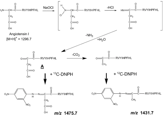
mass spectrometer after ZipTip purification (Fig. 1). The MALDI-TOF mass spectrum shown in Fig. 2a indi-cates that a major product peak was observed at m/z 1330.7 besides that of a protonated ion of angiotensin I (the amino acid sequence: DRVYLHPFHL) at m/z 1296.7. This mass di#erence of 34 Da corresponds to chlorination of the tyrosine residue. In addition to the monochlorination of tyrosine (m/z 1330.7), the ion cor-responding to dichlorination of the tyrosine residue in angiotensin I was also observed at m/z 1364.7. These observations indicate that the mono- and dichlorina-tion on the tyrosine residue occur preferentially with NaOCl.16)
Along with these products, the MALDI-TOF mass spectrum also shows four sets of doublet peaks having a mass di#erence of 6 Da (Fig. 2b). This result indicates that four kinds of carbonylated products of angiotensin I were generated by treatment with NaOCl.
3.2 Structure of 12C
6-DNPH-modified oxidized
angiotensin I
In general, the mass increase in the carbonylation of an amino acid and its subsequent DNPH-modification is estimated to be 194 Da (14 Da due to carbonylation and 180 Da due to DNPH-modification) on the basis of the molecular mass of the unmodified peptide. Since the molecular mass of angiotensin I is 1295.7, accord-ing to this calculation, the mass of the protonated ion of the DNPH-modified oxidized angiotensin I is 1490.7. However, this mass was not identical to any observed mass at m/z 1415.7 (peak 1), 1431.7 (peak 2), 1442.7 (peak 3), or 1475.7 (peak 4) in Fig. 2b. To elucidate these structures and the reaction mechanisms, tandem mass spectrometry is used. We first tried post-source decay analysis, but this did not work because of the extremely low quality of data.
Using an ESI-iontrap mass spectrometer, we success-fully observed ESI-tandem spectra of doubly charged
Fig. 3. ESI first generation product ion mass spectrum of12C
6-DNPH-modified angiotensin I. The ion at m/z 716.3 was
selected as the precursor corresponding to peak 2 (Fig. 2).
Fig. 4. a) ESI first generation product ion mass spectrum of12C
6-DNPH-modified angiotensin I. The ion at m/z 738.3 was
selected as the precursor corresponding to peak 4 (Fig. 2). b) ESI second generation product ion mass spectrum (738.3ῌ716.3) of12C
6-DNPH-modified angiotensin I. The precursor ion was selected for m/z 716.3 appeared in Fig.
4a.
Fig. 5. Reaction scheme for generating ions at m/z 1431.7 (peak 2 in Fig. 2) and m/z 1475.7 (peak 4 in Fig. 2).
ions at m/z 716.3 and 738.3 (data not shown), which correspond to peaks 2 and 4 in Fig. 2b, respectively. The ESI first generation product ion mass spectrum of the doubly charged ion, m/z 716.3 (corresponding to peak 2), is shown in Fig. 3. The fragment ions at m/z 269.1, 416.1, 513.1, 650.7, 763.4, and 956.5 are identical to the calculated masses of y2 to y7 of angiotensin I, respectively. The fragment ions at m/z 782.3, 919.3, 1016.4, 1163.5, and 1300.4 correspond to b5ῌ135 to b9 ῌ135 of the b-series ions at angiotensin I, respectively, indicating that the modification occurs near the N-terminal amino acids-aspartic acid, arginine, and valine. The ESI first generation product ion mass spectrum of the doubly charged ion at m/z 738.3 (peak 4) showed the ion at m/z 716.3 as the product ion corresponding to the decarboxylation from the precur-sor (Fig. 4a). Figure 4b shows the second generation product ion mass spectrum from the precursor ions at m/z 738.3 and 716.3, which is identical to the fragmen-tation profile of the first generation product ion mass spectrum of m/z 716.3 (Fig. 3). These fragmentations indicate that the structure giving peak 2 is identical to the decarboxylated form of the structure giving peak 4. Putting these results together, the reaction scheme to generate the products corresponding to peaks 2 and 4 can be summarized as shown in Fig. 5. The reaction of angiotensin I with NaOCl leads to the formation of chloramine6), 16), 17)
at the N-terminal, followed by hy-drolysis to the carbonyl group.18)
The carbonyl group is generated as 1,2-diketone (compound A, in Fig. 5); thereafter, the ketonization at the N-terminal side in-duces a reaction with DNPH (corresponding to m/z 1475.7, peak 4), and a partial decarboxylation, yielding the product giving peak 2 (corresponding to m/z 1431.7) in Fig. 2b.
The alternative mechanism for peaks 1 and 3 in Fig. 2b can be described as follows: the mass di#erence
between the calculated DNP-modified angiotensin I (1296.7ῌ180῍1476.7 Da) and peak 3 (1442.7 Da) was rationalized by the loss of two nitrogen-containing molecules. Therefore, 1,2-diketone, the compound A shown in Fig. 6, was susceptible to further loss of an amine group. The structure of peak 3 (1442.7 Da) is possibly accounted for by the elimination of H2NOH
(33 Da) from compound A via the intermediate struc-ture.19)
Subsequently, the elimination of HCN (27 Da) from the cyanamide structure was expected to give peak 1 (1415.7 Da).20)
Figure 6 summarizes the possible reaction mechanism for generating peaks 1 and 3.
By reacting with NaOCl, angiotensin I underwent chloramination at the N-terminal, which resulted in forming the carbonyl group after the loss of HCl and hydrolysis. The reaction of12C
6-DNPH and13C6-DNPH
with the carbonyl group lead to MALDI-TOF mass spectra showing four sets of doublet peaks consisting of a 6 Da di#erence. By using angiotensin I as the model peptide, we could develop the feature of 13C
6
-DNPH for the selective detection of the carbonylated product in a mixture of oxidized peptide products.
3.3 Carbonylated peptide from oxidized lysozyme We applied this method to other proteins to identify the carbonylated peptide from the digested protein. As shown in Fig. 1, oxidized lysozyme with NaOCl was divided into two aliquots, which were independently modified with12
C6-DNPH and13C6-DNPH, and mixed at
the ratios of 1 : 3, 1 : 1, and 3 : 1, respectively. These mixtures were digested with trypsin and then meas-ured with the MALDI-TOF mass spectrometer. Since a peptide mixture generated from a protein digest shows a complex profile in the mass spectrum, we searched the carbonylated peptide by the following two steps: using the superimposed spectra of the 1 : 3, 1 : 1, and 3 : 1 mixtures, first selecting doublet peak(s) having a mass di#erence of 6 Da, then, retrieving the pair of12C
6
-Fig. 7. MALDI-TOF mass spectra of tryptic peptide mixtures of12C
6-DNPH- and13C6-DNPH-modified oxidized lysozyme.
Portions of the mass spectra representing both the mass di#erence of 6 Da and peak height ratios of 3 : 1, 1 : 1, and 1 : 3 are shown as a, b, and c. The signals at m/z 1191.7 and 1207.7 were assigned as 12C
6-DNP and
oxidatively modified RHGLDNYR and CELAAAMKR, respectively. See the text for details. T. Kinumi et al.
DNPH- and 13C
6-DNPH-modified peptides by checking
the peak intensities indicating ratios of 1 : 3, 1 : 1, and 3 : 1.
We observed 9 peptides in the mass spectra among the 26 peptides theoretically predicted for trypsin-digestion of lysozyme. In addition to the observed 9 peptides, we found 2 carbonylated peptides that met the above criteria (Fig. 7). We could infer that cysteine and methionine residues were oxidized to cysteine sul-fonic acid and methionine sulfone after reacting with NaOCl.21), 22)
Glutamic acid and aspartic acid were decarboxylated as in the case of angiotensin I. By considering the above reactions, the ions at m/z 1191.7 and 1207.7 were assigned as follows: the ion at m/z 1191.7 was assigned as tryptic peptide RHGLDNYR resulting from decarboxylated aspartic acid and dichlorinated tyrosine. 12C
6-DNPH reacted with the
deguaniginated arginine at the ketonic carbonyl group.23) The ion at m/z 1207.7 was assigned as tryptic
peptide CELAAAMKR derived from cysteine sulfonic acid, methionine sulfone, and decarboxylated glutamic acid. 12C
6-DNPH reacted with the carbonyl group of
the carbonylated lysine.24) The intensity ratios
ob-served for the two peptides did not exactly match the theoretical ratios (3 : 1, 1 : 1, and 1 : 3). The exact match could be prevented by overlapping with other oxida-tion or digesoxida-tion products and errors caused during sample preparation.
The results indicate that the oxidation of peptide simultaneously induces several independent oxidation reactions. It was di$cult to identify carbonylated peptide in the digested protein because of the isolation of complex products by oxidation reaction and the complex spectra profile. Using the mass di#erence and the ratio of the intensities due to12
C6-DNPH- and13C6
-DNPH-modification obviates, to some extent, the di$-culty of determining the carbonylated peptides in a complex mixture.
4. Conclusion
This study illustrates a mass spectrometric method for determining the carbonylated peptide and the site of carbonylation using12C
6-DNPH and13C6-DNPH. The
peptides simultaneously generate several kinds of oxi-dation products by oxioxi-dation reaction. This complica-tion makes it di$cult to assign modified structure in the complex mixture from the mass spectrum. The method presented here can distinguished carbonylated peptide from the purified peptide as well as a peptide mixture. This approach could be applied to the quan-tification of a specific carbonylated peptide in a com-plex mixture.
Acknowledgement
The authors thank Dr. Mamoru Ohashi for his help-ful suggestions.
References
1) B. S. Berlett and E. R. Stadtman, J. Biol. Chem., 272, 20313 (1997).
2) R. Stocker and J. F. Keaney, Jr., J. Thromb. Haemost., 3, 1825 (2005).
3) R. Sultana, D. Boyd-Kimball, H. F. Poon, J. Cai, W. M. Pierce, J. B. Klein, M. Merchant, W. R. Markesbery, and D. A. Butterfield, Neurobiol. Aging, 27, 1564 (2006). 4) S. R. Danielson and J. K. Andersen, Free Radical Biol.
Med., 44, 1787 (2008).
5) J. Q. Guan and M. R. Chance, Trends Biochem. Sci., 30, 583 (2005).
6) E. R. Stadtman and R. L. Levine, Amino Acids, 25, 207 (2003).
7) J. Choi, A. I. Levey, S. T. Weintraub, H. D. Rees, M. Gear-ing, L. S. Chin, and L. Li, J. Biol. Chem., 279, 13256 (2004).
8) A. T. Nguyen and R. P. Donaldson, Arch. Biochem. Bio-phys., 439, 25 (2005).
9) T. A. Doser, S. Turdi, D. P. Thomas, P. N. Epstein, S. Y. Li, and J. Ren, Circulation, 119, 1941 (2009).
10) H. A. Headlam and M. J. Davies, Free Radical Biol. Med., 36, 1175 (2004).
11) R. L. Levine, D. Garland, C. N. Oliver, A. Amici, I. Cli-ment, A. G. Lenz, B. W. Ahn, S. Shaltiel, and E. R. Stadt-man, Methods Enzymol., 186, 464 (1990).
12) E. R. Stadtman and R. L. Levine, Ann. N. Y. Acad. Sci., 899, 191 (2000).
13) M. Y. Aksenov, M. V. Aksenova, D. A. Butterfield, J. W. Geddes, and W. R. Markesbery, Neuroscience, 103, 373 (2001).
14) H. Mirzaei and F. J. Regnier, J. Chromatogr. A, 1134, 122 (2006).
15) E. L. Thomas, Infect. Immun., 25, 110 (1979).
16) N. M. Domigan, T. S. Charlton, M. W. Duncan, C. C. Win-terbourn, and A. J. Kettle, J. Biol. Chem., 270, 16542 (1995).
17) C. L. Hawkins, D. I. Pattison, and M. J. Davies, Amino Acids, 25, 259 (2003).
18) M. N. Kumara, N. S. Linge Gowda, K. Mantelingu, and K. K. S. Rangappa, J. Mol. Catal. A: Chem., 309, 172 (2009).
19) G. C. Wallace and J. M. Fukuto, J. Med. Chem., 34, 1746 (1991).
20) A. I. Lesnikovich, O. A. Ivashkevich, G. V. Printsev, P. N. Gaponik, and S. V. Levchik, Thermochim. Acta, 171, 207 (1990).
21) L. Mouls, E. Silajdzic, N. Haroune, C. M. Spickett, and A. R. Pitt, Proteomics, 9, 1617 (2009).
22) C. Y. Yang, Z. W. Gu, M. Yang, S. N. Lin, A. J. Garcia-Prats, L. K. Rogers, S. E. Welty, and C. V. Smith, J. Lipid Res., 40, 686 (1999).
23) G. Xu, K. Takamoto, and M. R. Chance, Anal. Chem., 75, 6995 (2003).
24) A. Temple, T. Y. Yen, and S. Gronert, J. Am. Soc. Mass Spectrom., 17, 1172 (2006).
Keywords: Oxidation, Dinitrophenylhydrazine, Stable iso-tope-labeling, MALDI, Angiotensin