IRUCAA@TDC : Repeated adjustment of new dentures for Dysphagia.
9
0
0
全文
(2) Bull Tokyo Dent Coll (2012) 53(4): 173–180. Case Report. Repeated Adjustment of New Dentures for Dysphagia Hiromi Hotta, Yuki Kanai and Shuichiro Yamashita Department of Clinical Oral Health Science, Tokyo Dental College, 2-9-18 Misaki-cho, Chiyoda-ku, Tokyo 101-0061, Japan Received 29 February, 2012/Accepted for publication 13 July, 2012. Abstract When multiple tooth loss causes loss of occlusal-masticatory function, functional recovery is normally obtained with the help of removable dentures. After resection of the jawbone or tongue because of tumors, the movement of the tongue and its surrounding tissues is limited, and patients exhibit a more pronounced loss of chewing and swallowing than that observed in other cases of multiple tooth loss. In such cases, it is necessary to take extra care in determining the position of the mandible, arrangement of artificial teeth, and morphology of the palate. In the present case, the left lower jawbone was resected because of a gingival tumor, and when the new denture was manufactured, the intercuspal position was based on the resting position of the mandible. The stability of the lower complete denture was a priority and the artificial teeth were partially arranged on the lingual side. The new denture, however, caused insufficient closing of the mouth aperture and insufficient impact between tongue and palate, resulting in dysphagia. Therefore, the vertical dimension of occlusion was reduced multiple times to improve chewing and swallowing function. Key words:. Complete denture — Jawbone resection — Arrangement of artificial teeth — Dysphagia — Vertical dimension of occlusion. Introduction When missing teeth impair occlusal-masticatory function, swallowing, pronunciation, or esthetics, prostheses such as removable dentures can be used for functional recovery. In the case of edentulous jaws, several techniques are used to reproduce the lost intercuspal position. These techniques may be physiological or functional and be based on the resting position of the mandible or swallowing position, or they may be morpho-. logical and be based on the distance between facial parts. Furthermore, various methods are used to determine arrangements that improve the retention stability of artificial teeth and recover occlusal-masticatory function. Thus, various techniques have been developed for functional recovery in cases where the jaw is edentulous. However, the movement of the tongue and surrounding tissues is often limited in patients that have undergone partial jawbone or tongue resection because of oral tumors, which can exacerbate. 173.
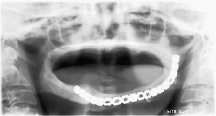
(3) 174. Hotta H et al.. Fig. 1 Panoramic radiograph showing reconstruction of left side of lower jaw using titanium plate. functional impairments such as pronunciation problems. Therefore, it is necessary to take extra care in such cases in determining the intercuspal position and arrangement of artificial teeth so that they match movement of the tongue and tissue function. Here, we report a completely edentulous patient in whom the left side of the lower mandible was resected because of a tumor. The intercuspal position was adjusted several times after a denture had been fitted as difficulties arose in eating. Dysphagia later occurred on further prosthetic treatment including application of complete dentures.. Case Report The patient was an 87-year-old man who first visited our department in October, 2006. His chief complaint was instability of the complete dentures and reduced ability to bite. His medical history revealed that he had undergone unilateral resection of the left lower jawbone because of a gingival tumor in the left lower mandible in 1981. Surgical procedures were subsequently performed three times between 1983 and 1985 and the mandible reconstructed using titanium plates. Upper and lower complete dentures were fitted in 1985, but repeated adjustment and denture relining were subsequently required.. In November 1999, elastic relining material was unsuccessfully attached to the lower denture, and further adjustment and additional attachment of the elastic relining material were repeatedly required1). Status on presentation: both the maxilla and mandible were edentulous. Maxillary alveolar ridge resorption was moderate and the morphology was mostly favorable. No alveolar ridge was observed on the left side of the mandible, which had been reconstructed using titanium plates after bone resection (Fig. 1), only a groove between the cheek and tongue. In contrast, some mandibular bone remained on the right side. The alveolar ridge was relatively smooth, although some alveolar resorption was observed. A small area had been resected from the left side of the tongue, but no reconstruction with mucosal grafting. Tongue movement was relatively good. However, the patient had some difficulty in poking his tongue into the right side of the mouth and then returning it to the palate. In addition, postoperative scars and the lack of a lower jawbone imposed a high level of strain on the left lower lip, which had recessed lingually. Figure 2 shows the complete dentures that were fitted in 1985 and are currently in use. A lack of significant problems in the arrangement of the artificial teeth was presumed as the upper alveolar ridge morphology was.
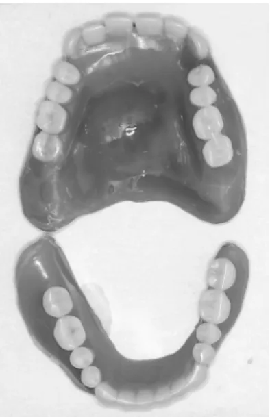
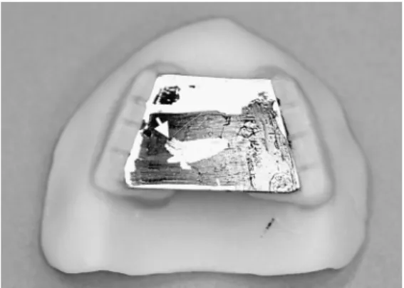
(4) Adjustment of New Dentures for Dysphagia. 175. Fig. 3 Lower denture in use: view of basal surface Artificial teeth before and after left canine positioned closer to labial side than buccal denture border.. Fig. 2 Upper and lower complete dentures currently in use. favorable and had been taken into account in manufacturing the upper dentures. Because the mandibular denture had been manufactured in accordance with the arrangement of the artificial teeth in the maxillary denture, the mandibular artificial teeth exceeded the buccal border of the denture base from the anterior teeth to the left canine and were located on the labial side (Fig. 3). This resulted in movement of the denture because the indented left unilateral lower lip placed right posterior pressure on the artificial teeth in the same region when the mouth was opened. The mandibular position was unstable due to movement of the lower denture during opening and closing of the mouth, causing the upper denture to also move during tapping, thereby lowering retention stability7). As a result, although the patient complained of difficulty in chewing, he did not report dysphagia. Treatment Strategy: adaptation of the complete dentures was insufficient, the occlusal relationship was unstable, and vertical dimension of occlusion (VDO) was reduced because of alveolar ridge resorption and attrition of. the artificial teeth. Therefore, the necessity of new upper and lower dentures was explained to the patient and his consent obtained. 1. Impression taking The morphology of the lower alveolar ridge was of an extremely inferior type, and the bearing ability was predicted to be extremely low. Thus, to widen the bearing area as much as possible, individual trays were used and an impression obtained using a rubber-based impression material after the morphology of the border of the base had been determined using border molding. Although the upper alveolar ridge conditions were favorable, a combined impression with the lower denture was obtained because it appeared that increased denture stability would additionally contribute to the stability of the corresponding lower denture. 2. Maxillo-mandibular registration2,3) The jaw position was determined on the basis of the resting position of the mandible3) and the horizontal jaw position was determined using Gothic arch tracing2) as VDO was reduced and the occlusal relationship unstable. 3. Artificial teeth arrangement The artificial teeth in the upper denture were arranged according to regulations and considering esthetic requirements4). However, in arranging the artificial teeth in the lower.
(5) 176. Hotta H et al.. denture it was noticed that the morphology of the alveolar ridge, which provides the foundation for denture support and retention stability, was extremely inferior. In addition, the lower dentures were matched with the surrounding tissues to increase stability due to overstressing and recession of the left lower lip. On the left side, in particular, the arrangement was based on the neutral zone of muscle pressure in the lower lip, cheek, and tongue rather than using the intercuspal relationship with the upper artificial teeth. 4. Fitting and adjustment The lower denture must be harmonized with the surrounding tissues, even if it has been polymerized and fitted according to regulations. Adjustment according to regulations dictates that if the patient is dissatisfied, functional impressions should be used to arrange the morphology of the basal plane and muscle pressure plane. The denture should then finally be completed with indirect rebasing. 5. Treatment progress 1) The beginning of November, 2006 — Impression taking Combined impressions were obtained for the upper and lower dentures using individual trays. 2) The middle of November, 2006 — Maxillomandibular registration Maxillo-mandibular registration was conducted using a bite plate. Maxillary face-bow transfer was performed at the same time2). The VDO was determined on the basis of the resting position of the mandible, and 2 mm of rest space was subtracted from the distance between the subnasal and submental points3,4,8) to allow for the decreased VDO arising from attrition of the current complete dentures and resorption of the jawbone. In addition, to improve denture stability, the cheek-tongue position of the occlusal rim of the mandible was lingually measured in the right upper denture, which demonstrated postoperative scar strain. The denture was set in a position that matched the pressure from. Fig. 4 Gothic arch tracing confirmed limitation of movement to right side caused by ablative surgery to left lower jaw Tapping area is shown (by an arrow).. the lower lip, buccal mucosa, and tongue. 3) The beginning of December, 2006 — Confirmation of horizontal jaw position The horizontal jaw position was determined using Gothic arch tracing2,8). Gothic arches (Fig. 4) were used to confirm alteration of horizontal momentum that appeared to have been caused by surgical resection of the lower jawbone. During tapping, a bite path from the right direction toward the apex that corresponded to the movement path of the right border was observed (Fig. 4, arrowhead). It appeared to have been caused by the patient habitually masticating on the right side where the jawbone remained. The denture had a strong bearing capacity. The horizontal jaw position was determined as being toward the end of this tapping path and adjacent to the apex. 4) The middle of December, 2006 — Trial application of wax model A trial application of wax dentures with arranged artificial teeth was performed. Favorable retention stability was achieved in the lower denture and hardly any movement was apparent during mouth opening. Regarding VDO, the patient stated, “It feels a little high, but isn’t uncomfortable.” 5) The end of December, 2006 — Insertion of new dentures New upper and lower dentures were.
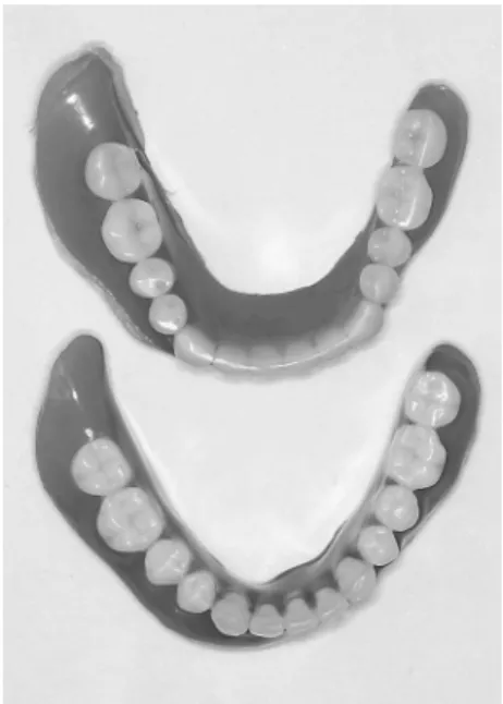
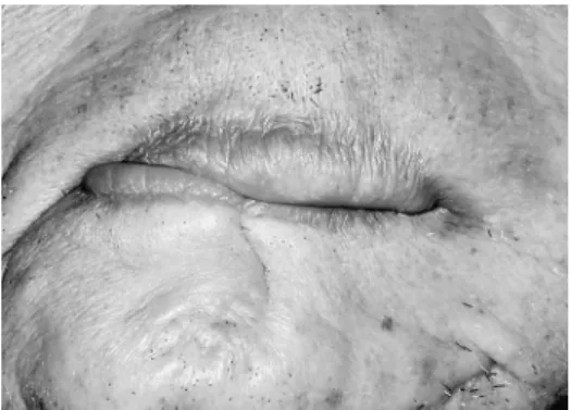
(6) Adjustment of New Dentures for Dysphagia. Fig. 5 New complete denture. Fig. 6 Incomplete closure of left side of lips. inserted (Fig. 5). No strong pain was observed in either jaw. Favorable retention stability and few problems were observed in the upper denture, which appeared to be functional. The lower denture was almost in harmony with the surrounding tissues and no movement occurred during mouth opening. Occlusal adjustment was also performed for the misalignment in the intercuspal position that was confirmed during the try-in.. 177. Fig. 7 Comparison of new and old lower dentures Above: old denture. Below: new denture. New denture arranged closer to lingual side around left canine than old denture.. 6) Three days after insertion — First adjustment The patient expressed dissatisfaction and stated, “There is no pain, but food and soup spill out of my mouth when I eat and I cannot swallow properly”6,9). During the first adjustment, recession in the left half of the lower lip and incomplete closure of the upper lip were confirmed (Fig. 6), indicating the probable cause spillage during mastication. It also appeared that the patient found it difficult to swallow a bolus as the tongue had an adequate impact on the palate due to the increase in the VDO5,6,10). The lower denture (Fig. 7) was adjusted to the right side of the left premolar region from the front tooth to prevent labial pressure with the aim of improving lower denture stability. This resulted in insufficient lip support for the left half of the lower lip. In addition, VDO had been determined on the basis of the resting position of the mandible, without taking the state of oral aperture closure or tongue contact with the palate into consideration. This appeared to be the reason lip.
(7) 178. Hotta H et al.. Fig. 8 Closure of lips was improved by reducing occlusal vertical dimension. support was insufficient. Therefore, to improve mouth closure and contact between the tongue and palate, an extra occlusal adjustment was performed to reduce VDO. 7) The middle of January, 2007 — Second adjustment Although the VDO had somewhat reduced since the previous adjustment, the patient still experienced difficulty in eating and complained of dysphagia. Therefore, for re-examination of VDO and artificial teeth arrangement, the artificial teeth in the mandibular denture were removed, an occlusion rim installed, and the interocclusal relationship re-registered. This caused loss of stability in the lower denture, so the artificial teeth arranged from the lower anterior region to the left premolar region could not be moved to the labial and buccal sides. Therefore, to prevent spillage on mastication, the VDO was reduced so that the mouth aperture could close naturally. After this procedure was performed, the patient found it easy to swallow saliva, suggesting that contact between the tongue and palate had improved. 8) The end of January, 2007 — Trial-in after correction of artificial teeth arrangement The VDO was reduced, arrangement of artificial teeth corrected, and the trial-in of a wax model conducted. The closing state of the left half of the upper and lower lips was favorable7) (Fig. 8), and the patient was able to drink water without complaining of any. difficulty. 9) The beginning of February, 2007 — Insertion of complete dentures after correction of artificial teeth arrangement The stability of the lower denture during mouth opening and the labial closed state during mouth closing were favorable. 10) The middle of February, 2007 — Adjustment after correction Although some tenderness occurred in the mucous membrane under the upper denture, it disappeared when the basal surface was adjusted. Furthermore, the dysphagia which had caused patient dissatisfaction prior to correction had almost completely disappeared. The patient indicated that his condition was completely favorable.. Discussion When the loss of numerous teeth results in functional impairment such as difficulty in chewing or swallowing, or loss of intercuspal position, occlusion is reconstructed with dentures to aid functional recovery. In particular, when large-scale dentures such as complete dentures are fitted, various factors such as the intercuspal position and the arrangement of artificial teeth must be considered. Normally, the intercuspal position is determined based on physiological and functional standards such as resting position of the mandible during swallowing or morphological standards determined by measuring facial parts3,4). Furthermore, in many cases, the position of the upper and lower jaw alveolar ridges and their corresponding relationship are used as standards for artificial teeth arrangement, while also taking esthetic elements into consideration in improving retention stability of the artificial teeth against occlusal-masticatory force. It is also necessary for the morphology of the palate to match the formation and movement of a bolus by the tongue. However, such movement is often impaired in cases that have undergone oral tissue or tongue resection because of oral tumors. Therefore, it is necessary to use different methods to.
(8) 179. Adjustment of New Dentures for Dysphagia. determine the intercuspal space, arrangement of artificial teeth position, and denture base morphology5,10). When difficulty is experienced in bringing the tongue into contact with the palate, the morphology of the upper denture base must be adjusted so that proper impact is restored. In the present case, the intercuspal position was determined on the basis of the resting position of the mandible, and the artificial teeth were given an unconventional arrangement to improve the stability of the lower denture. As a result, the rocking of the lower denture caused by opening and closing of the mouth was reduced and the intercuspal position stabilized. However, it appeared that when the artificial teeth were arranged on the lingual side of the right premolar region from the lower frontal tooth region, the strain on the lower lip, caused by surgical scars, and raised intercuspal position, caused by previous dentures, resulted in incomplete closure of the mouth aperture in the intercuspal position. This seemed to be the reason for food spillage. It also appeared that insufficient impact of the tongue on the palate caused by increased VDO6) resulted in increased dysphagia, because the morphology of the palate was identical to that of the previous dentures. Therefore, the VDO was first used to adjust the position of the artificial teeth to improve the incomplete closure of the mouth aperture. Rocking of the lower denture was observed during mouth opening when the artificial teeth were moved to the labial and buccal sides, so the position of the artificial teeth could not be changed. Therefore, incomplete closure of the mouth aperture was improved by reducing the VDO, which appeared to result in a more favorable impact of the tongue on the palate and improvement of dysphagia10). Therefore, to promote recovery of eating function, therapeutic strategies must address not only the function of the temporomandibular joint and masticatory muscles as well as the maxillofacial morphology but also the functional recovery of various tissues sur-. rounding the oral cavity such as the lips and tongue in determining the intercuspal and artificial tooth positions for dentures. Extra care is required in making major alterations to the VDO and arrangement of artificial teeth in comparison with previous dentures in elderly patients as this may cause unexpected problems. Therefore, it is necessary to proceed with correction of older dentures while carefully monitoring patient reaction.. Acknowledgements We would like to thank Associate Professor Jeremy Williams, Tokyo Dental College, for his assistance with the English of this manuscript.. References 1) Babiuc I, Păuna M, Maliţa MA, Ariton SG, Damian M, Ungureanu V, Petrini A (2009) Correct complete denture rehabilitation, a chance for recovering abused tissues. Rom J Morphol Embryol 50:707–712. 2) Davies SJ, Gray RMJ, McCord JF (2001) Good occlusal practice in removable prosthodontics. Br Dent J 191:491–494, 497–502. 3) Hall WA Jr (1958) Important factors in adequate denture occlusion. J Prosthet Dent 8: 764–775. 4) Mohindra NK, Bulman JS (2002) The effect of increasing vertical dimension of occlusion on facial aesthetics. Br Dent J 192:164–168. 5) Murayama R, Takebe J, Ito S, Tanabe N, Simazaki N, Ishibashi K (2011) A case report of functional recovery with palatal augmentation prosthesis for reconstruction after glossectomy. Maxillofacial Prosthetics 34:20–26. (in Japanese) 6) Nagao K, Kitaoka N, Kawano F, Komoda J, Ichikawa T (2002) Influence of changes in occlusal vertical dimension on tongue pressure to palate during swallowing. Prosthodont Res Pract 1:16–23. 7) Prakash V (2008) Prosthetic rehabilitation of edentulous mandibulectomy patient: A clinical report. Indian J Dent Res 19:257–260. 8) Shanahan TEJ (2004) Physiologic vertical dimension and centric relation. J Prosthet Dent 91:206–209. 9) Tamura F, Tomita K, Okano T, Mizukami M, Kikutani T, Mukai Y (2004) Effect of denture.
(9) 180. Hotta H et al.. wearing for lip closing pressure during swallowing on edentulous elderly people. J J Gerodont 19:169–173. (in Japanese) 10) Yamamoto C (2001) A case of prosthesis to consider the difficulty of swallowing. J Jpn Prosthodont Soc 45:442–443. (in Japanese). Reprint requests to: Dr. Hiromi Hotta Department of Clinical Oral Health Science, Tokyo Dental College, 2-9-18 Misaki-cho, Chiyoda-ku, Tokyo 101-0061, Japan Tel: +81-3-3262-3421 Fax: +81-3-3262-3420 E-mail: [email protected].
(10)
図
+2
関連したドキュメント
2 Combining the lemma 5.4 with the main theorem of [SW1], we immediately obtain the following corollary.. Corollary 5.5 Let l > 3 be
It is suggested by our method that most of the quadratic algebras for all St¨ ackel equivalence classes of 3D second order quantum superintegrable systems on conformally flat
This paper is devoted to the investigation of the global asymptotic stability properties of switched systems subject to internal constant point delays, while the matrices defining
In this paper, we focus on the existence and some properties of disease-free and endemic equilibrium points of a SVEIRS model subject to an eventual constant regular vaccination
Then it follows immediately from a suitable version of “Hensel’s Lemma” [cf., e.g., the argument of [4], Lemma 2.1] that S may be obtained, as the notation suggests, as the m A
Our method of proof can also be used to recover the rational homotopy of L K(2) S 0 as well as the chromatic splitting conjecture at primes p > 3 [16]; we only need to use the
Classical definitions of locally complete intersection (l.c.i.) homomor- phisms of commutative rings are limited to maps that are essentially of finite type, or flat.. The
Yin, “Global existence and blow-up phenomena for an integrable two-component Camassa-Holm shallow water system,” Journal of Differential Equations, vol.. Yin, “Global weak
