IRUCAA@TDC : An experimental study on the features of peri-implant epithelium: immunohistochemical and electron-microscopic observations
16
0
0
全文
(2) Bull. Tokyo dent. Coll., Vol. 44, No. 4, pp. 185⬃199, November 2003. 185. Original Article. AN EXPERIMENTAL STUDY ON THE FEATURES OF PERI-IMPLANT EPITHELIUM: IMMUNOHISTOCHEMICAL AND ELECTRON-MICROSCOPIC OBSERVATIONS MASATSUGU FUJISEKI, KENICHI MATSUZAKA*, MASAO YOSHINARI**, MASAKI SHIMONO and TAKASHI INOUE* Oral Health Science Center, Department of Pathology, Tokyo Dental College, 1-2-2 Masago, Mihama-ku, Chiba 261-8502, Japan * Oral Health Science Center, Department of Clinical Pathophysiology, Tokyo Dental College, 1-2-2 Masago, Mihama-ku, Chiba 261-8502, Japan ** Oral Health Science Center, Department of Dental Materials Science, Tokyo Dental College, 1-2-2 Masago, Mihama-ku, Chiba 261-8502, Japan. Received 16 September, 2003/Accepted for Publication 29 October, 2003. Abstract The purpose of this study was to investigate the immunohistochemical and the ultrastructural features of the implant circumference epithelium of the beagle dog using various types of antibodies. The peri-implant epithelium was at an acute-angle from the gingival epithelium and was arranged in parallel to the implant surface. With immunohistochemical staining, the peri-implant epithelium was strongly positive for KL-1, and weakly positive for CK4, CK8 and CK19. These positive reactions for keratins and also for PCNA and BM-1 were similar to those seen in the oral mucosa. In the periimplant epithelium, a plentitude of microvilli were observed at the periphery of cells at the implant sites, and bacteria were observed between the implant and the peri-implant epithelium without the formation of half desmosomes. There were many lipid-like vacuoles or lysosome-like granules. The intercellular space was wider than the junctional epithelium, and random migrations of large numbers of neutrophils could be seen. Taken together, the peri-implant epithelium is similar to that seen in the oral mucosa, and it is structurally different from the junctional epithelium. Key words:. Cytokeratin —Peri-implant epithelium— Immunohistochemistry — Transmission electron microscopy. This paper is a dissertation submitted in June 2003 by Masatsugu Fujiseki to the Graduate School of Tokyo Dental College for the degree of Doctor of Philosophy.. 185.
(3) 186. M. FUJISEKI et al.. INTRODUCTION. MATERIALS AND METHODS. The understanding of the implant-tissue interface is important for successful tooth replacement therapy. Much research has been performed on the bone interface, and the theory has been established that there should be no intervention of soft tissue between the implant and the bone tissue at the light microscopic level; this is called osseointegration1,8,9,11,13,27). Romanos et al.32,33) reported that the implant-connective tissue interface is thin; the thickness of type I collagen controls the stability of the tissue. However, type V collagen resists bacterial invasion by increasing in amount compared with the natural tooth. Dental implantation therapy creates an open wound, because the oral mucosa is penetrated along the implant surface and, as a result, an epithelium-implant interface is formed. Therefore, elucidation of the defense mechanisms, including those of the epithelium itself, becomes an important point, because the peri-implant tissue is always exposed to the possibility of inflammation16,21–23). In the junctional epithelium of the natural tooth, there are defense elements, such as bonding mechanical closures with hemidesmosomes, differentiation and proliferation capability, and phagocytic capability to resist stimuli. Furthermore, the random migration of neutrophils into the intercellular space of the junctional epithelium, which is continually washed by the crevicular exudates to the gingival sulcus, needs to be considered36,38). However, bacteria can accumulate around the implant circumference and more easily induce inflammatory destruction than around the natural tooth. Furthermore, pocket formation around the implant occurs earlier, and bone resorption also seems to be induced10,29,30). Thus, the epithelium-implant interface influences the fate of the implant2,3,5,7,17,18,28,39,40); however; the details of its morphological features are not well known. This study examined the immunohistochemical features of the peri-implant epithelium using various antibodies and also examined its ultrastructural features.. 1. Laboratory animals and experimental methods All studies conformed to the Tokyo Dental College laboratory animal facilities experimental guidelines. The laboratory animals used in these experiments were four one-year-old female beagle adult dogs, each weighing 10 kg. Prior to each experiment, general anesthesia was conducted using thiopental sodium (Nembutal 0.1 ml/kgbd). Premolar teeth of both sides of the lower jaw were extracted, and bone tissue was allowed to heal for two months. After the healing of the extracted socket, general anesthesia was again conducted using the same procedure described above, and local anesthesia was applied using xylocaine with 2% epinephrine. An incision was made on the alveolar ridge, and the mucoperiosteal flap was raised using a periosteal elevator. A bone cavity was generated using a round bur mounted on a dental handpiece cooled with phosphate buffered saline (PBS). A titanium plasma sprayed (TPS) ITI implant, 3.3 mm in diameter and 8 mm in length, were implanted and merged in the edentulous area. Finally, the peri-implant mucous membrane was closed with sutures. The gingival mucosa and junctional epithelium of the mandibular molar teeth of the same animals were used as controls. Plaque control was not carried out throughout the experimental periods. Each mandible received three implants and a total of 24 implants were inserted; however, three cases in which bone resorption occurred due to inflammation were excluded from the experimental groups. As results, 21 implants were examined in these experiments. 2. Light microscopy For paraffin sections without implants, perfusion fixation was carried out six months after the implantation via the carotid artery with formomethanol (37% formalin: 99% methanol: Distilled water⳱1:1:2). The mandibles with the implant and the surrounding tissues were removed, and 16 implants were.
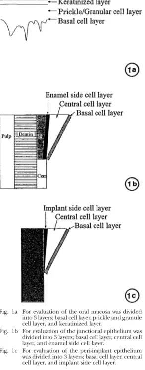
(4) THE FEATURES OF PERI-IMPLANT EPITHELIUM. 187. sliced along the extended shaft of the implant as bucco-lingual sections, using a rotary saw cooled with running water. They were then immersed in the same fixative solution for one week. The tissues were then decalcified with 10% EDTA for eight weeks at room temperature, and the implant body was mechanically removed. Specimens without implants were dehydrated in a graded ethanol series before being embedded in paraffin and cut in serial sections approximately 5m in thickness. Sections were then stained with either hematoxylin-eosin or periodic acid-Schiff (PAS) and observed in a light microscope. For ground sections with implants, five implants were embedded in methylmethacrylate as previously described14,29). The methylmethacrylate blocks were sectioned by use of a low speed saw equipped with a 0.3 mmthick diamond wafering blade. The sectioning technique was devised to provide bucco-lingual sections. The initial section thickness was approximately 200m and was reduced to approximately 80m by petrographic grinding techniques. The sections were then stained with toluidine blue and alizarin red. 3. Immunohistochemistry For immunohistochemical observations, the streptavidin-biotin peroxidase complex method was carried out using a Histofine kit according to the manufacturer’s instructions (Histofine Kit, Nichirei, Tokyo). Antibodies to cytokeratins, KL-1 (Immunotech, France, at a dilution of 1:150), CK4 (Progen Germany, at a dilution of 1:10), CK8 (DAKO, Denmark, at a dilution of 1:25), CK19 (Progen Germany, at a dilution of 1:10), and either PCNA (PC10, DAKO, Denmark, at a dilution of 1:100) or BM-1 (DAKO Denmark, at a dilution of 1:25) were employed as primary antibodies. Paraffin sections were deparaffinized with xylene and washed in 10 mM PBS (pH 7.2). Sections were then treated with 0.1% trypsin (in 10 mM Tris-HCl buffer, pH 7.6: Gibco BRL, USA) for 30 minutes at 37°C and then treated with 0.3% H2O2 in methanol at room temperature to block endogenous peroxidase.. Fig. 1a For evaluation of the oral mucosa was divided into 3 layers; basal cell layer, prickle and granule cell layer, and keratinized layer. Fig. 1b For evaluation of the junctional epithelium was divided into 3 layers; basal cell layer, central cell layer, and enamel side cell layer. Fig. 1c For evaluation of the peri-implant epithelium was divided into 3 layers; basal cell layer, central cell layer, and implant side cell layer.. After washing in PBS, the samples were incubated with primary antibodies at a dilution of 1:10 at 4°C overnight. Reactions were visualized using 0.05% diaminobenzidine (DAB) and 0.005% H2O2 in Tris-HCl buffer,.
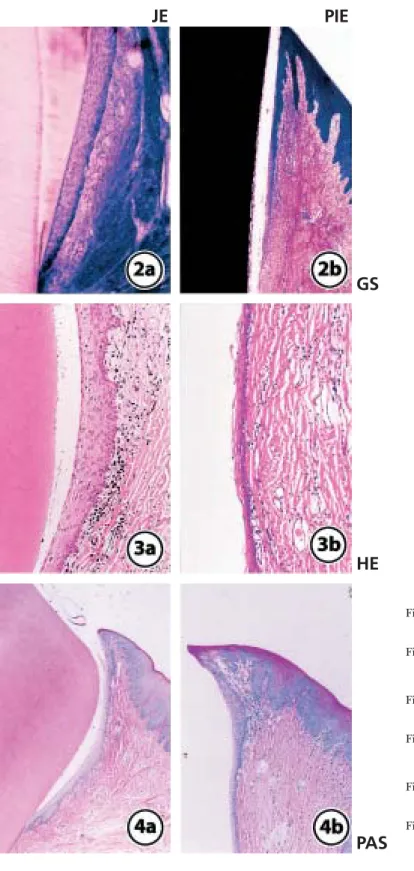
(5) 188. M. FUJISEKI et al.. and sections were finally counterstained with hematoxylin and observed using a light microscope. For evaluation of immunohistochemical activities, the oral mucosa was divided into three layers: basal cell layer, prickle and granular cell layer, and keratinized layer (Fig. 1a). The junctional epithelium was divided into three layers; basal cell layer, central cell layer, and enamel side cell layer (Fig. 1b). The peri-implant epithelium was also divided into three layers; basal cell layer, central cell layer, and implant side cell layer (Fig. 1c). For all the light microscopic observations, the middle parts of both the junctional and peri-implant epithelium were mainly compared. 4. Electron microscopy For transmission electron microscopic observations, part of buccal area of each implant circumference tissue used for ground sections was removed after the perfusion fixation and further fixed with Karnovsky’s fixative solution (at a final concentration of 2% paraformaldehyde and 2.5% glutaraldehyde in 0.24M Sorensen’s phosphate buffer) for 48 hours at room temperature. After the samples were postfixed in 2% osmium tetroxide for two hours, they were block stained with 2% uranyl acetate in 10% ethanol for 20 min. They were then dehydrated in a graded series of ethanol and embedded in Epon 812 using routine procedures. After ultra-thin sections were cut approximately 60 nm thick, specimens were stained with uranyl acetate and lead citrate and were observed using a Hitachi 7100 transmission electron microscope.. RESULTS 1. Macroscopic observations At the natural tooth circumference, a slight deposition of plaque was observed, but no periodontitis was evident. In three of the 24 implants, the periimplant tissue showed obvious inflammation, and there was clear absorption of the bone. Those three samples were excluded from the. study. The remaining 21 implants had slight plaque deposition, but bone resorption was not seen by X-ray. 2. Light microscopic observations The junctional epithelium adhered to the enamel from the bottom of the gingival crevice, was composed of about 10 cell layers, and was in the cuneus state. The cell population was decreased in the enamel-cement junction (Fig. 2a, 3a). No PAS positively staining cells were found in the junctional epithelium (Fig. 4a). The peri-implant epithelium was at an acute angle from the gingival epithelium and was arranged in parallel to the implant surface. A sharply narrow gap was evident between the implant body and the peri-implant epithelium. It was of a uniform thickness, and the number of component cells was less than that of the junctional epithelium. No obvious tendency toward cornification was observed (Fig. 2b, 3b). At the position where inflammatory cell infiltration could be seen in the connective tissue under the implant epithelium, the thickness of the peri-implant epithelium was increased, and the intercellular space became wider. The cells of the implant side and the central cell layers were weakly positive for PAS staining (Fig. 4b). The oral mucosal epithelium could be distinguished as the basal cell layer, prickle cell layer, granular cell layer, and keratinized layer. The keratinized layer was strongly positive for PAS staining and the cells of the prickle cell layer were weakly positive. 3. Immunohistochemical observations When using immunohistochemical staining for PCNA, the cells of the enamel side cell layer and the basal cell layer of the natural tooth (Fig. 5a), a few cells of the basal cell layer of the peri-implant epithelium (Fig. 5b), and some cells of basal cell layer of the oral mucosa (Fig. 5c) were positive for PCNA. However, the positive reaction of the periimplant epithelium was the weakest, and the positive cell number was the lowest of the three. When using immunohistochemical staining.
(6) THE FEATURES OF PERI-IMPLANT EPITHELIUM. JE. 189. PIE. GS. HE Fig. 2a Ground section of the junctional epithelium (low magnification) Fig. 2b Ground section of the peri-implant epithelium (low magnification) Fig. 3a HE staining of the junctional epithelium (middle magnification) Fig. 3b HE staining of the peri-implant epithelium (middle magnification). PAS. Fig. 4a PAS staining of the junctional epithelium (low magnification) Fig. 4b PAS staining of the peri-implant epithelium (low magnification).
(7) 190. M. FUJISEKI et al.. PCNA. BM-1. JE. PIE. OM. Fig. 5a Immunohistochemical staining of junctional epithelium for PCNA. The cells of enamel side cell layer and basal cell layer were positive. (middle magnification) Fig. 5b Immunohistochemical staining of peri-implant epithelium for PCNA. A few cells of the basal cell layer were positive. (middle magnification) Fig. 5c Immunohistochemical staining of oral mucosa for PCNA. Some cells of basal cell layer were positive. (middle magnification) Fig. 6a Immunohistochemical staining of junctional epithelium for BM-1. Central cell layer was positive. (middle magnification) Fig. 6b Immunohistochemical staining of peri-implant epithelium for BM-1. The cells of both the implant side cell layer and central cell layer were positive. (middle magnification) Fig. 6c Immunohistochemical staining of oral mucosa for BM-1. Keratinized layer was positive. (middle magnification).
(8) THE FEATURES OF PERI-IMPLANT EPITHELIUM. KL-1. 191. CK19. JE. PIE. Fig. 7a Immunohistochemical staining of junctional epithelium for KL-1. All the cells of the junctional epithelium were positive. (middle magnification) Fig. 7b Immunohistochemical staining of peri-implant epithelium for KL-1. All the cells of the junctional epithelium were positive. (middle magnification) Fig. 7c Immunohistochemical staining of oral mucosa for KL-1. All the cells of the junctional epithelium were positive. (middle magnification). OM. Fig. 8a Immunohistochemical staining of junctional epithelium for CK19. The cells of the central cell layer and enamel side cell layer were positive. (middle magnification) Fig. 8b Immunohistochemical staining of peri-implant epithelium for CK19. The cells of the basal cell layer were weakly positive. (middle magnification) Fig. 8c Immunohistochemical staining of oral mucosa for CK19. The cells of the basal cell layer of the oral mucosa were strongly positive. (middle magnification).
(9) 192. M. FUJISEKI et al.. CK8. CK4. JE. PIE. OM. Fig. 9a Immunohistochemical staining of junctional epithelium for CK8. The cells of the central cell layer and enamel side layer were positive. (middle magnification) Fig. 9b Immunohistochemical staining of peri-implant epithelium for CK8. The cells of the central cell layer and implant side cell layer were positive. (middle magnification) Fig. 9c Immunohistochemical staining of oral mucosa for CK8. The cells of the prickle cell layer of the oral mucosa were positive. (middle magnification) Fig. 10a Immunohistochemical staining of junctional epithelium for CK4. All the cells of the junctional epithelium were negative. (middle magnification) Fig. 10b Immunohistochemical staining of peri-implant epithelium for CK4. The cells of the central cell layer were positive. (middle magnification) Fig. 10c Immunohistochemical staining of oral mucosa for CK4. The cells of the prickle cell layer of the oral mucosa were slightly positive. (middle magnification).
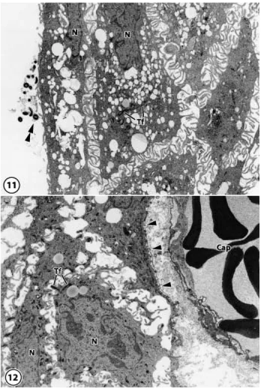
(10) THE FEATURES OF PERI-IMPLANT EPITHELIUM. Fig. 11–14 Electron-micrographs of the peri-implant epithelium Fig. 11 In the peri-implant epithelium, plenty of microvilli were observed at the periphery of cells at the implant sites, and bacteria were observed between the implant and the peri-implant epithelium. N: nucleus, Tf: tonofilament, Double arrowheads: bacteria (original magnification⳯1,600) Fig. 12 A basement membrane, which distinguishes the lamina lucida from the lamina densa, was observed at the connective tissue side. N: nucleus, Tf: tonofilament, Arrowheads: basement membrane, Cap: capillary (original magnification⳯1,600). 193.
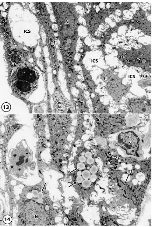
(11) 194. M. FUJISEKI et al.. Fig. 13 In the cytoplasm of cells in the peri-implant epithelium, a number of tonofilaments were observed diffusely with a few mitochondria and endoplasmic reticula. The intercellular spaces were wider than that of the junctional epithelium, and random migration of large numbers of neutrophils could be seen. N: nucleus, Tf: tonofilament, ICS: intercellular space (original magnification⳯2,000) Fig. 14 Many vacuoles and lysosome-like granules, N: nucleus, Tf: tonofilament, Arrows: vacuoles and lysosome-like granules (original magnification⳯2,000).
(12) THE FEATURES OF PERI-IMPLANT EPITHELIUM. for BM-1, the cells of the central cell layer of the junctional epithelium (Fig. 6a) and both the cells of the implant side cell layer and those of the central layer of the peri-implant epithelium (Fig. 6b) and the keratinized layer of the oral mucosa were positive for BM-1 (Fig. 6c). When using immunohistochemical staining for KL-1, all the cells of the junctional epithelium (Fig. 7a), peri-implant epithelium (Fig. 7b), and oral mucosa (Fig. 7c) were strongly positive. When using immunohistochemical staining for CK19, the cells of central and enamel side cell layers of the junctional epithelium were strongly positive, the cells of the basal cell layer were weakly positive (Fig. 8a), the cells of the central and basal cell layers of the periimplant epithelium were weakly positive (Fig. 8b), and the cells of the basal cell layer of the oral mucosa were strongly positive (Fig. 8c). When using immunohistochemical staining for CK8, the cells of central cell layer and enamel side layer of the junctional epithelium (Fig. 9a), the cells of the central cell and implant side cell layers of the peri-implant epithelium (Fig. 9b), and the cells of the prickle cell layer of the oral mucosa (Fig. 9c) were positive. When using immunohistochemical staining for CK4, all the cells of the junctional epithelium were negative (Fig. 10a). The cells of the central cell layer of the peri-implant epithelium (Fig. 10b) and the prickle cell layer of the oral mucosa (Fig. 10c) were slightly positive. 4. Electron microscopic observations In the natural tooth, the junctional epithelium contacted the enamel surface by half desmosomes, and on the connective tissue side, a basement membrane was present. Cells of the junctional epithelium had many micro-processes combined with desmosomes, and various degrees of tonofilaments were observed in the cytoplasm. In the peri-implant epithelium, many microvilli were observed at the periphery of the cells at the implant sites, and bacteria were. 195. present between the implant and the periimplant epithelium (Fig. 11). However, half desmosomes and the basement membrane had not formed because the peri-implant epithelium was not in close contact with the implant body. A basement membrane, which distinguishes the lamina lucida from the lamina densa, was observed at the connective tissue side (Fig. 12). In the cytoplasm of cells in the peri-implant epithelium, a number of tonofilaments were diffusely present, as were a few mitochondria and endoplasmic reticula, but many lipid-like vacuoles or lysosome-like granules were also observed. The intercellular space was wider than that of the junctional epithelium, and the random migration of large numbers of neutrophils could be seen (Figs. 13, 14).. DISCUSSION 1. Morphological features of the epithelia The normal gingival mucosa eventually differentiates into the stratum corneum, a layer of cells without nuclei but with tonofilaments and keratohyalin granules which arises following the division of basal cells; these cells are eventually discarded into the oral cavity36). The normal junctional epithelium arises from the deciduous enamel epithelium during the development of the dental germ and contains enamel and half desmosomes when the teeth erupt. The defense function of the epithelium compared with the cornified oral mucosa is weak. However, the protection is supplemented by the transudation and many neutrophils derived from the fenestrated capillaries, which exist at the side of the connective tissue38). In addition, it is known that bacteria are resisted by the secretion of defensin, an antibacterial peptide, by epithelial cells4). The epithelium observed at the circumference of the implant arose from the healed gingival stratified squamous epithelium, which became keratinized after the extraction of the tooth22,23). Many discussions have arisen about the kind of interface formed when.
(13) 196. M. FUJISEKI et al.. the implant penetrates this epithelium and whether they form half desmosomes and/or basement membranes14,15,18,21–24). The results of PAS staining, which reacts with glycogen in the cytoplasm, were negative in the cells of junctional epithelium. In contrast, a positive reaction was observed in both peri-implant epithelium and oral mucosa, suggesting that both epithelia are similar in terms of glycogen production. 2. Peri-implant epithelium Gould et al.14) reported that hemi-desmosomes and basement membranes formed on the titanium surface in vitro. However, Jansen et al.24) suggested that half desmosomes did not form on metals such as titanium. Ikeda et al.19) used implants made of titanium in rats and reported that the junctional epithelium structure exists only at the bottom of the implant circumference epithelium. They also reported that the peri-implant epithelium on the oral cavity side had many microprocesses and that they could not see any adhesion with the implant in that area. Takata et al.37) characterized the permeability of the junctional epithelium and the implant circumference epithelium. They dropped goldconjugated concanaban A into the gingival crevice and then observed that the tracer could through pass the intercellular space of the junctional epithelium and penetrate to the connective tissue. This means that the exogenous material easily penetrated from the gingival crevice to the connective tissue and that this route is probably involved in the initial stage of disease around the teeth. Ikeda et al.19) carried out a similar experiment in the implant circumference groove. They reported that many more particles could be found in the circumference epithelial cells of the implants than in the junctional epithelium. They further reported that the permeating material also reached deep into the connective tissue and that bacteria and toxins could also easily permeate the connective tissue further than the periodontal tissue of the natural tooth. From the above facts, the invasion of exogenous materials. and bacteria into the structure of the implant circumference epithelium should be easy, and the defense support from the connective tissue side is clearly important. Ikeda et al.20) reported that the endocytotic capacity of the cells of peri-implant epithelium was inferior to that of junctional epithelium because, intracellularly, the quantity of horseradish peroxidase which was found in the vesicles and granules of peri-implant epithelium was less than that of the junctional epithelium. The electron microscopic observations of the present study that the intercellular space of the peri-implant epithelium was enlarged further than that of the junctional epithelium, that microvilli were observed in the plane which meets in the implant well, and that clear desmosomes were not formed, suggest that the junctional epithelium differs in function and structure and support their opinions. 3. Functions of epithelial cells By having enamel and half desmosomes involved in the protection of the junctional epithelium, the following are considered to be important in addition to the mechanical closure: the capability to differentiate and proliferate to resist stimuli and to phagocytose the foreign materials of the epithelial cells. Inoue et al.21) reported that the proliferation capability of the junctional epithelium was greater than that of the peri-implant epithelium, that activity was due to exogenous stimulation, and that it contributed to the defense reaction. In this study, using PCNA which is a marker for proliferating activity and BM-1 which is known as marker for apoptosis, the positive reactions of PCNA and BM-1 produced opposite results; the cells of both the enamel side and the basal cell layer were positive for PCNA and the cells of the central cell layer were positive for BM-1 in the junctional epithelium. These results support the results of Inoue et al.21) and indicate that both the enamel side cells and the basal cells of the junctional epithelium metabolize to the central area. In contrast, only a few cells of basal cell layer were positive for PCNA and.
(14) THE FEATURES OF PERI-IMPLANT EPITHELIUM. the cells of central cell layer and the implant side cell layer were positive for BM-1 in the peri-implant epithelium; these were similar to the results for the oral mucosa. Ayasaka et al.6) reported that the lysosomal enzymes cathepsins B, D, and H exist locally in the junctional epithelium component cells, and Yamaza et al.41) reported that there is an intracellular resolution in the endosome/ lysosome system in the junctional epithelium. Further, elimination of xenobiotica by neutrophil phagocytosis in the intercellular space seems to supplement the defense of the junctional epithelium. In this study, many lysosome-like granules were seen in the cytoplasm of peri-implant epithelium, which had wide intercellular spaces, which seems to support this argument. 4. Keratin of epithelium component cells Cytokeratin, which is contained in various types of epithelial cells and helps form the cytoskeleton, is an intracytoplasmic fibrous constituent and is classified as one type of intermediate filament. Twenty subtypes of cytokeratins, which are classified into type I or type II, have been reported. Type I cytokeratins (CK9 to CK20) are of low molecular weight and have acidic isoelectric points, while type II cytokeratins (CK1 to CK8) are of high molecular weight and have neutral or basic isoelectric points. It has been demonstrated that the genesis of cytokeratins has regularity as well as the specific character of original tissue and cell even in tumor cells18,28,31,34,35). KL-1 is detectable in the stratum cell layer, which is the upper portion of the basal layer in the keratinized epithelium of gingiva, tongue, and palate. CK19 can also be expressed in the analogous layer12,28,34,35). In particular, it reacts positively with simple epithelium, but it is also immunostained in basal cells of the gingival stratified squamous epithelium, in dental lamina of odontogenic epithelium, in the enamel organ, and in epithelial rests of Malassez31,34,35). Basal cells of all epithelia, including oral, junctional, and peri-implant epithelium, which were also positive for CK19, and all cells (except basal cells) of those. 197. epithelia were positive for KL-1. The results of this study showed that the positive staining for CK19 and KL-1 in the peri-implant epithelium were similar to that seen in the oral mucosa. Comparable staining of CK4 and CK8 was seen except for the basal cells in the oral mucosa and in the peri-implant epithelium. In short, in the peri-implant epithelium, only the implant side cells were positive for CK4 and 8; the prickle cells in the oral epithelium were also positive for CK4 and 8. However, in the junctional epithelium, the enamel side cells showed results similar to those of the basal cells in the oral epithelium. This may indicate that the peri-implant epithelium does not have a keratinized layer but is similar to the prickle cells of the oral epithelium, which continue to the implant surface. Mackenzie and Tonetti26) reported that the intensity of the staining reactions for cytokeratins of peri-implant epithelium were different from that of junctional epithelium. It is possible that titanium-based implant materials could influence the phenotype of oral gingival epithelial cells differently (Lagneau et al.25)). In junctional epithelium, the degeneration tendency is strong, the intercellular space expands, and no clear cornification is observed. However, the staining of keratins was similar to that seen in the oral mucosa epithelia and it was structurally different from that in the junctional epithelium.. REFERENCES 1) Albrektsson, T., Branemark, P.I., Hansson, H.A. and Lindstrom, J. (1981). Osseointegrated titanium implant. Acta Orthop Scand 52, 155– 170. 2) Abrahamsson, I., Berglundh, T., Moon, I.S. and Lindhe, J. (1999). Peri-implant tissues at submerged and non-submerged titanium implants. J Clin Periodontol 26, 600–607. 3) Abrahamsson, I., Berglundh, T., Glantz, P.O. and Lindhe, J. (1998). The mucosal attachment at different abutments. An experimental study in dogs. J Clin Periodontol 25, 721–727. 4) Abiko, Y., Mitamura, J., Nishimura, M.,.
(15) 198. 5). 6). 7). 8) 9). 10) 11). 12). 13). 14). 15). 16). M. FUJISEKI et al. Muramatsu, T., Inoue, T., Shimono, M. and Kaku, T. (1999). Pattern of expression of betadefensins in oral squamous cell carcinoma. Cancer Lett 141, 1–7. Arvidson, K., Fartash, B., Hilliges, M. and Kondell, P.A. (1996). Histological characteristics of peri-implant mucosa around Branemark and single-crystal sapphire implants. Clin Oral Implants Res 7, 1–10. Ayasaka, N. and Tanaka, T. (1989). A cytochemical study of horseradish peroxidase uptake in rat junctional epithelium. J Dent Res 68, 1503–1507. Ayukawa, Y., Takeshita, F., Inoue, T., Yoshinari, M., Ohtsuka, Y., Murai, K., Shimono, M., Suetsugu, T. and Tanaka, T. (1996). An ultrastructural study of the bone-titanium interface using pure titanium-coated and pure titanium rod implant. Acta Histochem Cytochem 29, 243– 254. Berglundh, T. (1991). The soft tissue barrier at implants and teeth. Clin Oral Implants Res 2, 81–90. Buser, D., Schenk, R.K., Steinemann, S., Fiorellini, J.P., Fox, C.H. and Stich, H. (1991). Influence of surface characteristics on bone integratio of titanium implants. A histomorphometric study in miniature. J Biomed Mater Res 25, 889–902. Bauman, G.R., Raley, J.W., Hallmon, W.W. and Mills, M. (1993). The peri-implant sulcus. Int J Oral Maxillofac Implants 8, 273–280. Buser, D., Weber, H.P., Donath, K., Fiorellini, J.P., Paquette, D.W. and Williams, R.C. (1992). Soft tissue reactions to non-submerged unloaded titanium implants in beagle dogs. J Periodontol 63, 225–235. Carmichael, R.P., McCulloch, C.A.G. and Zarb, G.A. (1991). Quantitative immunohistochemical analysis of keratins and desmoplakins in human gingiva and peri-implant mucosa. J Dent Res 70, 899–905. Deporter, D.A., Watson, P.A., Pilliar, R.M, Howley, T.P. and Winslow, J. (1988). A histological evaluation of functional endosseous, porous-surfaced, titanium alloy implant system in the dog. J Dent Res 67, 1190–1195. Gould, T.R.L., Brunette, D.M. and Westbury, L. (1981). The attachment mechanism of epithelial cells to titanium in vitro. J Periodontal Res 16, 611–616. Gould, T.R.L., Westbury, L. and Brunette, D.M. (1984). Ultrastructural study of the attachment of human gingiva to titanium in vivo. J Prothet Dent 52, 418–420. Gitto, C.A., Plata, W.G. and Shaaf, N.G. (1994). Evaluation of the peri-implant epithelial tissue of percutaneous implant abutments. 17). 18). 19). 20). 21). 22). 23). 24). 25). 26). 27). supporting maxillofacial prostheses. Int J Oral Maxillofac Implants 9, 197–206. Hashimoto, M., Akagawa, H., Nikai, H. and Tsuru, H. (1989). Ultrastructure of the periimplant junctional epithelium on single-crystal sapphire endosseous dental implant loaded with functional stress. J Oral Rehabil 16, 261– 270. Heikinheimo, K., Hormia, M., Happornen, R.P., Virtanen, I. and Bosch, F.X. (1991). Cytoskeletal gene expression in normal and neoplastic human odontogenic epithelia. Lab Invest 65, 688–701. Ikeda, H., Yamaza, T., Yoshinari, M., Ohsaki, Y., Ayukawa, Y., Kido, M.A., Inoue, T., Shimono, M. and Tanaka, T. (2000). Ultrastructural and immunoelectron microscopic studies of the peri-implant epithelium-implant (Ti-6Al4V) interface of rat maxilla. J Periodontol 71, 961–973. Ikeda, H., Shiraiwa, M., Yamaza, T., Yoshinari, M., Kido, M.A., Ayukawa, Y, Inoue, T., Koyano, K. and Tanaka, T. (2002). Difference in penetration of horseradish peroxidase tracer as a foreign substance into the peri-implant or junctional epithelium of rat gingivae. Clin Oral Impl Res 13, 243–251. Inoue, T., Takeda, T., Chan, Y.L., Abiko, Y., Ayukawa, Y., Tanaka, T., Yoshinari, M. and Shimono, M. (1997). Immunolocalizaion of proliferating cell nuclear antigen in the periimplant epithelium. Bull Tokyo dent Coll 38, 187–193. Inoue, T., Shimono, M., Abiko, Y. and Kaku, T. (1994). Dental implant-tissue interface (endosseous titanium implant). Bull Kanagawa Dent Coll 22, 125–138. Inoue, T., Matsuzaka, K., Yoshinari, M., Abiko, Y. and Shimono, M. (1999). Implant-bone tissue interface. Bull Kanagawa Dent Coll 27, 132–141. Jansen, J.A., DeWijin, J.R., Wolters-Lutgerhorst, J.M.L. and Ban Mullem, P.J. (1985). Ultrastructural study of epithelial cell attachment to implant materials. J Dent Res 64, 891–896. Lagneau, C., Farges, J.C., Exbrayat, P. and Lissac, M. (1998). Cytokeratin expression in human oral gingival epithelial cells: In vitro regulation by titanium-based implant materials. Biomaterials 19, 1109–1115. Mackenzie, I.C. and Tonetti, M.S. (1995). Formation of normal gingival epithelial phenotypes around osseo-integrated oral implants in humans. J Periodontol 66, 11–24. Maniatopoulos, C., Rodriguez, A., Deporter, D.A. and Melcher, A.H. (1986). An improved method for preparing histological sections of metallic implants. Int J Oral Maxillofac Implantol.
(16) THE FEATURES OF PERI-IMPLANT EPITHELIUM. 1, 31–37. 28) Morgan, P.R., Shirlaw, P.J., Johnson, N.W., Leight, I.M. and Lan, E.B. (1987). Potential applications of anti-keratin antibodies in oral diagnosis. J Oral Pathol 16, 212–222. 29) Naknstirm, H.S., Fritz, M.E., Timmis, D.P. and Van Dyke, T.E. (1990). Osseointegrated treatment of a patient with rapidly progressive periodontitis: A case report. J Periodontol 61, 300–304. 30) Pag, R.C. and Schroeder, H.E. (1976). Pathogenesis of inflammatory periodontal disease. A summary of current works. Lab Invest 33, 235–249. 31) Pelissier, A., Ouhayoun, J.P., Sawaf, M.H. and Forest, N. (1992). Changes in cytokeratin expression during the development of human oral mucosa. J Periodontal Res 27, 588–598. 32) Romanos, G.E., Schroter-Kermani, C. and Weingart, D. (1995). Healthy human periodontal versus peri-implant gingival tissue: An immunohistochemical differentiation of the extracellular matrix. J Oral Maxillo Facial Imp 10, 750–758. 33) Romanos, G.E., Kermani, C.S., Nat, R. and Strub, J.R. (1996). Inflamed human periodontal versus peri-implant gingival tissues: An immunohistochemical differentiation of the extracellular matrix. Int J Oral Maxillofac Implants 11, 605–611 34) Sawaf, M.H., Ouhayoun, J.P. and Forest, N. (1991). Cytokeratin profiles in oral epithelia: A review and a new classification. J Biol Buccale 19, 187–198. 35) Schlegel, R., Banks-Schlegel, S. and Pinkus, G.S. (1980). Immunohistochemical localization of keratin in normal human tissues. Lab Invest 42, 91–96.. 199. 36) Squier, C.A. (1994). Oral mucosa. In Oral Histology. (A.R. Ten Cate ed.), 4th ed., pp.389– 431, Mosby, St. Louis. 37) Takata, T., Nikai, H., Ijuin, N. and Ito, H. (1988). Penetration and uptake of colloidal gold-labeled concanavalin A in the junctional epithelium of the rat. J Periodontol 59, 823–829. 38) Ten Cate, A.R. (1994). Development of the periodontium. In Oral Histology. (A.R. Ten Cate ed.), 4th ed., pp.257–275, Mosby, St. Louis. 39) Weber, H.P., Buser, D., Donath, K., Fiorellini, J.P., Doppalapudi, V., Paquette, D.W. and Williams, R.C. (1996). Comparison of healed tissues adjacent to submerged and nonsubmerged unloaded titanium dental implants. A histometric study in beagle dogs. Clin Oral Implants Res 7, 11–9. 40) Weber, H.P. and Fiovellini, J.P. (1992). The biology and morphology of the implant-tissue interface. Alpha Omegan 85, 61–64. 41) Yamaza, T., Kido, M.A., Kiyoshima, T., Nishimura, Y., Himeno, M. and Tanaka, T. (1997). A fluid-phase endocytotic capacity and intracellular degeneration of a foreign protein (horseradish peroxidase) by lysosomal cysteine proteinases in the rat junctional epithelium. J Periodontal Res 32, 651–660. Reprint requests to: Dr. Takashi Inoue Oral Health Science Center, Department of Clinical Pathophysiology, Tokyo Dental College, 1-2-2 Masago, Mihama-ku, Chiba 261-8502, Japan.
(17)
図
関連したドキュメント
The edges terminating in a correspond to the generators, i.e., the south-west cor- ners of the respective Ferrers diagram, whereas the edges originating in a correspond to the
H ernández , Positive and free boundary solutions to singular nonlinear elliptic problems with absorption; An overview and open problems, in: Proceedings of the Variational
The only thing left to observe that (−) ∨ is a functor from the ordinary category of cartesian (respectively, cocartesian) fibrations to the ordinary category of cocartesian
An easy-to-use procedure is presented for improving the ε-constraint method for computing the efficient frontier of the portfolio selection problem endowed with additional cardinality
Keywords: Convex order ; Fréchet distribution ; Median ; Mittag-Leffler distribution ; Mittag- Leffler function ; Stable distribution ; Stochastic order.. AMS MSC 2010: Primary 60E05
W ang , Global bifurcation and exact multiplicity of positive solu- tions for a positone problem with cubic nonlinearity and their applications Trans.. H uang , Classification
Let X be a smooth projective variety defined over an algebraically closed field k of positive characteristic.. By our assumption the image of f contains
It is suggested by our method that most of the quadratic algebras for all St¨ ackel equivalence classes of 3D second order quantum superintegrable systems on conformally flat
