Page 1 of 23 Article
Tissue Distribution of Suvorexant in Three Forensic Autopsy Cases
Brian Waters*, Kenji Hara, Natsuki Ikematsu, Mio Takayama, Aya Matsusue, Masayuki Kashiwagi, and Shin-ichi Kubo
Department of Forensic Medicine, Fukuoka University, 7-45-1 Nanakuma, Jonan-ku, Fukuoka 814-0180, Japan
*Author to whom correspondence should be addressed.
Email: [email protected]
Page 2 of 23 Abstract
Suvorexant (Belsomra®) is a relatively new insomnia medication that has been available in the USA and Japan since 2014. It is a dual orexin receptor antagonist that promotes sleep by inhibiting the binding of orexin neurons to the OX1R and OX2R receptors. In this report, we describe the detection and quantitation of suvorexant from the postmortem specimens of three separate autopsy cases handled by our department. Suvorexant was identified by fast gas chromatography/mass spectrometry during routine screening, and quantitated by a fully validated liquid chromatography-tandem mass spectrometry method. Quantitation was achieved by positive electrospray ionization in the selected reaction monitoring mode. Monitored transitions were m/z 451 > 186 for quantitation and m/z 451 > 104 for qualification. To our knowledge, this is the first instance of suvorexant being quantitated from actual autopsy cases. It is likely that this compound will be encountered more often by the forensic toxicology community going forward.
Page 3 of 23 Introduction
Suvorexant, also known by its trade name Belsomra, is a relatively new sedative hypnotic distributed by Merck & Co. for the treatment of insomnia (1). Suvorexant was approved for use by the U.S. Food & Drug Administration in 2014 and released in the United States and Japan in August and November of that year, respectively (2, 3).
Suvorexant is used to treat insomnia in patients by regulating the sleep-wake cycle. It is similar in usage to other insomnia medication like zolpidem (Ambien), eszopiclone (Lunesta) and zaleplon (Sonata); however, its mechanism of action is quite different.
Suvorexant promotes sleep through the binding inhibition of orexin A (hypocretin-1) and orexin B (hypocretin-2), neuropeptides that promote wakefulness (4). It accomplishes this by blocking the ability of these neurons to bind to the orexin-1 (OX1R) and orexin-2 (OX2R) receptors, effectively inhibiting the activation of arousal or wakefulness (5). The chemical structure, chemical formula, and molecular weight of suvorexant can be seen in Figure 1.
Suvorexant is prescribed in oral doses of between 5 and 20 mg. The onset of sleep typically occurs between 56 to 68 minutes after oral ingestion. Reported side-effects include somnolence, muscle weakness, abnormal dreams, and headache. Its oral bioavailability is 82% and peak plasma concentrations typically occur between 2 and 3 h after administration. Its mean volume of distribution (Vd) has been reported as between 49 and 105.9 L, it is 99% bound to plasma proteins, and its reported half-life (t½) is 12 hours (6). Metabolism is primarily performed by cytochrome P450 (CYP3A4), and over 10 metabolites have been reported, though none appear to be pharmacologically active (7).
Page 4 of 23
To our knowledge, there have been no published reports involving suvorexant detected from human postmortem samples. Several reports regarding the analysis of suvorexant in spiked samples, human clinical studies and animal studies have been published. Analysis of suvorexant has been accomplished by gas chromatography/mass spectrometry (GC-MS), high-performance liquid chromatography-tandem mass spectroscopy (HPLC-MS/MS), ultra-performance liquid chromatography-tandem mass spectroscopy (UPLC-MS/MS), and liquid chromatography-quadrupole/time-of-flight mass spectrometry (LC-Q/TOF-MS) (8-11).
In this study, we describe the identification of suvorexant by GC-MS(/MS) and LC-MS-MS, the validation of an LC-MS-MS quantitation method for suvorexant, and the tissue distribution in postmortem specimens from three autopsy cases.
Materials and Methods Chemicals and reagents
Suvorexant, as a solid powder in its free form, was purchased from MedChem Express (Monmouth Junction, New Jersey, USA). A methanolic solution (1 mg/mL) of diazepam- d5 was purchased from Cerilliant Corporation (Round Rock, Texas, USA) for use as the internal standard (IS). Acetonitrile and methanol, both HPLC-grade, were obtained from Sigma-Aldrich (St. Louis, Missouri, USA). Formic acid and n-propyl acetate were acquired from Kanto Chemical Co., Inc. (Tokyo, Japan). Solid tissues were homogenized using a µT-12 beads crusher (TAITEC, Saitama, Japan). Captiva Non-Drip (ND) Lipids filtration cartridges (3 mL) were purchased from Agilent (Santa Clara, CA, USA). Water was distilled using an Aquarius Automatic Water Distillation Apparatus (RFD230CA, Advantec, Ehime, Japan). Blank human whole blood was purchased from Biopredic
Page 5 of 23
International (Rennes, France). Blank urine was provided by a volunteer and tested prior to use to ensure the absence of interfering compounds.
Preparation of calibration samples
A stock solution of 1 mg/mL of suvorexant was prepared by weighing out solid powder and dissolving in a solution of acetonitrile:distilled water (1:1). The stock solution was serially diluted in acetonitrile to prepare working stock solutions. Blood and urine calibrators were prepared by spiking 10 L of the appropriate working standard into 200
L of blank sample for final concentrations of 1, 5, 10, 25, 50, 100, 250, and 500 ng/mL.
Tissue specimens were quantitated by using the standard addition method, by directly spiking into 0.2 g of homogenized case sample tissue for final concentrations of 0, 25, 50, 100, 250, and 500 ng/mL. A working IS solution was prepared by dilution of the 1 mg/mL stock solution with methanol to a concentration of 2,000 ng/mL.
Preparation of case samples
Whole blood, urine, and tissue specimens collected at autopsy were kept frozen at -30C until analysis. The preparation method for postmortem body fluid samples for the routine screening of drugs by fast GC-MS can be found in a previous report (12). For quantitation by LC-MS-MS, 0.2 mL of body fluid or 0.2 g of tissue was accurately weighed into a 5- mL plastic tube. Distilled water (0.2 mL) was added along with 10 L of the IS working solution. Solid tissues were fortified with suvorexant and homogenized in a beads crusher for 60 s at 3,200 rpm. After thorough vortexing and homogenization (tissues only), the addition of 1.2 mL of acetonitrile was added for the extraction of drugs and protein precipitation. After thorough vortexing and centrifugation, the supernatant was passed
Page 6 of 23
through a Captiva ND Lipids cartridge. The resulting filtrate was evaporated at 60C under a stream of nitrogen until dryness. The residue was reconstituted in 0.1% formic acid in water for analysis on the LC-MS-MS system.
GC-MS equipment and conditions
The GC-MS system was a GCMS-TQ8030 (Shimadzu, Kyoto, Japan). The column used was a tandem column consisting of a ZB-SemiVolatiles column (2 m x 0.18 mm i.d. x 0.5 μm; Phenomenex, Torrance, CA, USA) as the pre-column coupled to a BPX5 column (4 m x 0.15 mm i.d. x 0.25 μm; SGE Analytical Science Pty. Ltd, Melbourne, Australia) as the separation column connected by a SilTite® μ-Union connector (SGE) (13).
Electron impact (EI) ionization was employed at a voltage of 70 eV. The carrier gas was helium delivered at a constant flow of 2.32 mL/min. The oven temperature program was initially 70°C for 0.5 min, ramped to 200°C at 70°C/min, increased to 340°C at 50°C/min, and held for 4 min. The inlet temperature was 270°C, the interface temperature was 300°C, and the ion source temperature was 230°C. The injection was made in split mode with a split ratio of 1:22. The injection volume was one microliter and the total run time was 9.16 min.
For routine screening, mass spectral data were collected in the scan mode from m/z 40 to 550 at 5,000 amu/s. For targeting by MS/MS, detection was made in the multiple reaction monitoring mode (MRM). MRM conditions for the analysis of suvorexant can be found in Table I.
LC-MS-MS equipment and conditions
Page 7 of 23
The LC system was a Prominence liquid chromatograph (Shimadzu, Kyoto, Japan) with a binary pump and integrated degasser, autosampler, and heated column compartment.
The detection system was a TSQ Quantum Access MAX tandem mass spectrometer (Thermo Scientific, Waltham, MA, USA). The column utilized was a Hypersil GOLD PFP column (50 mm x 2.1 mm i.d., particle diameter of 0.5 μm; Thermo Scientific, Waltham, MA, USA).
The column temperature was set at 40°C and the injection volume was 10 μL. The mobile phase consisted of a gradient between 0.1% formic acid in water (mobile phase A) and 0.2% formic acid in acetonitrile (mobile phase B). The gradient was 5% B for 5.5 min, linear ramp to 100% B at 8.5 min, and held at 100% B for 4 min. The flow was set at 0.2 mL/min and the eluate was delivered to the MS/MS by electrospray ionization in the positive mode. The total run time was 15 min.
To determine the appropriate transitions to monitor for detection in the selected reaction monitoring (SRM) mode, a shooting standard of suvorexant at a concentration of 1 g/mL in mobile phase was infused into the MS/MS for determination of the optimal precursor and product ions and their respective collision energies. The optimum transitions and collision energies for suvorexant can be found in Table I. The SRM transitions determined by this method were consistent with the MS/MS conditions reported by publications referenced in this article (9-11).
Method validation
The LC-MS-MS quantitation method used in this report was validated with recommended guidelines (14). Bias and precision were assessed by the analysis of quality control (QC) samples in blank whole blood and blank urine prepared at concentrations of 3, 100, and
Page 8 of 23
400 ng/mL measured in triplicate over five different runs. Acceptable results were bias and precision (coefficient of variation, CV) within ±20%. The calibration model was evaluated by the least-squares linear regression method over five different runs. Carry- over was assessed by analyzing blank matrix extracts following the injection of the highest calibration level (500 ng/mL). Endogenous matrix interference was evaluated by extracting 10 different whole blood and urine sources that were negative for suvorexant and the IS. To evaluate interferences from other commonly encountered analytes, blank whole blood and urine samples were spiked with 20 commonly encountered drugs (7- aminoflunitrazepam, acetaminophen, caffeine, carbamazepine, codeine, dextromethorphan, diazepam, fluoxetine, methamphetamine, methadone, nordiazepam, oxazepam, phenobarbital, pseudoephedrine, salicylic acid, temazepam, tramadol, trazodone, valproic acid and zolpidem) at 1,000 ng/mL. Positive controls containing 100 ng/mL of suvorexant and IS, and negative controls containing no suvorexant or IS, were evaluated against the spiked samples to determine interferences. Ionization suppression/enhancement was assessed by a post-extraction addition approach by comparing 10 different blood and urine samples extracted in duplicate and spiked with suvorexant and IS post evaporation at 20 and 200 ng/mL. Ten samples of blank mobile phase were spiked at the same concentrations. Matrix effect (ME) was assessed by comparing the peak area ratios (analyte/IS) for the extracted whole blood/urine and the spiked mobile phase. Acceptable results were ME and CV values of less than 25% and 15%, respectively. Extraction recovery was measured by comparing the peak areas of suvorexant from samples extracted from spiked whole blood/urine to non-extracted samples at 100 ng/mL. Limit of detection (LOD) and limit of quantitation (LOQ) were determined by analyzing suvorexant spiked into three blank whole blood and urine
Page 9 of 23
samples from different sources at decreasing concentrations processed in three separate analyses. The LOD was determined to be the lowest concentration that produced a peak with a signal to noise ratio of greater than 3:1 at the expected retention time. The LOQ was determined to be the lowest concentration that produced a peak with a signal to noise ratio of at least 10:1 at the expected retention time and was within ±20% of the spiked concentration. Stability was measured by reanalyzing the QC samples after storage at 4°C at 6, 12, 18, and 24 h.
Application to Actual Autopsy Cases
Between October of 2016 and August of 2017, the Department of Forensic Medicine at Fukuoka University encountered three autopsy cases involving suvorexant.
Case 1
A female in her early forties was found washed up on a beach. She had a history of depression and was prescribed several medications, including Belsomra. An autopsy was conducted soon after her body was discovered. Autopsy findings revealed that water and white foam were present in the airway and the lungs displayed oedema aquosum.
There were no other remarkable wounds found on the body. Diatoms found in the lungs, liver, and kidneys were consistent with diatoms from the water near where she was found.
Toxicology results revealed the presence of several prescription medications in the blood.
The medications, excluding suvorexant, and their concentrations in cardiac blood can be found in Table II. Olanzapine, quetiapine, 7-aminoflunitrazepam and flunitrazepam were detected. Ethanol in the blood was negative. The cause of death was determined to be drowning.
Page 10 of 23 Case 2
A male in his mid-50s was found dead lying prone in his bedroom. He had many pharmaceutical prescriptions, including Belsomra. The decedent was autopsied soon after his death was discovered. Autopsy findings revealed no remarkable wounds, however the oral cavity, esophagus, tracheobronchi and stomach all contained a bluish- green substance. Rohypnol, a benzodiazepine hypnotic used to treat insomnia and infamous for its intense blue-green color once dissolved, was among the medications prescribed to the deceased. Ethanol was not detected, but toxicology testing revealed many drugs present in the blood and urine. The 15 other drugs, excluding suvorexant, and their concentrations in cardiac blood can be found in Table II. Toxic levels of promethazine and elevated levels of nifedipine, chlorpromazine and zotepine were detected in the blood and urine. Due to the number of medications, some of which were at toxic or elevated levels, and a lack of other relevant autopsy findings, the cause of death was determined to be combined drug intoxication.
Case 3
A male in his 80s was discovered by his son who came home and found him incompletely suspended by his neck from an extension cord in a workroom. The deceased had become blind from diabetes ~10 years ago. He had a history of acute myocardial infarction 25 years ago. At the time of death, he had been visiting the hospital regularly for diabetes and anxiety. Belsomra, triazolam and other medications were prescribed. An autopsy was performed less than 24 h of the discovery of the body. Autopsy revealed a deep, distinct furrow encircling the neck with subcutaneous bleeding and a fracture of the upper
Page 11 of 23
left corner of the thyroid cartilage. Ethanol in the blood was 0.02 mg/mL. The concentrations in cardiac blood of the other drugs found in his system are shown in Table II. The manner and cause of death was suicide by hanging.
Results and Discussion
Results of GC-MS(/MS) screening
The chromatogram and spectra from the fast GC-MS screening of suvorexant from the right heart blood of Case 1 can be seen in Figures 2 and 3, respectively. The low concentration of suvorexant in Case 2 made screening in the scan mode difficult, so an MRM mode method was developed to improve the sensitivity (Figure 4). In the fast GC- MS analysis utilized by our department, the retention time for suvorexant was 5.25 min.
Suvorexant has a high boiling point (669C) and is highly retained using gas chromatography (11). To demonstrate the high degree of retention, a shooting standard of suvorexant (10 g/mL) was analyzed using conditions that are commonly used for GC- MS screening. The resulting chromatogram can be seen in Figure 5. With a retention time of 23.7 min, this compound could easily be missed if data acquisition were not long enough or if the analyst did not know to search for it.
Results of the LC-MS-MS method validation
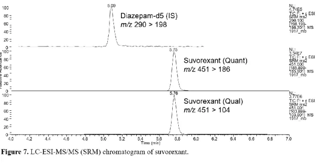
The LC-MS-MS product ion spectrum and chromatogram for suvorexant can be seen in Figures 6 and 7, respectively. A summary of the bias and precision of the quantitative method can be seen in Table III. The within-run CVs for blood and urine were 3-16% and 2-15%, respectively. The between-run CVs for blood and urine were 3-12% and 3-15%, respectively. Linear calibration curves were constructed using spiked blank whole blood
Page 12 of 23
or urine, or standard addition by spiking directly into the tissue homogenates. The average coefficient of determination (R2) for the blood and urine curves were 0.995 and 0.997, respectively. The R2 for the standard addition curves were >0.992 (0.992–0.996).
Carryover was not observed following the highest calibrator containing 500 ng/mL of suvorexant. Post-extraction addition for whole blood and urine was used to determine ME.
For whole blood, the mean ME was -13 and -12% at 20 and 200 ng/mL, respectively. The CV in whole blood was 5 and 3% at 20 and 200 ng/mL, respectively. For urine, the mean ME was -17 and -11% at 20 and 200 ng/mL, respectively. The CV in urine was 13 and 5% at 20 and 200 ng/mL, respectively. The average extraction recovery in whole blood and urine over 5 separate analyses at 100 ng/mL was 83 ± 7% and 92 ± 6%, respectively.
These are comparable to other recent publications (9-11). There were no interferences observed from either blank matrix or from 20 commonly detected analytes spiked into either matrix. LOD was 0.5 ng/mL in both whole blood and urine. LOQ was 1 ng/mL for both whole blood and urine. Stability at 4°C was confirmed after 6, 12, 18 and 24 h (92–
99%).
Quantitation results
The concentrations of suvorexant in the body fluids and tissues from all cases can be seen in Table IV. The concentration of suvorexant in the blood sources from Case 1 (421-491 ng/mL) suggested a level higher than the peak plasma levels reported from the recommended 10 or 20 mg oral dose (15-18). Sun et al. (16) studied the pharmacokinetics of suvorexant in healthy young men and reported that an oral dose of 50 mg resulted in an average peak plasma concentration of 392 ng/mL (range: 275-559 ng/mL). Based on
Page 13 of 23
police records, the decedent in Case 1 was prescribed 20 mg tablets of Belsomra, with instructions to take one tablet before bedtime in cases of insomnia.
The decedent in Case 2 was also prescribed 20 mg tablets of Belsomra to be taken once nightly. The suvorexant levels in the blood from Case 2 (11-17 ng/mL) were well below the reported peak plasma levels in any of the clinical studies that have been published. The concentration of suvorexant in the specimens of this case were much lower than in Case 1 (Table IV). A total of 16 drugs were found in the decedent’s postmortem blood, some at toxic levels, and thus the death was reported as combined drug intoxication.
The decedent in Case 3 also had a prescription for 20 mg tablets of Belsomra to be taken once nightly. The concentration of suvorexant in the blood of this case (138-155 ng/mL) were in between those of Cases 1 and 2 (Table IV). The cause of death in this case was determined to be hanging.
Discussion of tissue distribution
To our knowledge this is the first report of suvorexant being found in postmortem samples.
There are many factors that influence the discussion of postmortem toxicology findings in comparison to clinical studies. Interpretation of postmortem drug levels are often subject to postmortem artifacts, postmortem redistribution (PMR), pharmacogenomics and other factors (19). Thus, it is difficult to speculate on the amount of suvorexant consumed in these cases, and the effect on the deceased. More studies on suvorexant in postmortem samples are needed.
The distribution of suvorexant in the specimens of the three cases can be found in Table IV. Case 1 exhibited the highest levels of suvorexant, with the highest concentrations in the blood samples, followed by the fat, kidney and liver. Case 3 showed
Page 14 of 23
the highest level of suvorexant in the fat, followed by the kidney, liver, femoral vein blood and cardiac blood. Case 2 displayed the lowest concentration overall, with the highest level found in the lung, followed by the left heart blood, right heart blood, and femoral blood. While it is difficult to identify any significant pattern, it appears the drug readily accumulates in the blood, fat, kidney and liver. Blood/plasma (b/p) ratios have yet to be experimentally examined in humans, but with such high plasma protein binding (99%), it is reasonable to assume the b/p ratio would be around 0.5 or so (20). Plasma is often difficult to extract from postmortem whole blood, which was the case with these samples.
Suvorexant does not appear to exhibit marked PMR; central/peripheral (C/P) ratios ranged from 0.89 to 1.36 in these three cases. The Vd of suvorexant has been reported in several of the sources referenced in this publication, but some of them seem to disagree.
Yang et al. reports the Vd as 49 L, while Bennett et al. reports it as 105.9 L, and Sullinger et al. as 49 L/kg (2, 4, 11). The Vd is highly associated with the PMR that would be demonstrated in postmortem cases. For example, a Vd of 49 L/kg would be considered extremely high, and one would expect the result to be a high level of PMR. This is not what we saw in these cases, particularly with Case 1, where the highest levels were in the blood sources, and the C/P levels were close (right heart blood to femoral blood, 1.08).
In Case 2, the highest levels were in the fat, kidney, and liver, followed by the blood sources (C/P: 1.36). This would suggest a slighter higher level of PMR, but still not to the degree that 49 L/kg would predict. It is our estimation that the true value of Vd is somewhere in the range of 49 to 105.9 L, which is also more in agreement with the value reported in Baselt’s most recent edition of “Disposition of Toxic Drugs and Chemicals in Man” (11th edition, 2017), which lists the Vd as a range from 0.5 to 0.9 L/kg (20).
Page 15 of 23 Conclusion
A relatively new insomnia medication, suvorexant, was found in three recent forensic autopsy cases. Screening and confirmation was achieved by fast GC-MS and fast GC- MS-MS. A reference standard was procured, and quantitation performed on all collected specimens by a validated LC-MS-MS quantitation method. Although numerous metabolites have been identified in previous reports, none were detected in the GC-MS screening of this case. The lack of commercially available metabolites also made targeting of these compounds by LC-MS-MS prohibitive.
Suvorexant is similar to other sleep aids such as zolpidem, and may be found in more cases in Japan in the near future. It is possible that suvorexant may be used in cases of drug-facilitated sexual assault (DFSA), as is the case with zolpidem and other hypnotics. The forensic medicine and toxicology communities should be aware of and on the lookout for this compound in postmortem samples.
References
1. Merck & Co. (2014) Suvorexant Prescribing Information. http://www.merck.com/
product/usa/pi_circulars/b/belsomra/belsomra_pi.pdf (accessed 2017).
2. Yang, L.P. (2014) Suvorexant: First Global Approval. Drugs, 74, 1817-1822.
3. Sutton, E.L. (2015) Profile of suvorexant in the management of insomnia. Drug Design, Development and Therapy, 9, 6035-6042.
4. Bennett, T., Bray, D., Neville, M.W. (2014) Suvorexant, a dual orexin receptor antagonist for the management of insomnia. Pharmacy and Therapeutics, 39, 264- 266.
Page 16 of 23
5. Dubey, A., Handu, S., Mediratta, P. (2015) Suvorexant: The first orexin receptor antagonist to treat insomnia. Journal of Pharmacology & Pharmacotherapeutics, 6, 118-121.
6. Ali, S., Ahmed, Z., Ahmad, A., Khan, S.A., Husain, A. (2015) Pharmacological, pharmaceutical and safety profile of suvorexant: a dual orexin receptors antagonist for treatment of insomnia. International Educational Scientific Research Journal, 1, 26-30.
7. Cui, D., Cabalu, T., Lai Yee, K., Small, J., Li, X., Liu, B., et al. (2016) In vitro and in vivo characterisation of the metabolism and disposition of suvorexant in humans.
Xenobiotica, 46, 1-14.
8. Carson, M., Kerrigan, S. (2017) Quantification of suvorexant in urine using gas chromatography/mass spectrometry. Journal of Chromatography B, 1040, 289-294.
9. Breidinger, S.A., Simpson, R.C., Mangin, E., Woolf, E.J. (2015) Determination of suvorexant in human plasma using 96-well liquid-liquid extraction and HPLC with tandem mass spectrometric detection. Journal of Chromatography B, 1002, 254-259.
10. Iqbal, M., Ezzeldin, E., Khalil, N., Al-Rashood, S., Al-Rashood, K. (2017) Simple and Highly Sensitive UPLC-ESI-MS/MS Assay for Rapid Determination of Suvorexant in Plasma. Journal of Analytical Toxicology, 41, 114-120.
11. Sullinger, S., Bryand, K., Kerrigan, S. (2017) Identification of Suvorexant in Urine Using Liquid Chromatography-Quarupole/Time-of-Flight Mass Spectrometry (LC- Q/TOF-MS). Journal of Analytical Toxicology, 41, 224-229.
12. Hara, K., Waters, B., Ikematsu, N., Tokuyasu, T., Fujii, H., Takayama, M., et al.
(2016) Development of a preparation method to produce a single sample that can be
Page 17 of 23
applied to both LC-MS/MS and GC-MS for the screening of postmortem specimens.
Legal Medicine, 21, 85-92.
13. Waters, B., Hara, K., Kashiwagi, M., Matsusue, A., Sugimura, T., Hamasato, S., et al.
(2012) Combination of a short middle-bore capillary column with a thicker stationary phase and a short narrow-bore separation column with a thinner stationary phase for the rapid screening of non-volatile drugs by gas chromatography—mass spectrometry.
Forensic Toxicology, 31, 67-69.
14. Scientific Working Group for Forensic Toxicology (2013) Scientific Working Group for Forensic Toxicology (SWGTOX) Standard Practices for Method Validation in Forensic Toxicology. Journal of Analytical Toxicology, 37, 452-474.
15. Herring, W.J., Snyder, E., Budd, K., Hutzelmann, J., Snavely, D., Liu, K., et al. (2012) Orexin receptor antagonism for treatment of insomnia: a randomized clinical trial of suvorexant. Neurology, 79, 2265-2274.
16. Sun, H., Kennedy, W.P., Wilbraham, D., Lewis, N., Calder, N., Li, X., et al. (2013) Effects of Suvorexant, an Orexin Receptor Antagonist, on Sleep Parameters as Measured by Polysomnography in Healthy Men. Sleep, 36, 259-267.
17. Vermeeren, A., Sun, H., Vuurman, E., Jongen, S., Van Leeuwen, C., Van Oers, A., et al. (2015) On-the-Road Driving Performance the Morning after Bedtime Use of Suvorexant 20 and 40 mg: A Study in Non-Elderly Healthy Volunteers. Sleep, 38, 1803-1813.
18. Uemura, N., McCrea, J., Sun, H., Donikyan, M., Zammit, G., Liu, R., et al. (2015) Effects of the Orexin receptor antagonist suvorexant on respiration during sleep in healthy subjects. Journal of Clinical Pharmacology, 55, 1093-1100.
Page 18 of 23
19. Jones, G. R. (1998) Interpretation of postmortem drug levels. Drug Abuse Handbook, 970-985.
20. Baselt R.C. (2017) Suvorexant. Disposition of toxic drugs and chemicals in man, 11th Edition, Biomedical Publications, Seal Beach, CA, 2005-2007.
Legends to Figures and Tables
Table I. Chromatographic separation and mass spectrometry conditions for suvorexant Table II. Toxicology results from the three cases in this report, excluding suvorexant Table III. Bias and precision of the quantitative method
Table IV. Distribution of suvorexant in three autopsy cases N/A = not available.
Figure 1. Chemical structure of suvorexant.
Figure 2. Fast GC-EI-MS extracted ion chromatogram of suvorexant in the right heart blood of Case 1.
Figure 3. Full scan EI mass spectrum for suvorexant.
Figure 4. Fast GC-EI-MS/MS MRM chromatogram of suvorexant from the right heart blood of Case 2.
Figure 5. GC-EI-MS chromatogram of suvorexant using common screening conditions.
Figure 6. LC-ESI-MS/MS (SRM) product ion spectrum of a suvorexant shooting standard.
Figure 7. LC-ESI-MS/MS (SRM) chromatogram of suvorexant.
Page 19 of 23
Table I. Chromatographic separation and mass spectrometry conditions for suvorexant
Rt (min)
Prominent Ions (m/z)
Rt (min)
Prominent Ions (m/z)
Rt (min)
Precursor Ion (m/z)
Product Ions (m/z)
Collision Energy (eV)
Rt (min)
Precursor Ion (m/z)
Product Ions (m/z)
Collision Energy (eV)
104 18 186 26
77 24 104 54
GC-EI-MS/MS (fast) LC-ESI-MS/MS
5.75 451 5.25 186
23.7 186, 104, 221, 450
5.25 186, 104, 221, 450 GC-EI-MS (fast method) GC-EI-MS
(common method)
Page 20 of 23
Table II. Toxicology results from the three cases in this report, excluding suvorexant
Compound name Case 1 Case 2 Case 3
Cardiac blood Cardiac blood Cardiac blood
ng/mL ng/mL ng/mL
7-Aminoflunitrazepam 27 39
Biperiden 8
Bisoprolol 21
Chlorpromazine 159
N-Desalkylflurazepam 327
Diazepam 58
Etizolam 6
Flunitrazepam <5 <1
Ketoprofen <1
Nateglinide 68
Nifedipine 285
Olanzapine 776
Oxazepam 3
Promethazine 999
Quetiapine 150
Risperidone 55
Spironolactone 22
Triazolam 10
Valsartan 1,070
Zotepine 106
Page 21 of 23
Page 22 of 23
Page 23 of 23
Table III. Bias and precision of the quantitative method Nominal concentration
(ng/mL)
Calculated concentration (ng/mL, mean ± SD)
Bias (n = 15) (%)
Within-run precision (n = 5) (%)
Between-run precision (n = 15) (%)
3 2.9 ± 0.36 -3 6 12
100 97.7 ± 10.3 -2 16 11
400 401.7 ± 12.0 0.43 3 3
Urine 3 3.2 ± 0.36 7 2 11
100 99.5 ± 14.6 -0.5 15 15
400 401.3 ± 12.6 0.33 2 3
Whole blood
Table IV. Distribution of suvorexant in three autopsy cases
Specimen Case 1
ng/mL (g)
Case 2 ng/mL (g)
Case 3 ng/mL (g)
Right heart blood 455 15 138
Left heart blood 491 17 N/A
Left femoral blood 421 11 155
Urine N/A < 1 10
Liver 201 6 158
Kidney 280 4 232
Spleen 36 < 1 31
Pancreas 55 1 24
Lung 122 40 41
Muscle 93 <1 30
Fat 359 5 278
N/A = not available