鉄負荷によるラット実験肝癌の抑制
山下裕人,飯田芙佐枝,石井三和子 千馬正敬,板倉英世
長崎大学熱帯医学研究所病理学部門
The Inhibitory Effect of Iron-overload on the Chemically-induced Hepatocarcinogenesis in Rats.
Hiroto YAMASHITA, Fusae IIDA, Miwako ISHII, Masachika SENBA and Hideyo ITAKURA
(Department of Pathology, Institute for Tropical Medicine, Nagasaki University)
Abstract: Effects of iron-overload on the chemically-induced carcinogenesis of the rat liver were studied. The rats were given 2% of ferric amonium citrate in drinking water for 3 weeks, usual drinking water for 1 week and then 50 ppm diethylnitrosamine. The rats of the control group were given usual drinking water for 4 weeks and then 50 ppm diethylnitrosamine. Each paired group composed of treated rats and control rats given diethylnitrosamine for the same period was sacrificed simultaneously at the different expe- rimental periods. The hepatoma nodules and the size of each nodule were examined. Then the histopathological study of the liver was done. The comparison of the development of the hepatoma, estimated by the number and sizes of the nodules, were made in each paired group. Although 16 paired groups were tested, the comparison could be made only in 10 pairs. Nine pairs showed more developed hepatoma nodules in the control group than in the treated group and one pair showed the reversed result. Sign test was done and the calculated probability of the observed result was 0.0107. This result suggests that iron- overload inhibits the carcinogenicity of diethylnitrosamine in rats.
T
ropical Medicine, 22(3), 173-180, October,
は じ め に
我々は東アフリカ・ケニア国において種々の調査 を行い,この国が肝癌の多発地帯でありB型肝炎ウ ィルスや毒性真菌類の高浸産地帯であること,また 肝鉄沈着症が高率に存在することを報告してきた (長崎大学熱帯医学研究所病理学部門, 1980).この 鉄沈着症が肝に与える影響,特にそのfibrogenetic effectについては,それを肯定する報告(Bothwell and Bradlow, 1960)と否定する報告(Higginson
et al・, 1953)とがありまだ明確な結論は出ていな い・また人の特殊な肝硬変症である‑モクロマト‑
シス(肝硬変症+鉄沈着症)では一般の肝硬変症に くらべて肝癌の合併率が高い(Warren and Drake, 1950)ので鉄も肝発癌因子の一つと考えられてい る・しかし動物実験(Dunn, 1976, Yamamoto et al., 1971)や臨床的観察(Jacobs, 1979)では鉄 の肝発癌性に対して否定的な報告が多い・本研究で は,これら鉄が肝に与える種々の影響のうちで発癌 に対する影響に注目し鉄負荷が化学発癌に対し如何 長崎大学熱帯医学研究所業績 第1,008号
Received for publication, August 13, 1980
174
なる影響を及ばすかを調べた・そして鉄負荷が化学 発癌に対しむしろ抑制的に作用する結果を得たので
ここに報告する・
材料と 方法
動物:体重I50g前後のウィスター系のオスラッ トを用いた.飼料にほオリエンタル社製ラット用固
T
able 1. Number and size of cancer nodules observed in the rat treated with 2% iron and 50 ppm DEN
E
xperimental Duration of DEN No. of cancer Size of the each nodule
No. treatment (weeks) nodules (mm in diameter)
N
-lll 4 0
N-112 4 0
N-113 4 0
N-121 8 0
N-122 8 0
N-123 8 0
N-131 12 0
N-132 12 0
N-133 12 0
N-141 16 0
N-142 16 0
N-143 16 0
N-151 18 0
N-152 18 0
N-153 18 1 2
N-161 20 2 2, 2
N-162 20 1 3
N-163 20 3 2, 2
N-171 22 3 5, 2, 2
N-172 22 0
N-181 18 0
N-182 18 0
N-191 19 0
N-192 19 0
N-1101 20 0
N-1102 20 0
N-llll 21 0
N-1112 21 0
N-1121 22 0
N-1122 22 0
N-1131 23 6 10, 8, 5, 3, 2, 1
N-1132 died at 18 weeks of the DEN
treatment after begining
N-1141 24 0
N-1142 24 0
N-1151 25 0
N-1152 25 0
N-1161 26 5 20, 15, 10, 5, 5
N-1162 26 3 6, 1. 1
型飼料を使用した・
薬物:クエン酸鉄アンモニウム(石津)とジュテ ルニトロサミソ(DEN) (東京化成)を川いた・
実験方法:ラットを2%クエン酸鉄含有飲料水に て3週間飼育し, 1週間水道水にて飼育し,つぎに 50ppmのDEN含有水にて飼育したものを実験群
T
able 2. Number and size of cancer nodules observed in the rat liver treated with 50 ppm DEN
E
xperimental Duration of DEN No. of cancer Size of the each nodules
No. treatment (weeks) nodules (mm in diameter)
N
-211 4 0
N-212 4 0
N-213 4 0
N-221 8 0
N-222 8 0
N-223 8 0
N-231 12 0
N-232 12 0
N-233 12 0
N-241 16 1 4
N-242 16 0
N-243 16 3 2, 2, 2
N-251 18 0
N-252 18 2 9, 9
N-253 18 1 3
N-261 20 1 13
N-262 20 3 3, 3, 3
N-263 20 1 2
N-271 22 1 5
N-272 22 7 3, 3, 3, 3, 3, 2, 2
N-281 18 0
N-282 18 0
N-291 19 0
N-292 19 0
N-2101 20 0
N-2102 20 2 1, 1
N-2111 21 0
N-2112 21 0
N-2121 22 0
N-2122 22 1 15
N-2131 23 1 2
N-2132 23 0
N -2141 24 1 2
N-2142 24 1 3
N-2151 25 2 40, 20
N-2152 25 1 2
N-2161 26 7 28, 20, 15, 10, 10, 10, 10
N-2162 26 4 5, 5, 5, 2
176
とした.対照群ほ4週間水道水にて飼育し5週間目 から実験群と同様にSOppmのDEN含有水を与え た,つぎにTable 1, Tab①e2に示す間隔で動物を 屠殺した・同一時期に屠殺した実験群と対照群を対 として観察した・すなわち屠殺された個々のラット の肝における結節の個数,及びその結節の大きさを 調べ,各対問にて,実険群と対照群とについて癌の 発育生成度を比較した・つぎに肝の病理組織標本を 作成し組織像を調べた・
結 果
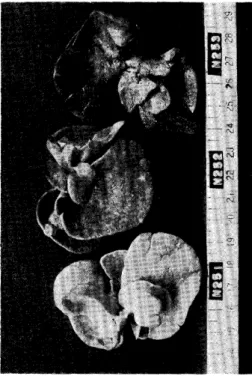
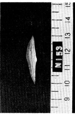
Table lに2%鉄とSOppmのDEN で飼育され た実験群のラットの肝に生じた癌結節の数と個々の 結節の大きさを, Table 2にSOppm DEN のみで 飼育された対照群ラットの肝の癌結節の数と個々の 結節の大きさを示した.代表的な癌結節の肉眼像を Figン l‑Fig. 4に,光顕像をFig・ 5‑Fig・ 7に 示してある・癌結節以外にも約1mmの黄白色砂粒 の病変や白斑,血腫の形成等がみられた・しかし,
これらの病変は癌でほないので考慮に入れていな い・また組織学的にほ痛か非痛かの判別がむずかし い組織もあるので肉眼的にはっきりとした結節を形 成し組織学的に癌と確認されたもののみを取りあげ た,また癌組織の構造異形,細胞異形からみた悪性 度の差ほ無視した・癌結節が最初に見出されるのは 実験群でほDEN 投与開始後18週目であり3匹中 の一匹にみとめられるのに対し対照群ではDEN投 与開始後16週目であり③匹中2匹にみとめられ対照 群の方が早く発癌している・つぎに同一時期に屠殺 された実験群と対照群のラット肝癌の進行度を比較 した.癌結節が肝を占める割合が大きいものほど痛 が早く進行しているものとした・ Table 3にその結 果を示す・ 16組の対のうち6組ほ判定不能であっ たン 判定可能であった残り1o組のうち9組は対照 群の方が癌の進行度が早く,残り1組は逝の結果 を示した・ この結果に基づいて符号検定を行うと PH(S, ≧9) ‑Pn(Sl ≦1) ‑0・0107となり,実験群 と対照群の間にほ,癌の進行度において有意の差が
Fig. 1. Macroscopic appearance of livers of one experimental-group-rats. In this group only one nodule seen in Fig. 3 was counted as the cancer nodule.
Fig・ 2・ Macroscopic appearance or the rat
①iver of the control group paired with
the group seen in Fig. 1・ In this
group, 3 nodules were counted as the
cancer nodules.
Fig. 3. Cross section of the liver lobe having the largest nodules in this experimental group.
Fig. 4. Cross section of the liver lobe having the largest nodules in this control group.
Fig. 5. Microscopic appearance of the cancer nodules seen in Fig, 3.
Hepatocellular carcinoma is seen in the right side of this picture.
Slightly unclear distinction between carcinoma tissue and normal
tissue. x200, H. E. stain.
178
Fig. 6. Microscopic appearance of the cancer nodule in Fig. 4. Hepato- celluar carcinoma occupies right side of this picture. Clear distinction between carcinoma tissue and normal tissue.
xlOO. H. E. stain.
Fig. 7. Reticulin stain reveal clear-cut distinction between carcinoma tissue and normal tissue. x200, Reticulin stain.
あり,実験群の方が癌の進行が遅れており,したが って鉄負荷が発癌に抑制的に作用するものと考えら れた・
考 案
鉄ほtissue cultureにおいて細胞障害性のある こと(Richmond, 1961) ,その作用はchemical carcinogenに類似していること(Vasiliev and Guelstein, 1963),ラットの皮下に肉腫を形成する ことが報告されている. (Richmond, 1959)・また
犬を用いての実験で鉄の大量長期投与によって人の へモクロマト‑シスに類似した病変を生じさせたと の報告もある(Lisboa, 1971)・ Jacobs (1977)は 頻回の輸血で鉄が大量に負荷された患者に肝癌が発 生した報告がないと述べ,また‑モクロマト‑シス の患者から治療によって鉄を抜き取っても,肝癌の 発生率は,未治療の患者の肝癌発生率と有意の差が なく(Powell, 1970),またはむしろ高い(Bomford and Wi①Hams, 1966)・これらのことから鉄そのも
のには肝発癌性はないと考えられる.
T
able 3. Comparison of the development of cancer nodules in each group between controls (DEN only) and treated (iron and DEN)
Paired groups Direction of o-
treated. control*1 difference*2 1Ön
N
-ll and N-21 N-ll =N-21 0
N-12 and N-22 N-12 =N-22 0
N-13 and N-23 N-13 =N-23 0
N-14 andN-24 N-14<N~24 +
N-15 andN-25 N-15<N-25 +
N-16 andN-26 N-16<N-26 +
N-17 andN-27 N-17<N-27 +
N-18 andN-28 N-18 =N-28 0
N-19 andN-29 N-19 =N-29 0
N-110and N-210 N-110<N-210 +
N-lll and N-211 N-111=N-211 0
N-112and N-212 N-112<N-212 +
N-113andN-213 N-113>N-213 -
N-114and N-214 N-114<N-214 +
N-115and N-215 N-115<N-215 +
N-116and N-216 N-116<N-216 +
SI Rats of paired groups were treate with DEN for the same period and sacrificed at the same day.
*2 Comparison of cancer development was based on the number and size of cancer nodules observed in each group.
Willson (1977)ほ鉄がfree‑radical反応を触媒 し,その反応が病原性物質を活性化すると述べ鉄が co‑carcinogen として作用する可能性を示唆した.
Dunn (1967)は etnionine と鉄負荷併用群と ethionine単独投与群との比較において肝癌の発生 率に有意の差がない事を報告した・ Yamamoto ら (1971)ほ肝に急速に鉄沈着症を起す8‑Hydroxyー quinolineをN・2‑fluorenylacetamideと併用投与 すると肝癌の発生率がN‑2‑fluoreny①acetamide 単独投与群よりも低くなることを報告し鉄負荷が発 癌過程に抑制的に働くと述べている.本実験でもク エン酸鉄アンモニウム投与後,肝細胞ならびにクッ パ‑細胞内に鉄が沈着していることを確認しており 鉄負荷が発癌過程に抑制的に作用していると考えら
れる・この結果は‑モクロマト‑シスでほ他の肝枚 '文六よりも発S!率が高い(Warren and Drake, 1950, Shikata et a①, 1977)という報告と矛盾しているよ
うに思わ/れる・じかし原因別からみた肝硬変桔では 萎縮性肝硬変(Purtilo et al, 1973)や肝炎性肝触 餐(Shikata et al., 1977)の方が, ‑モクロマ ト‑シスよりも肝癌の合併率が高い・ MacSween (1974)は‑モクロマト‑シスの患者のほとんどが 男性である(MacDonald and Mallory, 1960)とこ ろから,肝硬変患者の男性の肝癌発生率と‑モクロ マトーシスの患者の肝癌発生率とを比較したところ 両者に有意の差ほみとめられなかったと報告してい る・肝癌を合併した‑モクロマト」シス患者の剖検 年令はWarren and Drake (1950)によれば,最 若年者で59才,最高令老で79才,平均65・5才であり 日本での肝癌を合併した肝硬変患者の剖検年令が4o 才台から5o才台にかけてピークに達する(松下, 1970)ことと比べると, ‑モクロマト‑シスでほ癌 の発育がかなり遅れるものと考えられる.これも鉄 沈着が発癌に抑制的に作用する為であると思われる.
鉄負荷が肝発癌を抑制する機構は不明である.し かし,肝鉄沈着症と肝癌の合併例でほ鉄ほ組織学的 に正常の組織に認められるが,癌結節ならびに前 痛結節にほ染め出されないことが知られている (Wil①iams and Yamamoto, 1972)・また肝癌患者 では血中にferritin (含鉄蛋白)が増加することも 知られている(高彼ら, 1976) ・肝癌細胞が鉄を取 り込むことほすでに報告してある(Yamashita, 1979),したがって癌細胞ほ正常細胞に比べて比較 的容易に鉄を細胞外へと放出するものと考えられ る・鉄ほ細胞内でほFe4 の形で存在しFe4 の形 で細胞外へ放出される(Underwood, 1975).鉄が 電子を「個受け取って細胞外‑放出されることが 発癌の抑制と関係があるのかも知れない(Szen十 Gyorgyi, 1979).我々ほ鉄が前癌結節においても染 め出されないことから,その機構ほ不明であるが, 鉄の存在自体が発癌を抑制するものと考えている.
すなわち鉄を負荷された細胞が癌化する為にほ,質 ず鉄を細胞外に排出することが必要であり, Lたが って,それだけ発癌がおくれるのではないかと考え ている.
18o
文 献
1) Bomford, A., & Williams, R・ (1976): Long term results of venesection therapy in idiopathic
hemochromatosis・ Q・ J・ Med・ 45, 611‐623・
2) Bothwe①1, Tン H・, & Bradlow, B・ A・ (1969) : Siderosis in the Bantu・ A combined histopatho・
①ogical and chemical study・ Arch Path., 70, 279一292・
3) Dunn, W・ L・ (1967): Iron‑loading, fibrosis and hepatic carcinogenesis・ Arch Path・, 83, 258‑
266・
4) Jacobs, A. (1977): Ciba foundation sym. Iron Metabolism. 51, 351・
5) Higginson, J・, Gerristen, T・, & Walker, A・ R・ P. (1953): Siderosis in the Bantu of Southern Africa. Am・ J・ Path., 29, 779‑815・
6) Lisboa, P・ E・ (1971): Experimental cirrhosis in dogs caused by chronic massive iron overload.
Gut, 12, 363‑318・
7) MacDona①d, R・ A・, & Mallory, G・ K. (1960) : Hemochromatosis & hemosiderosis・ Study of
211 autopsied cases. Arch. Internン Med・ 105, 686‑700・
8) MacSween, R. N. M. (1974): A clinico‑pathological review of 100 cases of primary malignant tumor of the liver. J・ Clin・ Pathol・, 27, 669・692・
9)松下 寛(1975) :日本の肝硬変と肝癌.臨床科学, ll, 275‑286・
1o)長崎大学熱帯医学研究所病理学部門(1980) :東アフリカ・ケニア共和国における肝臓疾患の疫学と病理.
文部省科学研究費海外学術調査報告書.
ll) Powe①1, L. W・ (1970): Tissue damage in haemochromatosis: an analysis of the roles of iron and
alcoholism・ Gut, ll, 980・
12) Purtio, D・ T・, & Gott①ieb, L・ S・ (1973): Cirrhosis and hepatoma occuring at Boston City Hos‐
pital・ (1971"1968)・ Cancer, 32, 458‑462.
13) Richmond, H・ G・ (1995): Induction of sarcoma in the rat by iron‑dextran complex. Br・ Med・
J・ ll, 947・949・
14) Richmond, HンG. (1961): The toxic effects of iron‑dextran complex on mammalian ce①Is in tissue
culture. Br. J. Cancer, 15, 549‑606.
15) Snikata, T. , Yamazaki, S・ Uzawa, T・ (1977): Hepatocellular carcinoma & chronic persistent hepatitis・ Acta Pathol・ Jpn・, 27, 297‑3o4.
16) Szent‑Gyorgyi, A. (1979): Ciba foundation symp. Submolecular biology and cancer, 52, 3‑18.
17)高後 裕,新津洋司郎,渡辺直樹,大塚 忍,小閑純ラ 柴田恵子,漆崎一朗(1976):血清ferritinの radioimmunoassay法とその消化器疾患における臨床的応用に関する研究・日・消・病・誌, 73, 75‐88・
18) Underwood, E・ J.,日本化学会訳編(1975):微量元素一栄素と毒性‑, 15ー56,丸善
19) Vasiliev, J・ , & Guelstein, V・ (1963)‥ Sensitivity of normal and neoplastic cells to the damaging
action of carcinogenic substances: A review. J・ Nat①・ Cancer lnst・ , 1123・1143.
20) Warren, S., & Drake, W. L. (1950) : Carcinoma of the liver in hemochromatosis. Am. J.
Pathol・, 27, 573‑609・
21) Wi①①iams, G・ M・, & Yamamot。, R・ S・ (1972) : Absence of stainable iron from preneoplastic
and neoplastic lesions in rat liver with 8‑Hpdroxyquinoline‑induced siderosis. J. Natl. Cancer lnst., 49, 685‑692.
22) Willson, R. L. (1977): Ciba Foundation sym. Iron Metabolism, 51, 331‑334.
23) Yamamoto, R・ S・, Williams, G・ M・, Frankel, H・ H・, & Weiburger, J・ H・ (1971)‥ 8‑Hy‑
droxyquinoline‥ chronic toxicity and inhibitory effect on the carcinogenicity of N・2‑F①uorenyl acetamide・ Toxicology and app①led Pharmacology, 19, 687‐698・
24) Yamashita, H. (1979): Radio‑iron uptake of rat hepatoma・ Gann, 70, 593‑6oo・