Basic fibroblast growth factor (FGF-2) promotes angiogenesis and bone regeneration in rat calvarial bone defects
Risa Kigami
Nihon University Graduate School of Dentistry, Major in Periodontology
(Directors: Prof. Bunnai Ogiso and Assoc. Prof. Shuichi Sato)
Table of Contents
Page
1 Abstract
3 Chapter 1: Effect of basic fibroblast growth factor on angiogenesis and bone regeneration in non-critical-sized bone defects in rat calvaria
8 Chapter 2: FGF-2 angiogenesis in bone regeneration within critical-sized bone defects in rat calvaria
12 Conclusions
12 Acknowledgements
13 References
16 Tables
17 Figures
1
Abstract
Basic fibroblast growth factor (FGF-2) is a strong inducer of angiogenesis. Angiogenesis is a fundamental process for osseous formation or regeneration, and an early vascular response is the progression of fracture healing. FGF-2 is reported also to increase the number of osteoblasts and chondroblasts in vitro. Furthermore, FGF-2 induced differentiation and proliferation of mesenchymal stem cells in periodontal ligaments, resulting in periodontal tissue regeneration. Recently, the multicenter study for periodontal tissue regeneration using FGF-2 has been performed in Japan. FGF-2 applied to into the bone defects maybe a reasonable approach for engineering bone constructs to induce bone regeneration. The objective of this study was to evaluate the effect of FGF-2 on angiogenesis and bone regeneration in rat calvarial bone defects using in vivo micro computed tomography (micro-CT) and histological sections.
In the first chapter, the effect of angiogenesis preceding bone repair withFGF-2 within non-critical-sized bone defects in rat calvaria. Non-critical-sized bone defects (each 2.7 mm in diameter) were trephined into the parietal bone on each side of the sagittal suture. An absorbable collagen sponge (ACS) permeated with saline or with FGF-2 (0.1% or 0.3%) was placed on each side. The rats were assigned to the following treatments: control group (saline), 0.1% FGF-2 group, 0.3% FGF-2 group. Micro-CT and histological sections were used to obtain newly generated blood vessels and bone. Blood vessel volume (BVV) and bone volume (BV) were calculated. In micro-CT analysis, blood vessels began to appear newly around the edges of bone defects on day 7 in FGF-2 groups. Newly generated bone appeared on day 14. Newly generated blood vessels and bone filled the bone defects on day 28 in FGF-2 groups. On the other hand, newly generated blood vessels and bone filled about a half of the bone defects in the control group. BVV and BV gradually increased until day 28 in all groups. BVV and BV were significantly greater in the 0.3% and 0.1% FGF-2 groups than in the control group, and greater in the 0.3% FGF-2 group than in the 0.1% FGF-2 group, on days 14, 21 and 28. In histological sections, the number of new blood vessels were more in the FGF-2 groups than in the control group on day 28. On day 28, as compared with the 0.1%
FGF-2 group, the number of new blood vessels and the area of generated bone were more in the 0.3% FGF-2 group. Newly generated bone formed near the defect margins by day 28 in both FGF-2 groups, and new bone achieved better closure than control group.In addition, as compared with the control group, there were more osteoblast- and osteoclast-like cells in the FGF-2 groups on day 28.These observations indicate that, FGF-2 enhanced angiogenesis and bone regeneration.
In the second chapter, the effect of angiogenesis preceding bone repair withFGF-2 within critical-sized bone defects in rat calvaria. Experimental designs and data analyses were carried
2
out as described in chapter 1. Critical-sized bone defects (each 5 mm in diameter) were created in rat calvaria. In micro-CT analysis, blood vessels began to appear newly around the edges of bone defects on day 14 in FGF-2 groups. New blood vessels filled about two thirds of the bone defects in FGF-2 groups, and newly generated bone was observed only a little in defects margins on day 28. In control group, blood vessels began to appear newly around the bone defects on day 21 and increased until day 28, and newly generated bone was not observed. BVV and BV gradually increased until day 28 in all groups. BVV and BV were significantly greater in the 0.3% and 0.1% FGF-2 groups than in the control group, and greater in the 0.3% FGF-2 group than in the 0.1% FGF-2 group, on days 21 and 28. In histological sections, the number of new blood vessels were more in the FGF-2 groups than in the control group on day 28, and the number of new blood vessels and the area of generated bone were more in the 0.3% FGF-2 group than in the 0.1% FGF-2 group. Newly generated bone formed near the defect of margins by day 28, but no significant differences were found in defect closure in the FGF-2 groups. The number of osteoblast- and osteoclast-like cells increased in the FGF-2 groups by day 28. These results suggest that FGF-2 promotes angiogenesis and the subsequent bone regeneration.
Within the limitation of the present study, local administration of FGF-2 to rat calvarial bone defects induces angiogenesis and stimulates regenerative bone repair.
3
Chapter 1: Effect of basic fibroblast growth factor on angiogenesis and bone regeneration in non-critical-sized bone defects in rat calvaria
Risa Kigami, Shuichi Sato, Noriko Tsuchiya, Nobuaki Sato, Daigo Suzuki Yoshinori Arai , Koichi Ito, Bunnai Ogiso Journal of Oral Science 2014 (in press)
Introduction
Several growth factors are attracting interest because of their ability to actively regulate various cellular functions in angiogenesis and bone regeneration (1). Basic fibroblast growth factor (FGF-2) is a highly potent mitogen for mesenchymal cells and induces angiogenesis, which is essential for normal wound healing (2,3). In addition, in vivo studies have demonstrated that topical exogenous FGF-2 enhances healing of bone fractures (4,5).FGF-2 has been applied in periodontal tissue regeneration, and it is considered to be highly appropriate to be using bone regeneration (6).
Bone is a highly vascularized tissue that remodels and repairs itself. New bone regeneration is facilitated by angiogenesis; ie, the formation of new microvessels from existing vessels (7,8). Angiogenesis and osteogenesis are important factors in orthopedic healing. Angiogenesis is an important process in bone formation and repair, and early vascular response is essential in fracture healing (9,10).
In this study, the effect of angiogenesis-driven bone reconstruction was examined in the presence of FGF-2 in rat calvarial non-critical-sized bone defects using in vivo micro computed tomography (micro-CT) and histological sections.
Materials and Methods
Animals
Forty male Fischer rats (weight 130-160 g) were housed in cages in an animal room (temperature, 22°C; humidity, 55%; 12/12-h light/dark cycle), with free access to food and water. The Animal Experimentation Committee of the Nihon University School of Dentistry, Tokyo, Japan approved this study (AP11D006).
Surgery for animals
Sodium pentobarbital (somnopentyl, 0.5 mL/kg body weight; Schering-Plough, Munich,
4
Germany) was administered intramuscularly as general anesthesia, the skin of the surgical area was cleaned with 70% ethanol, and 0.5 mL lidocaine (xylocaine, 1:8 dilution; Astra Zeneca, Osaka, Japan) was injected intraperitoneally.
The dorsal cranial skin was incised along the sagittal suture. Two bone defects (width, 2.7 mm) were made in the dorsal cranial skin parietal bones on both sides of the sagittal suture.
The sagittal suture was not included in the bone defects, to eliminate any effects it might have on bone healing. The surgery was performed carefully, to avoid injuring the dura mater and sagittal sinus.
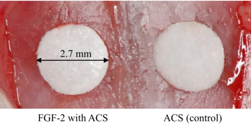
After washing out all bone fragments, an absorbable collagen sponge (ACS, Teruplug;
Terumo Co., Tokyo, Japan) with or without FGF-2 was implanted into both bone defects (Fig.
1) and the skin was closed with silk sutures (Ethicon, Somerville, NJ, USA). The day of surgery is described as day 0.
Preparation of ACSs with and without FGF-2
FGF-2 (Kaken Pharmaceutical Co. Ltd. Tokyo, Japan) was frozen and dried. Each ACS was filled with 0.1% or 0.3% FGF-2. Control ACSs were prepared using 20 μL of aseptic saline solution (Fig. 1).
Contrast perfusion by micro-CT
Ten rats were perfused with iopamidol radiographic contrast medium (Iopamiron; Bayer HealthCare AG, Leverkusen, Germany) on days 7 to 28. The front limbs were incised to the xiphoid process. One side of the sternum was cut using scissors, and the rib cage was retracted laterally. The descending aorta was clamped, and an angiocatheter was used to penetrate the left ventricle. The inferior vena cava was incised, and 20 mL heparinized saline (100 U/mL at 2 mL/min) was immediately perfused using a syringe pump. After perfusion with aseptic saline, the iopamidol solution was perfused to euthanize the animals. The perfusion was administered slowly, to reduce animal suffering.
Imaging system
Imaging was performed using the in vivo micro computed tomography (micro-CT) (R_mCT;
Rigaku, Tokyo, Japan) equipped with a microfocus X-ray tube with a focal point of 7 µA (L9181S; Hamamatsu Photonics, Hamamatsu, Japan) and an X-ray sensor with a 10.16 cm image intensifier. The X-ray source and image intensifier were connected by a basal plate, and the I-arm rotated in a vertical plane, driven by a direct-drive motor. Images of the areas of interest were captured. Repeated micro-CT imaging was performed from day 0 until euthanasia.
5
Micro-CT analysis
Micro-CT was used to assess bone regeneration and angiogenesis in specimens. Blood vessel formation was quantified based on perfusion of the vasculature with iopamidol. The exposure parameters were 90 kV and 88 µA. On day 0, we positioned a screen cylinder to overlap the initial bone defect. Measurement was performed in blood vessel volume (BVV) and bone volume (BV) within the cylinder on voxel images, using software (Kitasenjyu Radist Dental Clinic, i-View Image Center, Tokyo, Japan) that calculates gray values and corresponding numbers of voxels in regions of interest. A histogram of the X-ray absorption rate (x-axis) versus CT voxel number (y-axis) was constructed for the CT field of view. Histograms of the X-ray absorption rate showed peaks for hard and soft tissues, and the threshold was set at the value representing the trough between these peaks. The number of voxels that exceeded the threshold for the X-ray absorption rate was counted. Change in BVV was calculated by subtracting the value obtained from an image taken before angiography from that obtained from angiographic images of the bone defect each week. The space ratio filled with the BVV was measured for each bone defect (Fig. 2). BV was calculated as the number of bone-associated voxels multiplied by voxel volume. BVs in the regions of interest were measured weekly, under the same conditions, starting on day 0. BV gain was calculated by subtracting the day 0 value from each subsequent value. The defect reossification ratio was evaluated on days 7, 14, 21 and 28.
Tissue preparation and light microscopy
The experimental and control sites were evacuated, along with the surrounding bone and soft tissue, and fixed in 10% formalin on day 28. The specimens were then decalcified by soaking them in a 10% formic acid-sodium citrate solution and embedded in paraffin. The sections were cut through the center of the bone defects and stained with hematoxylin and eosin.
Histological sections were observed under a light microscope connected to a personal computer. Defect closure rate was defined as the distance between the margins of the defect and is presented as the percentage of total defect width. Newly generated bone (%) was defined as all tissues within the boundaries of the newly generated bone (Fig. 3).
The average numbers of osteoblast- and osteoclast-like cells in the new bone area were counted manually under light microscopy at 100× magnification.
Statistical analyses
The Kruskal-Wallis one-way analysis of variance by ranks test was used to assess reossification and BVV values obtained over time. A P value of < 0.05 was considered to indicate statistical significance.
6
Results Micro-CT analysis
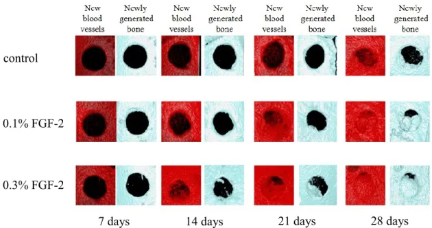
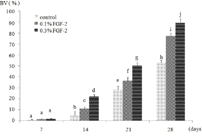
New blood vessels began to appear around the edges of bone defects on day 7, and BVV increased until day 28 in all groups. BVV and BV were significantly greater in the 0.3% and 0.1% FGF-2 groups than in the control group, and in the 0.3% FGF-2 group versus the 0.1%
FGF-2 group, on days 14, 21, and 28 (Figs. 4-6).
Light microscopy
Figure 7 shows histological sections of calvarial bone defects at 28 day after surgery. More new blood vessels were observed in the FGF-2 groups than in the control group on day 28. On day 28, as compared with the 0.1% FGF-2 group, more new blood vessels and generated bone were observed in the 0.3% FGF-2 group, and newly generated bone achieved better closure.
Bone regeneration induced by ACS with FGF-2 began at the edge rather than the center of the defect.
Histomorphometric analysis
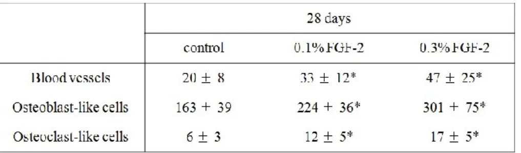
There was significantly more newly generated bone in the FGF-2 groups than in the control group (Table 1). In addition, as compared with control there were more new blood vessels and osteoblast- and osteoclast-like cells in the 0.1% and 0.3% FGF-2 groups on day 28. Numerous osteoclast-like cells were observed in the 0.3% FGF-2 group on day 28 (Table 2).
Discussion
Bone defects larger than a certain, or "critical," size cannot be repaired via bone augmentation (11). Non-critical-sized bone defects models are spontaneous closure and repairable. Udagawa et al. (12) analyzed angiogenesis-driven bone repair in both critical-sized and non-critical-sized defects and suggested that capillary bed formation is important in understanding the difference in bone regeneration between critical-sized and non-critical-sized bone defects. The present study showed that new blood vessels began to appear around the edges of bone defects on day 7 after surgery in the FGF-2 groups. The capillary bed was formed on day 14 in the 0.3% FGF-2 group. On day 21, capillary bed formation was observed in both the 0.3% and 0.1% FGF-2 groups. The capillary bed connected one side of the defect to the other in the FGF-2 groups at a very early stage, on day 21. Blood vessel and capillary bed formation occurred earlier than in critical-sized defects.
Capillary bed connection accelerates blood flow, thus facilitating bone regeneration (7).
7
Newly generated bone was observed at the edge rather than the center of defects, probably because precursor cells of osteoblasts are necessary to initiate bone formation.
Precursor cells in the periosteum differentiate into osteoblasts to induce bone formation (13).
In this study, bone repair began from the periosteum, at the outer and inner surfaces near the defect, not from the surface of the bony edge around the original defect. Osteoblasts and their progenitors in the periosteum on both sides participated in forming projections of regenerated bone. FGF-2 induces angiogenesis in the periosteum.
A collagen carrier is used as a scaffold, because collagen-based biomaterials are effective delivery vehicles for FGF-2. Collagen is also a good carrier for tissue regeneration. Murakami et al. (14) showed that FGF-2 delivered in a collagen or β-tricalcium phosphate collagen matrix stimulated healing in canine long bones.
The previous studies investigated the effects of other growth factors on animals in a GBA (guided bone augmentation) model (15,16). However, the characteristics of bone in those studies were inadequate for implant placement. Recently, the effect of local FGF-2 application on periodontal regeneration was investigated in a multicenter randomized clinical trial. The results indicated that FGF-2 was effective in periodontal tissue regeneration. Therefore, the use of FGF-2 may be an effective technique for bone regeneration in implant placement. A future study should examine the effects of FGF-2 in a GBA model.
In summary, FGF-2 induced angiogenesis and stimulated bone repair; its introduction into non-critical-sized bone defects may be a suitable means of inducing rapid bone regeneration in conjunction with scaffolds that facilitate bone construction.
8
Chapter 2: FGF-2 angiogenesis in bone regeneration within critical-sized bone defects in rat calvaria
Risa Kigami, Shuichi Sato, Noriko Tsuchiya, Tomohiro Yoshimaki, Yoshinori Arai, Koichi Ito Implant Dentistry 22, 2013, 422-427.
Introduction
Bone augmentation procedures are required for the treatment of many bone defects. Guided bone regeneration, a bone augmentation technique, has been employed to provide sufficient bone around defects (17-19). Bone is a highly vascularized tissue with a unique ability to remodel and repair itself. Restoration is achieved by the formation of new microvessels from existing vessels, termed angiogenesis (8,9). Angiogenesis and osteogenesis have been established as important factors in skeletal healing. Angiogenesis is a fundamental process for osseous formation and repair, and an early vascular response is essential for the normal progression of fracture healing (7).
FGF-2 belongs to the fibroblast growth factor family, a polypeptide family responsible for controlling proliferation and differentiation of various cell types (2). FGF-2 is one of the most effective osteogenic and angiogenic growth factors involved in recruiting mesenchymal and osteogenic cells. It promotes angiogenesis and supports bone formation, thereby accelerating bone healing (20). Because FGF-2 induces angiogenesis and stimulates bone repair, its introduction into a bone defect may be a suitable approach to induce rapid bone regeneration with scaffolds for the engineering of bone constructs.
In this study, the dynamics of angiogenesis preceding regenerative bone repair with FGF-2 was examined in rat calvarial critical-sized bone defects using micro-CT and histological sections.
Materials and methods
Animals
Forty 7-week-old male Fischer rats, weighing 130-160 g each, were used. The animals were housed in same condition as the chapter 1. The Animal Experimentation Committee of the Nihon University School of Dentistry, Tokyo, Japan approved this study.
9
Surgical procedure
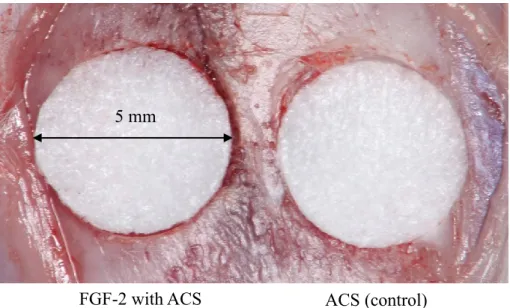
Surgical procedure was carried out as described in chapter 1. Symmetrical critical-sized bone defects (5 mm in diameter) were created in the dorsal parietal bones on each side of the midsagittal suture. ACS with or without FGF-2 was then implanted into each bone defect (Fig. 8).
Surgical procedure Preparation of ACSs with and without FGF-2
Preparation of ACS with or without FGF-2 carried out as described in the chapter 1.
Imaging system
Micro-CT system was used as described in the chapter 1. Repeated micro-CT imaging was performed on 7, 14, 21 and 28 days after surgery.
Micro-CT analysis
Micro-CT analysis was used as described in the chapter 1.
Histological analysis
Histological analysis was carried out as described in the chapter 1.
Statistical analysis
The Statistical analysis was carried out as described in the chapter 1.
Results
Micro-CT analysis
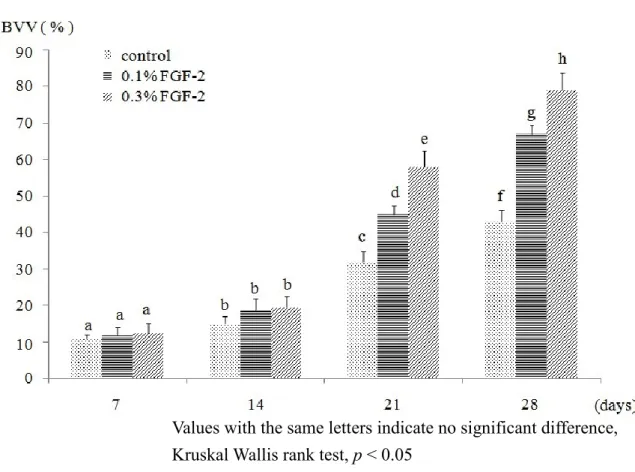
New blood vessels began to appear around the edges of bone defects on day 14 after surgery, and BVV increased until day 28, in the FGF-2 groups. In the control group, new blood vessels began to appear around the bone defects on day 21 and increased until day 28 (Fig. 9). BVV was significantly greater in the 0.3% FGF-2 groups compared with the control group on days 21 and 28 (Fig. 10). On day 28, BV was significantly greater in the 0.3% FGF-2 groups compared with the control group (Fig. 11). In the control groups, little new generated bone was observed in the part of defect margins on day 28.
Histological analysis
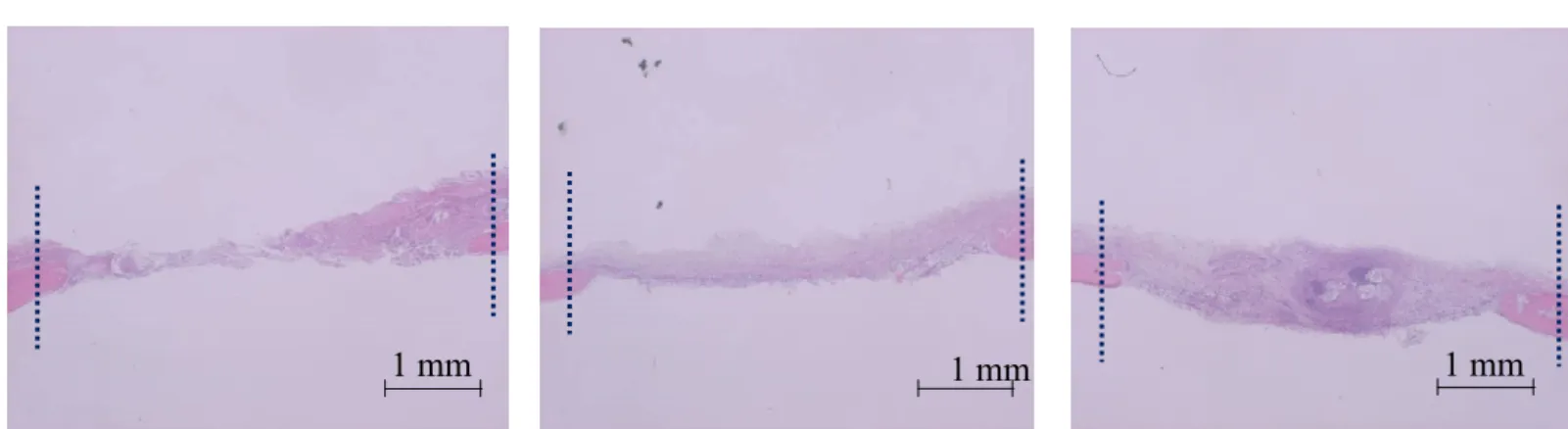
More new blood vessels were observed in the FGF-2 groups than in the control group on day 28 after surgery. On day 28, more new blood vessels and generated bone were observed in the
10
0.3% FGF-2 group than in the 0.1% FGF-2 group (Fig. 12).
Histomorphometric analysis
Newly generated bone areas were significantly increased in the FGF-2 groups compared to control groups. However, no significant difference was found in defect closure on day 28 (Table 3). New blood vessel formation and the presence of osteoblast- and osteoclast-like cells increased in the FGF-2 groups by day 28. Many osteoclast-like cells were observed in the 0.3% FGF-2 group on day 28 (Table 4).
Discussion
The previous studies showed that FGF-2 enhanced angiogenesis and bone regeneration in non-critical sized bone defects (12, 20, 21). Results of the latter study showed that angiogenesis in non-critical sized bone defects led to full bone formation. The critical-sized bone defect is a well-established model for the study of bone augmentation (11). Therefore, the effects of anigiogensis-driven bone repair were compared in the presence of FGF-2 in rat critical-sized bone defects.
Norrby (22) and Kodama et al. (23) reported that FGF-2 caused angiogenesis in a dose-dependent manner in rats. In this study, it was confirmed that 0.3% FGF-2 induced more blood vessel formation than 0.1% FGF-2 in critical-sized bone defects in rats. New blood vessels began to appear on day 14 in the FGF-2 groups, and BVV increased significantly on day 21. Kobayashi et al. (24) found many new blood vessels in the connective tissue of bone defects at FGF-2-treated sites during the early observation period (7-14 days). This discrepancy is likely attributable to differences in the bone defect size or scaffolds.
BV was significantly greater in the 0.3% FGF-2 group compared with the control group.
FGF-2 has been shown to promote the proliferation of endosteal osteoblasts and to accelerate bone formation (4, 25). Among all experimental groups, the 0.3% FGF-2 group showed the greatest increase in BV. These results were confirmed in histological sections. In a clinical trial of periodontal regeneration, bone height was significantly greater in the 0.3% FGF-2 group compared with the placebo group (26). However, this is difficult to explain why the total BV was very small. Further studies are needed to confirm this result. BV was significantly higher in the FGF-2 groups compared with the control group on day 28, and BVV increased significantly in the FGF-2 groups on day 21. Thus, bone regeneration was induced after angiogenesis.
Many new blood vessels and osteoblast- and osteoclast-like cells were observed in histological sections in the FGF-2 groups, especially in those from the 0.3% FGF-2 group.
11
These results are consistent with those of Kobayashi et al (24), who suggested that FGF stimulated osteoclasts and enhanced bone maturation. In this study, lamellar bone was observed in histological sections from the 0.3% FGF-2 group.
The present study evaluated the degree of angiogenesis and bone regeneration achieved with the combined use of FGF-2 and ACSs in cranial bone defects in rats. These results revealed that FGF-2-loaded ACSs promoted the differentiation of osteoblast- and osteoclast-like cells, and significantly stimulated angiogenesis and bone regeneration, compared with the effects of ACSs without FGF-2.These findings suggest that angiogenesis is facilitated by the application of FGF-2 via ACSs, which induced bone regeneration. FGF-2 has a short biological half-time in vivo after intravenous administration (27), but collagen scaffolds are able to bind to FGF-2 and show a biphasic release profile in vivo. After 28 days of incubation in vitro, the cumulative release of FGF-2 from collagen scaffolds was only 64%
(28). Many investigators have used collagen as a scaffold material in tissue engineering applications because of its good biocompatibility (29-33). Following results of the present study, BVs of the FGF-2 groups were about 2-2.5 fold compared with the control group on day 28. Newly generated bone was observed on defect margin along the parent bone, and it did not fill the defects fully. Otherwise, BVVs of the FGF-2 groups were significantly greater compared with control group on day 28.
These results strongly suggest that the continuous angiogenesis stimulated by FGF-2 after day 28 could promote bone repair with newly generated bone, although that the critical-sized bone defect is not completely repaired by itself. Therefore, further studies for long term observation are needed.
12
Conclusions
Within the limitation of the present study, the following conclusions were drawn:
1. FGF-2 enhanced angiogenesis and bone regeneration at an early stage in non-critical-sized bone defects in rat calvaria.
2. FGF-2 enhancedangiogenesis at an early stage, but no significant bone regeneration was observed in critical-sized bone defects in rat calvaria.
Conclusively, local administration of FGF-2 induces angiogenesis and stimulates the subsequent regenerative bone repair in rat calvarial bone defects
Acknowledgements
I would like to express my deepest gratitude to directors Prof. Bunnai Ogiso, Prof. Koichi Ito and Assoc. Prof. Shuichi Sato for their support and guidance throughout the research. I would also like to express my deep appreciation to Prof. Yoshinori Arai as a technical advisor on micro-CT analysis.
This doctoral work was supported by Grant from Dental Research Center, Nihon University School of Dentistry for 2011, 2012 and 2013 to RK, and supported in part by a Grant-in-Aid for Scientific Research C (Grant No. 22592197 to SS) from the Ministry of Education, Culture, Sports, Science, and Technology of Japan and by a grant from the Dental Research Center, Nihon University School of Dentistry (2011), (Grant No. 25463056) from the Japanese Society for the Promotion of Science and by a Grant-in-Aid for Scientific Research B from the Dental Research Center, Nihon University School of Dentistry.
13
References
1. Gerber HP, Ferrara N (2000) Angiogenesis and bone growth. Trends Cardiovasc Med 10, 223-228.
2. Bikfalvi A, Klein S, Pintucci G, Rifkin DB (1997) Biological roles of fibroblast growth factor-2. Endocr Rev 18, 26-45.
3. Kurokawa I, Hayami J, Kita Y (2003) A therapy-resistant chronic leg ulcer treated successfully with topical basic fibroblast growth factor. J Int Med Res 31, 149-151.
4. Mayahara H, Ito T, Nagai H, Miyahara H, Tsukuda R, Takemori S, Mizoguchi J, Kato K (1993) In vivo stimulation of endosteal bone formation by basic fibroblast growth factor in rats. Growth Factors 9, 73-80.
5. Radomsky ML, Thompson AY, Spiro RC, Poser JW (1998) Potential role of fibroblast growth factor in enhancement of fracture hearing. Clin Orthop Relat Res 355, 283-293.
6. Kitamura M, Akamatsu M, Machigashira Y, Hara R, Sakagami T, Hirofuji T, Hamachi K, Maeda M, Yokota J, Kido T, Nagata H, Kurihara S, Takashiba T, Sibutani M, Fukuda T, Noguchi K, Yamazaki H, Yoshie K, Ioroi T, Arai T, Nakagawa K, Ito S, Oda Y, Izumi Y, Ogata S, Yamada H, Shimauchi K, Kunimatsu M, Kawanami T, Fujii Y, Furuichi T, Furuuchi T, Sasano E, Imai M, Omae S, Yamada M, Watanuki S, Murakami S (2011) FGF-2 stimulates periodontal regeneration: results of a multi-center randomized clinical trial. J Dent Res 90, 35-40.
7. Carano RAD, Filvaroff EH (2003) Angiogenesis and bone repair. Drug Discov Today 8, 980-989.
8. Lambova SN, Müller-Ladner U (2010) Capillaroscopic pattern in systemic sclerosis – an association with dynamics of processes of angio- and vasculogenesis. Microvas Res 80, 534-539.
9. Schmid J, Wallkmm B, Hämmerle CH, Gogolewski S, Lang NP (1997) The significance of angiogenesis in guided bone regeneration. A case report of a rabbit experiment. Clin Oral Implants Res 8, 244-248.
10. Glowacki J (1998) Angiogenesis in fracture repair. Clin Orthop Relate Res 355, 82-89.
11. Carles B, Birte M, Karin V (1998) Importance of the critical-size bone defect in testing bone-regenerating materials. J Craniofac Surg 9, 310-316.
12. Udagawa A, Sato S, Hauike A, Kishida M, Arai Y, Ito K (2013) Micro-CT observation of angiogenesis in bone regeneration. Clin Oral Implants Res 24, 787-792.
13. Honma T, Itagaki T, Nakamura M, Kamakura S, Takahashi I, Echigo S, Sasano Y (2008) Bone formation in rat calvaria ceases within a limited period regardless of completion of defect repair. Oral Dis 14, 457-464.
14
14. Murakami S, Takayama A, Kitamura M, Shimabukura Y, Yanagi K, Ikezawa K, Saho T, Nozaki T, Okada H (2003) Recombinant human basic fibroblast growth factor (bFGF) stimulates periodontal regeneration in classⅡfurcation defects created in beagle dogs. J Periodontal Res 38, 97-103.
15. Hasegawa Y, Sato S, Takayama T, Murai M, Suzuki N, Ito K (2008) Short-term effects of rhBMP-2-enhanced bone augmentation beyond the skeletal envelope within a titanium cap in rabbit calvarium. J Periodontol 79, 348-354.
16. Tsuchiya N, Sato S, Kigami R, Yoshimaki T, Arai Y, Ito K (2013) Effects of platelet-derived growth factor on enhanced bone augmentation beyond the skeletal envelope within a plastic cap in the rat calvarium. J Hard Tissue Biol 22, 221-226.
17. Lekholm U, Adell R, Lindhe J, Brånemark PI, Eriksson B, Rockler B, Lindvall AM, Yoneyama T (1986) Marginal tissue reactions at osseointegrated titanium fixtures. (Ⅱ) A cross-sectional retrospective study. Int J Oral Maxillofac Surg 15, 53-61.
18. Becker W, Becker BE (1990) Guided tissue regeneration for implants places into extraction sockets and for implant dehiscences: surgical techniques and case report. Int J Periodont Rest Dent 10, 376-391.
19. Buser D, Dula K, Belser U, Hirt HP, Berthold H (1993) Localized ridge augmentation using guided bone regeneration. I. Surgical procedure in the maxilla. Int J Periodont Res Dent 13, 29-45.
20. Kigami R, Sato S, Tsuchiya N, Yoshimakai T, Arai Y, Ito K (2013) FGF-2 angiogenesis in bone regeneration within critical-sized bone defects in rat calvaria. Implant Dent 22, 422-427.
21. Kigami R, Sato S, Tsuchiya N, Sato N, Suzuki D, Arai Y, Ito K, Ogiso B (2013) Basic fibroblast growth factor (FGF-2) angiogenesis in bone regeneration within non-critical-sized bone defects in rat calvaria. J Oral Sci (in press).
22. Norrby K (1994) Basic fibroblast growth factor and de novo mammalian angiogenesis.
Microvasc Res 48, 96-113.
23. Kodama N, Nagata M, Tabata Y, Ozeki M, Takagi R (2009) A local bone anabolic effect of rhFGF-2-impregnated gelatin hydrogel by promote cell proliferation and coordinating osteoblastic differentiation. Bone 44, 699-707.
24. Kobayashi N, Miyaji H, Sugaya T, Kawanami M (2010) Bone augmentation by implantation of an FGF-2-loaded collagen gel-sponge composite scaffold. J Oral Tissue Engin 8, 91-101.
25. Nakamura T, Hanada K, Tamura M, Shibanushi T, Nigi H, Tagawa M, Fukumoto S, Matumoto T (1995) Stimulation of endosteal bone formation by systemic injections of recombinant basic fibroblasts growth factor in rats. Endocrinology 136, 1276-1284.
26. Kitamura M, Nakashima K, Kowashi Y, Fujii T, Shimauchi H, Sasano T, Furuuchi T,
15
Fukuda M, Noguchi T, Shibutani T, Iwayama Y, Takashiba S, Kurihara H, Ninomiya M, Kido J, Nagata T, Hamachi T, Maeda K, Hara Y, Izumi Y, Hirofuji T, Imai E, Omae M, Watanuki M, Murakami S (2008) Periodontal tissue regeneration using fibroblast growth factor-2: randomized controlled phase 2 clinical trial. PLos ONE 3, e2611.
27. Whalen GF, Shing Y, Folkman J (1989) The fate of intravenously administreted bFGF and the effect of heparin. Growth Factors 1, 157-164.
28. Piper JS, Hafmans T, van Wachmem PB, van Luyn MJ, Brouwer LA, Veerkamp JH, van Kuppevelt TH (2002) Loading of collagen-heparan sulfate matrices with bFGF promotes angiongensis and tissue generation in rats. J Biomed Mat Res 62, 185-194.
29. Park SN, Park JC, Kim HO, Song MJ, Suh H (2002) Characterization of porous collagen/hyaluronic acid scaffold modified by 1-ethyl-3-(3-dimethylaminopropyl) carbodiimide cross-linking. Biomaterials 23, 1205-1212.
30. Aigner T, Stöve J (2003) Collagens-major component of the physiological cartilage matrix, major target of cartilage degeneration, major tool in cartilage repair. Adv Drug Deliv Rev 55, 1569-1593.
31. Galois L, Hutasse S, Cortial D, Rousseau CF, Grossin L, Ronziere MC, Herbage D, Freyria AM (2006) Bovine chondrocyte behavior in three-dimensional type I collagen gel in terms of gel contraction, proliferation and gene expression. Biomaterials 27, 79-90.
32. Zeugolis DI, Paul GR, Attenburrow G (2009) Cross-linking of extruded collagen fibers-A biomimetic three-dimensional scaffold for tissue engineering applications. J Biomed Mate Res A89, 895-908.
33. Yuan T, Li K, Guo L, Fan H, Zhang X (2011) Modulation of immunological properties of allogeneic mesenchymal stem cells by collagen scaffolds in cartilage tissue engineering. J Biomed Mate Res A98, 332-341.
16
Table 1. The amount of newly generated bone and defects closure (non-critical-sized defect)
n = 10, unit: %, * Kruskal Wallis rank test, p < 0.05
Table 2. Number of blood vessels, osteoblast- and osteoclast-like cells in the newly generated tissue (non-critical-sized defect)
n = 10, unit: cells, * Kruskal Wallis rank test, p < 0.05
Table 4. Number of blood vessels, osteoblast- and osteoclast-like cells in the newly generated tissue (critical-sized defect)
n = 10, unit: cells, * Kruskal Wallis rank test, p < 0.05 Table 3. The amount of newly generated bone and defects closure (critical-sized defect)
n = 10, unit: %, * Kruskal Wallis rank test, p < 0.05
17
Fig 1. ACS with or without FGF-2 in critical-sized (diameter: 2.7 mm) calvarial bone defects.
2.7 mm
FGF-2 with ACS ACS (control)
Fig 2. Observation area by micro-CT.
Fig 3. Schematic drawing of calvarial defects, showing histomorphometric analysis.
18
Fig 5. Blood vessel volume in non-critical-sized bone defects
Fig 4. Blood vessels: Angiographic images of non-critical-sized calvarial bone defects.
Bone: Newly generated bone images.
control
0.1% FGF-2
0.3% FGF-2
7 days 14 days 21 days 28 days
Values with the same letters indicate no significant difference, Kruskal Wallis rank test, p < 0.05
19
Fig 6. Bone volume in non-critical-sized bone defects.
Fig 7. Representative bone healing in non-critical-sized defects.
Hematoxylin-and-eosin-stained tissue sections on day 28.
Dotted lines indicate edges of bone defects.
0.3% FGF-2
1 mm 1 mm 1 mm
control 0.1% FGF-2
Values with the same letters indicate no significant difference, Kruskal Wallis rank test, p < 0.05
20
Fig 8. ACS with or without FGF-2 in critical-sized (diameter: 5 mm) calvarial bone defects.
5 mm
ACS (control) FGF-2 with ACS
Fig 9. Blood vessels: Angiographic images of critical-sized calvarial bone defects.
Bone: Newly generated bone images.
control
0.1% FGF-2
0.3% FGF-2
7 days 14 days 21 days 28 days
21
Fig 11. Bone volume in critical-sized bone defects
Values with the same letters indicate no significant difference, Kruskal Wallis rank test, p < 0.05
Values with the same letters indicate no significant difference, Kruskal Wallis rank test, p < 0.05
Fig 10. Blood vessel volume in critical-sized bone defects
22
Fig 12. Representative bone healing in critical-sized defects.
Hematoxylin-and-eosin-stained tissue sections on day 28.
Dotted lines indicate edges of bone defects.
control
1 mm 1 mm0.3% FGF-2
control 0.1% FGF-2
1 mm