Case Report
Two Cases of Multiple Carcinoid Tumors and Gastric Enterochromaffin-like Cell Hyperplasia, Dysplasia and Neoplasia in Type A Gastritis
Gabit ALIPOV1), Masahiro NAKASHIMA1), Toshiyuki NAKAYAMA2), Eiichiro FUKUDA2), Daisuke NIINO2), Hirotoshi FUKUDA3), Masafumi YAMADA4), Hiroki KISHIKAWA4), Ichiro SEKINE1), 2)
1) Tissue and Histopathology Section, Division of Scientific Data Registry, Atomic Bomb Disease Institute, Nagasaki University School of Medicine
2) Department of Molecular Pathology, Atomic Bomb Disease Institute, Nagasaki University School of Medicine, Nagasaki, Japan 3) Department of Surgery, Inoue Hospital
4) Department of Surgery, Koseikai Hospital
We describe two cases of multiple carcinoids and hyperplasia, dysplasia and neoplasia of enterochromaffin-like (ECL) cells associated with atrophic gastritis type A in the stomach.
Multiple polypoid lesions measuring 1 cm showed upper gastroendoscopic features. They were all found in the upper body of the stomach. All polypoid lesions with carcinoid foci were observed from the deeper layers of the propria mucosa to the submucosa and were surrounded by ECL cells. In one case, the serum gastrin level which was as high as 1700 pg/mi, returned to normal range (17 pg/ml) after gastrectomy.
It is suggested that longstanding hypergastrinemia may have played a causative role in the development of multiple gas- tric carcinoid tumors. A total gastrectomy was considered essential for treatment of aggressive multiple carcinoid tu- mors with hypergastrinemia.
ACTA MEDICA NAGASAKIENSIA 47: 171-175, 2002
Key words: gastric carcinoid, enterochromaffin-like (ECL) cells, type A gastritis
hindgutl'. Foregut carcinoids, encompassing bronchial, gastric, and duodenal carcinoids, may cause an atypical carcinoid syndrome with severe generalized flush, diarrhea, cutaneous edema, lacrimation, and bronchoconstriction.
Three main variants of human gastric carcinoid tu- mors have been identified: types 1, 2 and 32-8'. Type 1 gastric carcinoids originate from the mucosal ECL cells which can synthesize and store histamine. These tumors are localized to the fundus, are mainly multi- ple, and are associated with type A gastritis. The pathogenesis of these tumors is claimed to be the trophic effect of the hypergastrinemia on the ECL cells in the fundus9-11'. Gastric carcinoid tumors associ- ated with gastritis type A are usually small (less than 1 cm), may be single or multiple, and rarely progress to metastatic disease1214'. We report here on two cases of gastritis type A with multiple carcinoid tumors which were observed post-gastrectomy.
Case report Introduction
Patient 1 Carcinoid tumors are traditionally divided into endo-
crine growths originating from foregut, midgut and
Address Correspondence: Gabit Alipov, M.D.
Tissue and Histopathology Section, Division of Scientific Data Registry, Atomic Bomb Disease Institute,
Nagasaki University School of Medicine 1-12-4 Sakamoto, Nagasaki 852-8523, Japan TEL: +81-95-849-7124 FAX: +81-95-849-7130
E-mail: [email protected]
Patient 1 was a 51 year old male who had been di-
agnosed with diabetes mellitus 11 years ago and had
lost 20 kg of weight during the last 2 months. Gastric
endoscopy confirmed multiple 1 cm gastric polyps
within the body of the stomach. Histological examina-
tion of the gastric mucosal biopsies of the corpus and
fundus showed atrophic gastritis with focal intestinal
metaplasia. Biopsies of the gastric polyps were consis-
tent with the multiple carcinoid lesions in the body of
the stomach. Laboratory data are shown in Table 1.
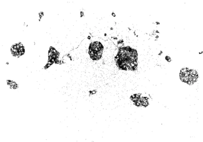
Total gastrectomy was performed because gastric carcinoid tumors showed infiltrative growth of ECL cells to the submucosa and lymph vessel invasion. In the corpus and fundus a total of 36 carcinoid lesions were found, ranging in size from 0.1 to 8.0 mm.
Table 1. Laboratory results of Cases 1 and 2.
Laboratory results Case 1 Case 2
WBC 7200/mm' 2770/mm'
RBC 386mm3 288mm3
Hb 11.5g/dl 9.7g/d/
Ht 33.8% 26.2%
Plate 28.0x 10⁴/mm3 15.0x 10⁴/mm3
Parietal cell antibody (+) ND
LDH 161 lU/l 456 lU/1
Gastrin 1700pg/ml ND
Serotonin 66ng/ml ND
Urine5-HIAA 1.9mg/day ND
CEA ND 2.2mg/ml
CA15-3 ND 17 U/ml
ND; not done
Patient 2
Patient 2 was a 50 year old female who had a mas- tectomy for stage IIIa left breast cancer. Six months later she complained of epigastric discomfort. Gastric examination showedmultiple gastric polyps in the body of the stomach. Biopsies suspected poorly differentiated adenocarcinoma in the body of the stomach. Multiple carcinoid tumors were confirmed in histological and immunohistochemical studies following total gastrectomy.
Thirty-one multiple carcinoid lesions ranging in size from 0.1 to 7.4 mm were found focal histologicaly in- vestigated specimens of the stomach.
Material and Methods
The resected stomachs were fixed in 10% neutral buffered formalin and then examined in serial cross- sections. The tissue blocks were dehydrated and rou- tinely embedded in paraffin. Sections of 3-micron
thickness were cut and stained with hematoxylin and eosin, Periodic acid-Schiff (PAS) and Grimelius and Fontana-Masson stains.
For immunohistochemistry, the standard avidin-biotin- peroxidase immune complex method was employed with a Vectastain ABC Kit (Vector Laboratories, Burlingame, CA) on the formalin fixed and paraffin embedded tissue sections. Immunohistochemical staining was performed using antibodies against the following: Chromogranin A, gastrin, serotonin, somatostatin, and synaptophisin.
Morphological examination
In both cases the fundic regions showed an atrophic change associated with a proliferation of mucous cells and moderate degree of intestinal metaplasia. ECL cells separated from the atrophic fundic glands or carcinoids were detected in the deep lamina propria, muscularis mucosa, or submucosa of the fundic re- gions showing the atrophic changes. A total of sixty- seven lesions of ECL cell hyperplasia or Endocrine cell micronest (ECM) and distinct microcarcinoids, ranging in size from 0.1 to 8.0 mm in size (36 in Case 1 and 31 in Case 2) were detected.
Of the 36 lesions in Case 1 of the ECL cells and microcarcinoids measuring from 0.1 to 8.0 mm in di- ameter (average 1.4 mm), there were 25 lesions from 0.1 to 8.0 mm (average 1.7 mm) in the mucosa, and 11 lesions from 0.1 to 6.0 mm (average 1.0 mm) in the submucosa.
In Case 2, the 31 lesions of the ECL cells and microcarcinoids measuring from 0.1 to 7.4 mm in di- ameter (average 0.94 mm) were found only in the mucosa of the stomach corpus.
Morphologically, of the 36 ECL or ECM and carcinoids, 14 (39%) showed trabecular or ribbon-like structures (Fig. 1A) and 22 (61%) had solid, anastomosing trabecular, ribbon-like, glandular and/or acinar structures with fibrovascular stroma (Fig. 1B). There were various de- grees of large cells with large atypical nuclei, which were about twice the size of lymphocyte nuclei. Carcinoid tu- mors showed an expansive growth and a conspicuous infiltration into the muscularis mucosae and submucosa.
They often showed a perivascular arrangement and focal infiltration of lymphatic vessels.
In both cases all carcinoid tumors were positive for argyrophil and argentaffin with Grimelius and Fontana- Masson silver stains.
Immunohistochemistry
The immunohistochemical findings of carcinoid tu-
mors are summarized in Table 2. All carcinoid lesions
showed diffuse positivity for Chromogranin A (Fig.
2A). In the antral region, there was a diffuse intra- glandular increase in the number of gastrin positive cells in the middle zone of the pyloric glands (Fig. 2B).
Synaptophisin and serotonin were scattered positive in carcinoid tumors.
Figure 1. (A) Carcinoid tumors illustrating a trabecular or al- veolar pattern with ribbon-like structures. Tumors are com- prised of small, round cells with single and round-to-oval nu- clei in case 1.
Figure 2. (A) Hyperplastic chromogranin A-positive endo- crine cells from lines in a focus of mucous neck hyperplasia and numerous micronodules in the deep lamina propria.
Figure 1. (B) Carcinoid showing an expansive growth and solid, anastomosing trabecular structure with a fibrovascular stroma, composed of large, atypical cells intermingled with small cells in case 2 (H&E; A, magnification x80; B, mag-
nification x80).
Table 2. Immunohistochemical results on carcinoid tumors
Immunohistochemical results Case 1 Case 2
Chromogranin A + +
Neuron-specific enolase + +
Gastrin - -
Serotonin + +
Somatostatin + -
Synaptophism + +
Figure 2. (B) Proliferation of gastrin positive cells in the antral region. Scattered gastrin positive cells are present even in the foveolar epithelium (A, magnification x80; B, magnification x80).
Discussion
Gastric carcinoids compose only 0.3% of all gastric tumors and 2.2% of all carcinoid tumors"'. Gastric carcinoids are derived from ECL cells present in the body of the stomach"'. In patients with or without pernicious anemia, antral G cells secrete enormous quantities of gastrin in response to achlorhydria re- sulting in proliferation of ECL cells" 16'1" The spectrum
of these proliferative changes include hyperplasia,
dysplasia, and neoplasia18'.
Although the preoperative clinical examinations in- dicated hypergastrinemia in the two cases presented here, the histological and immunohistochemical find- ings were consistent with the characteristics of gastric carcinoid tumors comprised of argyrophilic cells. In the rat, hypergastrinemia by itself has been reported to stimulate the development of gastric argyrophilic or ECL cell carcinoid tumors"'. However, in patients with Zollinger-Ellison syndrome (ZES), hypergastrinemia simply leads to a proliferation of argyrophilic or ECL cells within the confines of the fundic mucosa20'. In the current cases, there was no history of diarrhea, symp- toms of carcinoid syndrome and ZES, evidence of mul- tiple endocrine neoplasia type 1 (MEN-1), or pernicious anemia.
Autoimmune diseases such as hypothyroidism and diabetes mellitus may coexist with gastric multiple carcinoid tumors. Clinical management is still undefined"'.
In the current study, ECL cell hyperplasia or ECM nodules were localized in the lower third of the mucosa and showed scattered staining for the hor- mone serotonin but none for gastrin. However, in the 25 carcinoid lesions observed in this study, each carcinoid was accompanied by a large number of pe-
Case 1. Total gastrectomy
ripheral ECL cells or ECM, which usually were pre- sent adjacent to proliferating argyrophilic cells in multifocal pyloric gland metaplasia of atrophic gastri- tis. Thus, the origin and development of multiple gas- tric carcinoid tumors and ECM may be due to the proliferating argyrophilic cells in multifocal pyloric gland metaplasia, which in turn is caused by atrophic gastritis as well as to the trophic action of excess gastrin in the serum.
The finding of diffuse ECL cell hyperplasia or ECM within pseudopyloric metaplasia in gastric biopsies from fundus mucosa is a characteristic reported only for carcinoids with atrophic gastritis type A. In this study, the size of carcinoid tumors was less than 1 cm with focal lymph vessel invasion and scattered tumor infiltration of the mucosa and submucosa. Though it may be possible to misinterpet lymphatic invasion, true invasion of lymph vessels can be clearly distin- guished by reticulin fibers between argyrophil cells that are continuous with reticulin fibers of surround- ing tissue"'. The ECL cell hyperplasia or ECM may fi- nally expand to larger, infiltrating neoplasms with a significant risk of metastases"'. Carcinoid tumors measuring less than 1 cm in diameter apparently bear only a very low risk of metastases and can be re- moved by polypectomy or circumscribed local exci- sion. Subsequent endoscopic follow-up of these pa- tients has been recommended. Large carcinoid tumors may require local excision and antrectomy, whereas invasive tumors may need a total gastrectomy2s-25'.
Therefore, clinicians caring for patients with atrophic gastritis type A and hypergastrinemia must remain aware of the potential for the development of gastric carcinoid tumors.
References
Case 2. Total gastrectomy