Acta Med. Nagasaki 46 : 63-67
Case Report
Juvenile Idiopathic Nonspecific Interstitial Pneumonia.
Case Report and Review of Literature
Hiroshi ISHII 1), Jun-ichi KADOTA 2), Yoko OBATA 1), Tomoyuki KAKUGAWA 1), Towako NAGATA 1), Hideyuki KAIDA 1), Hiroshi MUKAE 1), Shigeru KOHNO 1)
1) Second Department of Internal Medicine, Nagasaki University School of Medicine 2) Second Department of Internal Medicine, Oita Medical College
We describe a case of juvenile idiopathic nonspecific in- terstitial pneumonia (NSIP). This is the first report of a Japanese patient with idiopathic NSIP aged 27 years. A computed tomographic scan of the chest showed ground- glass opacities and reticular opacities in subpleural distri- bution. Bronchoalveolar lavage fluid revealed no specific finding except for decreased CD4/CD8 ratio of lymphocyte subset. Histopathological features on examination of thoracoscopic lung biopsy specimens were consistent with those of NSIP group III. The patient was treated with corticosteroids and immunosuppressants, but no clinical im- provement was noted and the general condition has gradu- ally worsened. Although the prognosis is generally consid- ered to be good in patients with NSIP, some patients die as a result of progression of the disease. The prediction of prognosis based on histopathological, radiologic, and bronchoalveolar lavage cell findings in NSIP seems to be difficult at present.
ACTA MEDICA NAGASAKIENSIA 46 : 63-67, 2001
Key Words: juvenile onset, idiopathic nonspecific interstitial pneumonia
Introduction
Nonspecific interstitial pneumonia/fibrosis (NSIP) was first described by Katzenstein and Fiorelli in 1994 as a diffuse interstitial pneumonia with a patho- logic pattern distinct from usual, desquamative, and acute interstitial pneumonia". NSIP is characterized by an interstitial inflammatory cell infiltrate with or
Address Correspondence: Hiroshi Ishii, M.D.
Second Department of Internal Medicine,Nagasaki University School of Medicine, 1-7-1 Sakamoto, Nagasaki 852-8501, Japan TEL: +81-95-849-7273 FAX: +81-95-849-7285
without fibrosis; however, the most characteristic find- ing in NSIP is the lack of temporal heterogeneity, which is a cardinal feature of usual interstitial pneu- monia (UIP)' - 6' . The prognosis in NSIP is generally good compared with UIP' -"), however it is reported that patients with NSIP group III have a worse prog- nosis relative to those with NSIP groups I and II ' , 6 , 7 1 NSIP occurs mainly in middle-aged adults 2' 3' 6 - 9>, like most other interstitial pneumonias. According to clini- cal reports from Japan, the average age of patients at onset in NSIP is 56.4 to 57.7 years' 10'. We report here the first Japanese patient with early onset idio- pathic NSIP group III.
Case Report
A 27-year-old Japanese man was admitted to our hospital in June 1997 for further investigation of non- productive cough and progressive exertional dyspnea.
The medical history included bronchial asthma and atopic dermatitis during childhood. The father died of the progressive idiopathic pulmonary fibrosis (IPF) and diagnosed as UIP at autopsy. The uncle also died of IPF. The patient was a tobacco smoker (40 ciga- rettes a day) for 8 years, but reported no environ- mental exposure to chemicals.
Physical examination was otherwise normal apart from bilateral basilar inspiratory fine crackles on chest examination. Although reticulonodular opacities were detected on the chest X-ray film in November 1996, the patient was not admitted to the hospital.
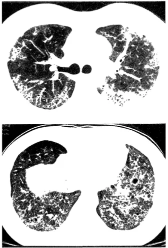
Chest X-ray film on admission showed diffuse reticulonodular opacities in both middle and lower lung fields (Fig. 1). A computed tomographic scan of the chest showed ground-glass opacities and reticular opacities in subpleural distribution (Fig. 2). The rele-
vant laboratory results included lactic dehydrogenase
Figure 1. Chest X-ray taken at the time of diagnosis in 1997 showing diffuse reticulonodular opacities in both middle and lower lung fields.
459 IU/L (normal range 202-435) and C-reactive pro- tein 0.32 mg/dl (normal; negative). Antinuclear anti- bodies were positive (X40, speckled type), while rheu- matoid factor and other specific antibodies for collagen vascular diseases were negative. Arterial blood gases on room air at rest were PaO 2 87.5 mmHg, PaCO 2 45.5 mmHg, and pH 7.376.
Pulmonary-function tests included vital capacity of 3.39 L (80.3 percent of predicted), FEV i of 2.98 L (89.5 percent of predicted), DLCO/VA of 4.092 ml/M/mmHg/
L, and %DLCO 46.8 percent. Bronchoalveolar lavage (BAL) was performed using 200 ml of physiological saline. Examination of BAL fluid showed 4.1 X 10 5 cells/ml including 82.6 percent macrophages, 0.3 per- cent neutrophils, 0.4 percent basophils, 3.8 percent eosinophils, and 12.9 percent lymphocytes. The CD4/CD8 ratio of lymphocyte subset was 0.44.
To establish a definite diagnosis, lung biopsy was performed by video-assisted thoracoscopic surgery on June 16, 1997. Pathological diagnosis was made as
Figure 2. A computed tomographic scan of the chest at the time of diagnosis in 1997 showing ground-glass opacities and reticular opacities in subpleural distribution.
Figure 3. Specimen obtained by thoracoscopic lung biopsy showing a mild degree of intra-airspace organization, the presence of minimal fibrosis, and subpleural honeycombing with loss of alveolar structure. Compared to UIP, temporal heterogeneity of the fibrous tissue was absent (HE stain, A
X5, BX33).
NSIP group Ill, based on the findings that lung tissue obtained from the left lower lobe showed a mild de- gree of intra-airspace organization, the presence of minimal fibrosis, and subpleural honeycombing with loss of alveolar structure. Compared to UIP, temporal heterogeneity of the fibrous tissue was absent (Fig. 3).
Since dust inhalation and collagen vascular disease were ruled out, the diagnosis was idiopathic NSIP group 1H.
Oral prednisolone was administered at 1 mg/kg/- day. Although such therapy was initially effective, de- terioration of the clinical condition and worsening of
interstitial pneumonia occurred following tapering of prednisolone. Accordingly, the patient was treated with a high dose of methyl prednisolone (1 g/day in- travenously for three days) followed by oral corticosteroids, combined with oral immunosuppressant
Figure 4. Chest X-ray taken in 2000 showing diffuse reticu- lar opacities and elevation of both domes of the diaphragm indicative of lung volume loss.
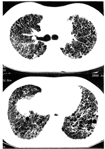
Figure 5. High resolution computed tomography of the chest taken in 2000 showing diffuse ground-glass opacities with honeycomb formation, local traction bronchiectasis, and inter- lobular septal thickening.
Figure 6. Summary of clinical course.
Therapeutic intervention, arterial blood gases, pulmonary function tests, and schema of chest X-ray films. BAL:
bronchoalveolar lavage, VATS: video- assisted thoracoscopic surgery, HOT:
home oxygen therapy, PSL: prednisolone, m-PSL: methyl prednisolone, AZP:
azathioprine. CYA: cvclosporin.
(either azathioprine 50 mg/day, or cyclosporin 200 mg/day). However, at that stage, NSIP showed resis- tance to treatment. Follow-up examination in March 2000 showed arterial blood gases on room air of Pa0 2 of 48.1 mmHg, PaCO 2 of 39.8 mmHg and pH of 7.443, and serum KL-6 of 2,320 U/ml. Chest X-ray film showed diffuse reticular opacities in both lung fields and lung volume loss (Fig. 4), and high resolution computed tomography of the chest showed diffuse
ground-glass opacities with honeycomb formation, local traction bronchiectasis, and interlobular septal thickening (Fig. 5). At present, the patient is on home oxygen therapy and on the waiting list for lung trans- plantation. Figure 6 provides a summary of the clini- cal course of this case.
described) of Nakamura et al." (Table 1). In our insti- tution, the average age at onset of patients with idio- pathic NSIP is similar to those of the above two Japanese studies (52.9 years, range, 27 to 65 yr) (Table 1). However, there is no report so far that described the clinical course of juvenile idiopathic NSIP. To our knowledge, therefore, this is the first report of a Japanese patient with early onset idiopathic NSIP.
Although the present case was suspected to be a he- reditary interstitial lung disease, this could not be confirmed because of lack of detailed genetic examina- tions.
Table 1. Age at Onset of Patients with Idiopathic Nonspecific Interstitial Pneumonia/Fibrosis (NSIP) reported in Japan.
Discussion
Nonspecific interstitial pneumonia/fibrosis (NSIP) was first defined by Katzenstein and Fiorelli in 1994'), and the name has gained broad acceptance 4 , ". The prognosis is generally good in NSIP patients, but some patients, especially those of group II and III of NSIP die as a result of progression of the disease"""'.
Recently, Travis et al.4 proposed that NSIP should be separated into cellular and fibrosing patterns because these histological patterns are associated with disease characteristics and prognosis. Based on the proposed category of NSIP, the cellular pattern generally corre- sponds to group I and fibrosing pattern to group II and III of NSIP, initially described by Katzenstein and Fiorelli ". They also reported that only patients with idiopathic cellular NSIP has an excellent long-term prognosis, and that the majority of patients with idio- pathic NSIP fibrosing pattern die mostly within 5 and 10 years4'. However, the prediction of prognosis based on histopathological, radiologic or bronchoalveolar lavage cell findings in NSIP seems to be difficult at present. Future studies need to evaluate this issue clinicopathologically in a large population of NSIP.
NSIP occurs most often in middle-aged adults""').
Although the disease could affect individuals at any age, cases of juvenile NSIP are comparatively rare.
Several juvenile patients with NSIP were reported by Katzenstein and Fiorelli' ), while connective-tissue dis- eases were also included in their report. According to recent reports of idiopathic NSIP, the average age at onset was 57 years (range, 40 to 73) in the study of Bjoraker et al.') and 43 years (range, 31 to 66) of Daniil et al.'). Furthermore, the age of affected Japanese patients is 57.7 years (range, 40 to 69) ac- cording to Nagai et al.') and 56.4 years (range, not
Reference Overall Group I Group I/II Group 11/RI Group III
Nagai et al.' 57.7±8.2* 46.50t 57.7±8.0* 58.4±8.0* 57.8±8.7*
(n=31) (n=2) (n=16) (n=29) (n=15)
Nakamura et al.' 56.4±9.97* 65.8±8.89* ND 54.0±8.75 ND
(n=30) (n=6) (n=24)
Our cases 52.9±12.9* 57t 52.9±12.9* 51.7± 14.6*
(n=9) (n=0) (n=2) (n=9) (n=7)
*: Mean ± SD, t: average, ND: not described
Acknowledgement
The authors thank Dr. Masanori Kitaichi, Department of Laboratory Medicine, Kyoto University Hospital for his advice in the pathological diagnosis, and Dr. F.G.
Issa, (Word-Medex, Sydney, Australia), for his assis- tance in editing the manuscript.
References
1) Katzenstein AA, Fiorelli RF. Nonspecific interstitial pneumonia/fi-
brosis. Histologic features and clinical significance. Am J Surg Pathol 18: 136-147, 1994.
2) Daniil ZD, Gilchrist FC, Nicholson AG, et al. A histologic pattern of nonspecific interstitial pneumonia is associated with a better
prognosis than usual interstitial pneumonia in patients with
cryptogenic fibrosing alveolitis. Am J Respir Crit Care Med 160:
899-905, 1999.
3) Cottin V, Donsbeck A-V, Revel D, et al. Nonspecific interstitial pneumonia: Individualization of a clinicopathologic entity in a se-
ries of 12 patients. Am J Respir Crit Care Med 158: 1286-1293,
1998.
4) Travis WD, Matsui K, Moss J, et al. Idiopathic nonspecific intersti- tial pneumonia: Prognostic significance of cellular and fibrosing
patterns: Survival comparison with usual interstitial pneumonia
and desquamative interstitial pneumonia. Am J Surg Pathol 24:
19-33, 2000.
5) Myers JL. NSIP, UIP, and ABCs of idiopathic interstitial pneumonias. Eur Respir J 12: 1003-1004, 1998.
6) Nagai S, Kitaichi M, Itoh H, et al. Idiopathic nonspecific intersti- tial pneumonia/fibrosis: Comparison with idiopathic pulmonary fi-
brosis and BOOR Eur Respir J 12: 1010-1019, 1998.
7) Nakamura Y, Chida K, Suda T, et al. A comparative study of the prognosis for Japanese patients with idiopathic interstitial pneu-
monia or BOOP based on histopathologic subsets. Nihon Kokyuki
Gakkai Zasshi 38: 659-664, 2000 (in Japanese, Abstract in
English).
8) Bjoraker JA, Ryu JH, Edwin MK, et al. Prognostic significance of histopathologic subsets in idiopathic pulmonary fibrosis. Am J
Respir Crit Care Med 157: 199-203, 1998.
9) Katzenstein AA, Myers JL. Idiopathic pulmonary fibrosis: Clinical relevance of pathologic classification. Am J Respir Crit Care Med
157: 1301-1315, 1998.
10) Fujita J, Yamadori I, Bandoh S, et al. Clinical features of three fatal cases of non-specific interstitial pneumonia. Intern Med 39:
407-411, 2000.
11) Katzenstein AA, Myers JL. Nonspecific interstitial pneumonia and the other idiopathic interstitial pneumonias: Classification and di-
agnostic criteria. Am J Surg Pathol 24: 1-3, 2000.