Yamanashi Med. J. 4 (4), l95i'-l99, 1989
Case Report
A Case ef
Renal Cell
withCarcinoma with Lung Metastases
Adoptive Immunetherapy
Treated
Noboru
DePartment
YABusAKii), Hideki KoMATsui), Akira UENoV,
and Kachio TAsAKAL')
of Urologyi>, DePartment' of Parasitolog:y & lmmunolog>}2),
Yamanashi Medical College
Abstract: A patient with renal cancer and ltmg metastases who was treated with
lymphokine-activated killer cells (LAK cells) is reported. A 52-year-old male was admkted because of asymptomatic hematuria in August l986. Computed tomography showed a large renal tumor on the right side and the chest X-ray film revealed multiple lung metastases.
Right nephrectomy was perfermed in September 1986. Kistologically, the ttimor was
diagnosed as a renal cell carcinoma. Infusion of vinblastin via £he broncl}ial aTtexies ha(l no effect on the metastatic lesiens. LAI< therapy was started in Apri.l l987. Peripheral blood lymphocytes were purified £rora the patient's leukocytes coilected by leul<apheresis and then cu}tured at a coRcentration of l-2xl06' cellsfml with S-6 Ulml of .human re-combinant interleukin-2 iR cornplete R.PMI }640 medium for 3-8 days. In the initial two treatments, LAK cells were infused intravenously, bat from the third treatment they were in£used via the bronchiai arteries. The number of LAK cells infused was 4×10⁸-g.2×109 per treatment and l6.1xlO" in a total of ll treatments. LAK therapy was stopped because o£ a toxic reaction which cleveleped duiring the llth infusion. The cause of this reaction was not obvious, but it was apparently an allergic mechaRism. During LAK therapy the metastatic lesions slightly increased in size.
Key words: Iymphokine-activated killer cell, interleukin-2, adoptive immimotherapy, lung metastases from renal cancer, plasma exchange
Since radiotherapy and chemotherapy have lit£le effect on reltal cell carcinoma,
surgical excision has been tkeught oE as
the only effective method for treating this
tumor. No method has been shewn to be effective for disseminated renal ce}l carci-noma. In l985, Resenberg and his
col-IeaguesQ) reported the use o£ adoptive immunotherapy with lymphokine-activated
killer (LAK) cells for patients with advaRced caltcer in NNThom other treatments had
proven ineffective. Renal cell carciRorna was reported to be one ef the best
respond-Tamaho, Nakakoma, Yamanashi 409-88, Japan
Received July 20, 1989 Accepted September 14, I989
ers to this therapy5). For this reason we treated a patient with lung metastases from
renal cell carcinoma by infusion of LAK
cells via the broRchial arteries.
CAsE IREpoRT
A 52-year-old man was hespitalized iR September 1986 for a right-sided renai tumor. Asymptomatic gross hematuria had
occured l raonth previously, the tumor x47as
detected by computed tomography (CT) at
another hospital, and he was then referred
to us. On physical examinatioR a large
mobile mass was palpable in the right
upper quadrant. CT showed a large yight
X96 N. Yabusaki, H. Komatsu, A,
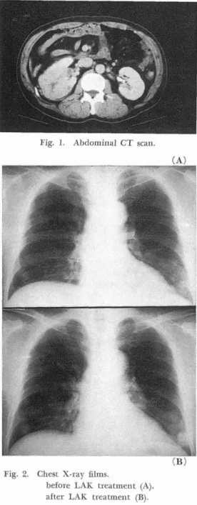
Fig. I. Abdominal CT scan.
(A)
・・;(・#)
Fig. 2. Chest X-ray films.
before LAK treatmen£ (A), after LAK treatment (B).
Right renal arteriography demonstrated a hypervascular tumor occupying the iower pole of the right kidney. The chest X-ray
film revealed multiple coin lesions in both
lungs (Figure 2-A). On September 22nd, 1986, right nephrectomy was performed.
Histologically, the tumor was diagnosed as
a renal cell carcinoma, stage pT2b, pNO,
Ueno, and K. Tasaka
Ml (the original tumor was in the renal
capsule without lymph node metastasis, but with distant metastasis). On October 27th vinblastine was infused via the bronchial
arteries for treatment of the metastatic
pul-monary lesions. Since vinblastine had no
effect on the pulmonary metastases,
adop-tive immunotherapy with LAK cells was started in April I987.
ADOPTIvE IMMuNoTHERApy wlTH LAK CELLs
Recombinant Interleukin-2 (rlL-2)
The rlL-2 used was TGP-8, which was
kindly supplied by Takeda Pharmaceutical
Co. Ltd., Osaka, Japan.
Induction of LAK cells
The lymphocyte-rich fraction was
ob-tained from the peripheral blood of the patient using a blood cell separator (CS-8000, Travenol, Co. Ltd., Tokyo, Japan). From this fraction lymphocytes were
fur-ther purified by Ficoll-Hypaque (Pharmacia
Fine Chemicals, Uppsala, Sweden) gradient
centrifugation, and were cultured at a con-centration of I-2×10⁶/ml in roller bottles
(Falcon, Becton Dickinson Co. Ltd., Ox-nard, CA., USA) under 5% C02 at 870C in a humidified atmosphere for 8 to 8 days.
Complete RPMI 1640 with 5% autologous plasma or 5% human fresh frozen plasma containing 8-6 U/ml of human rlL-2 was
used as the culture medium. After 8 to 8
days of cell culture, lymphocytes were
col-lected and suspended in 20 ml of physio-logical saline with l,OOOU of rlL-2. This suspension was then administered to the
patlent.
Examination of the activity of LAK cells The activity of the LAK cells was
de-termined by a 5iCr release assay, as
previ-ously reportedi). Fhe targets were Daudi
cells (which are resistant to natural killer cells and sensitive to LAK cells), K562 cells (which are sensitive to natural killer cells),
Adoptive
the human reRal cell carciRoma line CAKI-1, aRd autologous peripheral blood lympho-cytes (PBL). After target cells were
radio-labeled, effector and target cells were cu}-tured together for 4 1}ours at the ratios of
6/1, 12/l, 25/I, and 50/l in a C02
incu-bator (Tabai Espec, Co. Ltd., Osaka, Japan).
Cytotoxic activity was calculated by the
following equation:
% Killng
release
ww Experimental release-Spontakeous
imraunotherapy for lung metastases from renal cell
Maximum release---Spontaneous Telease × 100
Figure 8 shows the cy£otoxic activity of LAK cells induced from the patient's PBL
in July 1987. At an E/T ratio of 50/1, 80%
of Daudi or K562 cells and 50% of CAKM
cells were killed, but LAK celis showed no cytotoxicity agaiRst autologous PBL. InfzLsion of LAK cells
(%) 1eo 80 60 40 20 P[asma: lised in culture medium Target:Daudi
O--.-O E/T ratiom 50/1 A---A E/T ratio= 25/1
PE PE PE
" ii
LAK
tient Ilto Julte
the LAK ceil
venously,
infused it via bronchial arteries contact method of bronchial follows. PE PE ,
lst. 2nd. 3rd. 6th. 7th. 8th. 9th. 10th. 11th. autologeus FFP FFP FFP FFP FFP FFP autologous FFP FFP )E:'(;FEplXIBeSsR"FrEoX2Cehna"pg£sraa)
Fig. g. Cytotoxicity off LAK cells.
cells were administered to the times in the period from April I987
1988. The first aRd second times
suspension was infused
but from the third treatmeltt we
to mcrease with the metastatic tumor. The
artery infusion was as A Seldinger catheter was inserted
carcmoma 197
into the patient's £emoral artery aRd thetip of the catheter was selectively advaRced
to a bronchial artery. Then 40 ml of the LAK cell suspeRsiolt was infused over 3e
minutes by syringe pt}nLp. Each time 2000
U/day of rlL-2 was giveR for 5 days. The
LAK cell activity in eur patieRt decreasecl gradually from treatinent to treatment. We presumed that this was due to the preseRce o£ inhibiting factors agaiRst LAK activity which have been previevisly demonstrated2). ThereEore, we stopped using the patient's
plasma in the culture medium.
Further-more, we performed plasmapheresis the day before administration of LAK cells. Despite
£he use of fresh frozelt plasma in the cuilture
medium, LAK cell activity fel} to a
mini-mum at the 6th treatment, although from the 7th infusion it recovered slightly (Figure 4). This suggests tl}at
plasma-pheresis remeved the iRhibiting factors to
some extent. The number of traRsfered
LAK cells at one time raRged from 4xl08
to 8.2×10⁹, and a total e£ 16.1×10⁹ cells
were administered in the ll treatments.
Fever (880C-890C) develeped a£ter each
infusion, and sllbsided three to four days after the infusion wi£hout treatment. At
tl}e llth treatment symptoms such as chills,
shivering, high blood pressure (280/140 mmHg), an acute rise of body temperature (88.70C), altd tachycardia (l20/min) oc-curred }5 minutes after the beginning of
LAK cell infusioll via a bronchial artery. XfNie stepped the infusioR immediately. The
symptoms disappeared soon after intra-venous injectioR o£ methylprednisolone. The LAK cell s"speRsioR was examined for
sterility by bacterial culture and for
cofi-taminatien with endotoxin by the Limulus
test (VVako Chemicals, Osaka, Japan), bL}t they were beth llegative. Thelt we investi-gated allergy to rlL-2. VVe injected rlL-2 intraciitaneously into the forearm and
198 1OO 80
6e
o
.E 'M'4Q
20 e N. Yabusaki, M. Komatsu, Target: C>---Q Davdi MAutologous PBLHK 562
xx CAKI -1 ty---elCts---tCN----・---A5CVI 25/1 1271 6/1
Effector/-rarget ratioFig. 4, Kine£ics of LAK activity.
o£ which was dependent on the dose in-jected. Accordingly, we stopped £he LAK
treatment.
Figure 2-B shows the chest X-ray film
after ll LAK treatments. The multiple
coin lesions were increased in size com-pareel with before LAK treatment. TIrhe
total of the diameters o£ five evalt}able metastatic lesiens increased from 6} mm to 78 mm, about a 28% increase in size. This chaltge was evaluated as evideRce o£
pyo-gressive disease (PD). DIscussloN
Ilt 1987, Rosenberg and his colleagues
reported that they trea£ed I08 patients with
metastatic caRcer by using LAK cells aRd huraan rlL-2, aRd achieved 8 complete
re-missions and 15 partial rere-missions5)
.Over-all 22% of the patients responded to the treatment. Among them metastatic lesions
froma renal cancer respoftded particularly
well to this treatment. Four complete
re-missions aRd 8 partial remissiolts were
ob-tained among 86 metastatic renal cancer patients, a 38% respense rate. Compared
A. Uen,o, and K Tasaka
to tl}eir excelleltt result, the results of LAK
treatment in Japan have been much poorer.
According to a cumulative oR the results of LAK treatment at many institutions in
Japan6>, arnoRg K4 patients eRIy 6 partial
remissioi}s were reported (a 4% response
rate). The difference in response rates may
be due to the frequency of LAK cell
in-£usioR, the number o£ LAK cells infused, or the dose o£ rlL-2. For example,
RoseB-berg scl}edt}Ied the patients to receive five
daily leukapkeresis treatments, and infused
all the induced LAK cells day by day for the £ellowing four days. The cumulative doses of LAK cells and rlL-2 amounted to 5-IOxlOiO cells and l-2xl06U/kg
respec-tively. One unit o£ TGP-8 is reported to
be equivaleRt to 300-400 iknits of the rlL-2 used by Rosenberg (personal
communica-tion). So l-2×10⁶U/kg o£ rlL-2 equals
about 8,eOO to 6,OOO U/kg of TGP-3. rl'heir method required hundreds of li£ers of
cul-ture medium and an eRormous amount ef
IL-2 which produced maRy side effects. Thus, their original method was modified
for this patient.
A second reason why LAK therapy was
not se effective in this case may relate to anatomical considerations. Generally, LAK
therapy shows its best resvtlts in the braiR,
and we have reported one case of complete
remission of a brain tumor3). The reason
is that the brain is a confiked space, so LAK cells calt more easily reach the target and remain in its vicinity longer thaR in
other organs. In this case, the frequency
of infusion and the contact with the lesion
may have been iRsufficient, even though
LAK cells were injected selectively via the
broRchial arteries.
As side effects of LAK treatment,
Rosen-berg alld his coworkers reported chills,
hyperbilirubiRemia, and the so-called
capil-lary permeability leak syRdrome which in-duces elevation of creatiRine level,
Adoptive immunetherapy for lung
tension, weight gain, respixatory distress,
and arrhythmias5). However, they did not
report a toxic reaction to LAK therapy
with IL-2 or to IL-2 alone. NiVe presgmed that some antibody against TGP-8
(maltu-factured rlL-2) was the cattse of the
symp-toms in our patient, although we failed to demonstrate an anti-IL-2 aRtiboafy by
E LISA.
[REFERENCES
1) Iizuka E[, Naganuma IIf, Yabusaki N, et al. Study on Lymphokine-Activated Killer (LAK>
Cells. I. Improved LAK celHRduction in vitro.
Yamanashi Med J 1988; 3: 97-108.
2> Naganuma H, I<imura R, Sasaki A, et al.
hancernent of lymphokine-activated killer tivity by depleting adherent mononuclear cells.
metastases from renal cell cayciRoma 199
Neuxoimmunological Research 1988; 1: 2e5-211. 3) NagaRuma III, Kimura RL, Sasaki A, et ag. plete remission of recuyrent glioglas£oma £orme following infusions of
vated killer celis. Act Neurochir l989; 99:
l57-160.
4) Rosei}berg SA, Lotze MT, Muul LM, et al.
servatiens on the systemic administration of
autologous lymphokine-activated killer ce}ls and recombinant interleukiii-2 to patients with tastatic cancer. N Engl J Med i985; 313: 1492.
5) Rosenberg SA, Lotze Mr[', Muul LM, et al. A
progress report on the treatment of l57 patients
with advanced cancer using
vated killer cel}s and interleukin-2 alone. N
Engl J Med l987; 316: 889-897.
6) Confidential of Takeda Pharmaceutical Co. Ltd.
for the meeting of TGP-g adoptive