IRUCAA@TDC : Influence of free-hand vs uniform irradiation on tensile bond strength in Er:YAG-lased dentin
6
0
0
全文
(2) by N ht. Q ui. ot. n. fo r. Influence of Free-hand vs Uniform Irradiation on Bond Strength in Er:YAG-Lased Dentin. pyrig No Co t fo rP ub Tensile lica tio n te ss e n c e. Koya Aizawaa/Atsushi Kameyamab/Junji Katoc/Yutaka Odad/Yoshito Hiraie. Purpose: To compare effects of free-hand and mechanically applied uniform irradiation on tensile bond strength of 4META/MMA-TBB resin to Er:YAG laser-irradiated dentin using an X-Y moving stage. Materials and Methods: Three different laser conditions were evaluated: 1.0 W, 100 mJ/pulse, 10 pps; 1.0 W, 50 mJ/pulse, 20 pps; 1.0 W, 33 mJ/pulse, 30 pps. Samples of bovine dentin were set on a moving stage and mechanically irradiated with an Er:YAG laser at a scanning speed of 1.0 mm/s or subjected to free-hand irradiation. The lased dentin surfaces were acid conditioned with 10% citric acid/3% ferric chloride for 15 s, rinsed, and dried. The surfaces were bonded to PMMA rods with 4-META/MMA-TBB resin, and mini-dumbbell-shaped bonded specimens were prepared. Tensile bond strength was measured and compared with the results for free-hand irradiated samples obtained under the same conditions in an earlier study. SEM observations of fractured surfaces after tensile bond testing were also performed. Results: A significant difference was found in tensile bond strength among the 3 laser settings in both free-hand and mechanically uniform irradiations. A comparison of free-hand laser irradiation and mechanically performed uniform laser irradiation revealed no significant difference under either laser condition (p > 0.05). SEM observation of the fractured surfaces showed no difference between the two irradiation methods under all laser conditions. Conclusion: No significant difference was found in tensile bond strength between free-hand and mechanically irradiated dentin. Significant differences were found, however, with changes in laser setting in both groups. Keywords: Er:YAG laser, dentin bonding, 4-META/MMA-TBB resin, laser energy, pulse frequency, output energy. J Adhes Dent 2008; 10: 295-299.. R. esin bonding to Er:YAG laser-irradiated dentin has already been examined.5 Most studies have found a decrease in bond strength to lased dentin compared with unlased dentin. This is probably due to a combination of struc-. a Graduate Student, Department of Operative Dentistry, Tokyo Dental College,. Chiba, Japan. b Assistant Professor, Department of Operative Dentistry, Tokyo Dental College,. Chiba, Japan. c Senior Assistant Professor, Department of Operative Dentistry, Tokyo Dental. College, Chiba, Japan. d Professor and Head, Department of Dental Materials Science, Tokyo Dental. College, Chiba, Japan. e Professor and Head, Department of Operative Dentistry, Tokyo Dental Col-. lege, Chiba, Japan. Correspondence: Dr. Atsushi Kameyama, Department of Operative Dentistry, Tokyo Dental College, 1-2-2 Masago, Mihama-ku, Chiba 261-8502, Japan. Tel: +81-43-270-3958, Fax: +81-43-270-3959. e-mail: [email protected]. Vol 10, No 4, 2008. Submitted for publication: 02.10.06; accepted for publication: 26.07.07.. tural and morphological changes in the lased surface. The Er:YAG-lased dentin surface has many unique characteristics: no smear layer, open dentinal tubules, and a flaky and rough surface. While no change in crystal composition has been observed, organic components have been demonstrated to be denatured or disappear.2,9,16,22,23 Several factors are believed to affect the quality of adhesion to lased dentin, including output energy,26,35 the adhesive system used,4,6,29,34 acid etching,17,21 and additional priming.19,35 Few of these studies employed uniform laser irradiation with the dentin specimen fixed to a moving stage.36 However, these studies have focused on the effect of uniform irradiation in detail, even though some have investigated the influence of focal distance from the tip and the tooth surface.8,12,29 Our previous studies on resin bonding to Er:YAG-lased dentin1,17-19 also employed free-hand irradiation. This study investigated the effect of mechanically controlled uniform irradiation at a constant output energy/rep295.
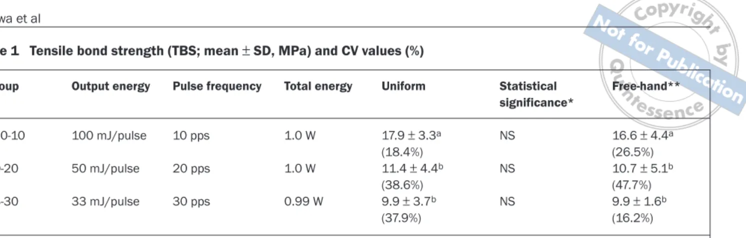
(3) Total energy. Uniform. Statistical significance*. 100-10. 100 mJ/pulse. 10 pps. 1.0 W. NS. 50-20. 50 mJ/pulse. 20 pps. 1.0 W. 33-30. 33 mJ/pulse. 30 pps. 0.99 W. 17.9 ± 3.3a (18.4%) 11.4 ± 4.4b (38.6%) 9.9 ± 3.7b (37.9%). NS NS. fo r. Pulse frequency. n. Output energy. Q ui. Group. ot. Table 1 Tensile bond strength (TBS; mean ± SD, MPa) and CV values (%). pyrig No Co t fo rP ub lica tio Free-hand** n te ss e n c e. by N ht. Aizawa et al. 16.6 ± 4.4a (26.5%) 10.7 ± 5.1b (47.7%) 9.9 ± 1.6b (16.2%). * Difference between mechanically uniform and free-hand at same laser setting (p < 0.05). Same superscript letters indicate no significant difference (p < 0.05) ** According to Aizawa et al1. etition rate on tensile bond strength to Er:YAG laser-irradiated dentin, and compared the results with those obtained by free-hand irradiation in an earlier study employing the same laser conditions.1 The null hypotheses in this study were that there was no difference in tensile bond strength between mechanically uniformly irradiated and free-hand irradiated dentin, and that laser setting had no effect on tensile bond strength in mechanically uniformly irradiated dentin.. MATERIALS AND METHODS Laser Device and Group Setting This study used a prototype Er:YAG laser device (J. Morita; Kyoto, Japan) set to emit a wavelength of 2.94 μm. The output energy of this device can be adjusted within a range of 30 to 250 mJ, and the pulse repetition rate can be adjusted within a range of 1 to 30 pulses per second (pps). The total energy delivered by the end of the probe, however, can only reach about 1.2 W. The pulse duration of this laser was set at approximately 400 μs. The teeth were divided into 3 lased groups, each subjected to a different output energy and pulse frequency (see Table 1). The total energy of each lased group was approximately 1.0 W. A 600-μm diameter straighttype contact probe was used. The energy levels were measured periodically with a power meter (Lasermate-P, Coherent; Santa Clara, CA, USA). Specimen Preparation Twenty-four bovine teeth were extracted, frozen to maintain freshness, then defrosted and cut at the cervix immediately before use in the experiments. The coronal sides of the cut surfaces were sequentially abraded under a stream of water with SiC paper (180-, 400-, and 600-grit) to prepare flat dentin surfaces. Next, they were randomly divided into 3 groups of 8 teeth each. The ground dentin surfaces were irradiated uniformly with an Er:YAG laser under a fine water spray. The tip of the laser was placed in light contact with the dentin surface to allow free movement, and the sample was fixed to an X-Y table (D-212, Suruga Seiki; Shizuoka, Japan) 296. set to travel at a scanning speed of 1.0 mm/s. The dentin surfaces in each group were then conditioned with 10 wt% citric acid solution containing 3 wt% ferric chloride (Green activator, Sun Medical; Moriyama, Japan; batch no. ES1) for 15 s, rinsed with distilled water for 30 s, sufficiently dried, and bonded to square PMMA rods (8.0 x 8.0 x 8.0 mm) using 4-META/MMA-TBB resin (Superbond C&B, Sun Medical; C&B Metabond, Parkell; Farmingdale, NY, USA) by the brushdip method. The batch numbers of this resin were GK2 (Polymer; Clear), KG2 (Monomer), and KE43 (TBB-O catalyst). The specimens were then subjected to the tests and observations detailed below. Tensile Bond Testing The specimens were kept at room temperature for 60 min immediately after preparation and then stored for 24 h in water maintained at 37°C. The bonded teeth were then serially sectioned vertically using a low-speed diamond saw (Isomet, Buehler; Lake Bluff, IL, USA) to create 2.0-mm-thick bonded dentin slabs. Each bonded slab was trimmed using a diamond point (FG #211 Regular, Shofu; Kyoto, Japan) high-speed air turbine handpiece under copious air-water spray to create a mini-dumbbell-shaped test specimen with a 3.0- x 2.0-mm cross section at the adhesive interface. After fixing the prepared specimens to a disposable PMMA jig, tensile strength was measured using a universal testing machine (Tensilon RTC-1150-TSD; Orientec; Tokyo, Japan) at a crosshead speed of 0.5 mm/min. Mean tensile bond strength was evaluated by one-way and two-way ANOVA followed by the Fisher’s PLSD test at a 95% level of confidence using StatView 5.0J (SAS institute; Berkeley, CA, USA). SEM Observation of Fractured Surface After tensile bond testing, each fractured specimen was placed on an aluminum stub and coated with Au-Pd using a Cool Sputter Coater (SC500A; VG Microtech; East Sussex, UK). They were then examined under a field-emission scanning electron microscope (SEM: ERA-8900FE, Elionix; Tokyo, Japan) to determine mode of failure. The Journal of Adhesive Dentistry.
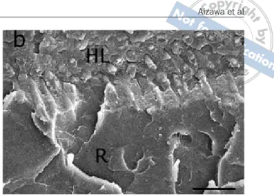
(4) Q ui. by N ht. pyr Co et ialg No Aizawa t fo rP ub lica tio n te ss e n c e. ot. n. fo r. Fig 1a SEM micrograph of fractured dentin side after tensile bond strength testing (uniform irradiation at 100-10). Mixed failure mainly in non-resin-impregnated dentin (D) and cured resin (R). Bar = 20 μm.. Fig 1b SEM micrograph of fractured rod side surfaces after tensile bond strength testing (uniform irradiation at 100-10).Mixed failure, mainly in cured resin (R) and partially hybrid layer (HL). Bar = 20 μm.. Fig 2a SEM micrograph of fractured dentin side after tensile bond strength testing (uniform irradiation at 50-20). Same as in 100-10 group, mixed failure revealed; however, area of cohesive failure in resin (R) was smaller than that in Fig 1. HL = hybrid layer. Bar = 20 μm.. Fig 2b SEM micrograph of fractured rod side surface after tensile bond strength testing (uniform irradiation at 50-20). Same as in 100-10 group, mixed failure revealed; however, area of cohesive failure in resin (R) was smaller than that in Fig 1. HL = hybrid layer. Bar = 20 μm.. RESULTS. 0.05). The coefficient of variation (standard deviation/mean x 100) was also calculated, but no difference was found.. Tensile Bond Strength Tensile bond strength and coefficient of variation (CV) in each group are shown in Table 1. The two-way ANOVA revealed a significant difference for “setting of laser” at p < 0.05, but no significant difference for “irradiation method” or interaction between these factors at p > 0.05. Bond strength in the 100-10 group was significantly higher than that in the 50-20 and 33-30 groups (p < 0.05) with both uniform and free-hand irradiation. However, no significant differences in bond strength were observed between the 50-20 and 33-30 groups (p > 0.05). No significant difference was found between uniform irradiation and free-hand irradiation at each laser setting (p > Vol 10, No 4, 2008. Features of Fractured Surfaces SEM observations of the fractured surfaces after tensile bond testing are shown in Fig 1. Cohesive failure in the cured resin was mainly observed in the 100-10 group, with partial failure within the hybrid layer and non-resin-impregnated dentin (Fig 1). In the 50-20 and 33-30 groups, failure in the cured resin was almost nonexistent, whereas failure within/at the bottom of the hybrid layer was extensive (Figs 2 and 3). When comparing the results of the previous1 and present study, no differences were observed between free-hand and uniform irradiation at each laser setting.. 297.
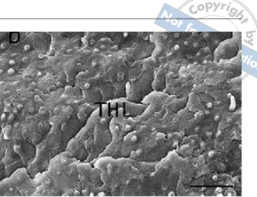
(5) pyrig No Co t fo rP ub lica tio n te ss e n c e n. ot. Q ui. by N ht. Aizawa et al. fo r. Fig 3a SEM micrograph of fractured dentin side surface after tensile bond strength testing (uniform irradiation at 33-30). Cohesive failure in resin almost nonexistent. Most failure occurred at interface between resin and top of hybrid layer (THL). Bar = 20 μm.. Fig 3b SEM micrograph of fractured rod side surface after tensile bond strength testing (uniform irradiation at 33-30). Cohesive failure in resin almost nonexistent. Most failure occurred at interface between resin and top of hybrid layer (THL). Bar = 20 μm.. DISCUSSION. Er:YAG-lased enamel and dentin. While they found no significant differences among 80 mJ-1 Hz, 80 mJ-2 Hz, 80 mJ3 Hz, and 80 mJ-4 Hz for enamel, increased frequency significantly decreased bond strength to dentin. This study compared bond strength to high-pulse frequency Er:YAGlased dentin in 3 groups, in which the total energy used was adjusted to 1.0 W at the same moving speed. Among the 3 groups, the 100-10 group yielded significantly higher tensile bond strength than the 50-20 or 33-30 groups (p < 0.05), between which there was no significant difference (p > 0.05). These results also allow us to reject the second null hypothesis that laser setting had no effect on tensile bond strength. A difference was also found in fracture pattern after tensile bond testing among the 3 groups. In the 100-10 group, there was a mixture of failure, mainly in the cured resin and partially within the resin-impregnated or laser-modified dentin. On the other hand, the 50-20 and 33-30 groups showed failure mainly between the cured resin and the resin-impregnated or laser-modified dentin. These results suggest that the denatured dentin layer in the 100-10 group was of a different chemical or physical nature to that in the other two groups. The heat accumulation effect of the pulsed laser is determined by the relationship between pulse duration, pulse interval, and thermal relaxation time.10,13,27 Irradiation applied at the same pulse duration with a high pulse frequency and lower pulse interval might easily lead to a greater accumulation of heat compared to irradiation at low pulse frequency. Physically speaking, pulsed laser irradiation at a high pulse repetition may easily denature tissue via heat accumulation, provided that the total energy is the same. Thus, the size of the denatured layer produced by the Er:YAG laser may depend on output energy, while the quality may depend on pulse frequency. This study revealed that a high pulse frequency resulted in an adverse effect on bond strength in Er:YAG laser-irradiated dentin. Both the size and depth of the abrasions in the 33-30 and 50-20 groups were previously reported to be significantly smaller than those in the 100-10 group.14 Al-. In this study, we used 4-META/MMA-TBB resin as the adhesive system. This resin is not usually used in a clinical setting for Er:YAG laser-prepared cavities. However, this adhesive has often been used in the investigation of dentin bonding over the last 20 years. It has been used to study the effects of different acid conditioners,15 additional application of monomer,20,24,32 bond durability with fluoride-containing polymer,30 and bond strength to heated dentin. Kameyama et al have also reported bond strength to Er:YAG laser-irradiated dentin with this resin, especially regarding the effect of different acid conditioners and the additional application of HEMA or glutaraldehyde.17-19 Several factors are believed to affect bond strength to lased dentin, including laser condition; experience with bonding procedures25,31 and laser irradiation, individual dentin substrates,29 and remaining dentin thickness.7,33,37 In this study, all the specimens in the 6 groups were prepared by the same investigator (K.A.) to exclude the influence of operator experience. Furthermore, to exclude the factor of depth of bonded dentin, we used cross-sectioned labial dentin perpendicular to the tooth axis at the cervix, including superficial to deep dentin, as the bonded dentin surface.15,24 A comparison of mechanical and free-hand irradiation under the same laser settings yielded no significant differences. No difference was found in the CV3 between free-hand and mechanical irradiation at each setting. CV has been reported to be an indicator of consistency of bond strength;3 therefore, these results allow us to reject the first null hypothesis. The influence of output energy and pulse frequency on bond strength has been reported. Monghini et al26 found no significant differences among specimens treated with 60 mJ-2 Hz, 80 mJ-2 Hz, and 100 mJ-2 Hz; however, an increase in laser output energy resulted in an increase in cratered surfaces, regardless of acid-etching association. Gonçalves et al11,12 compared the influence of pulse frequency in both 298. The Journal of Adhesive Dentistry.
(6) by N ht. 15.. Q ui. n. 16.. fo r. CONCLUSION In conclusion, this study found no significant difference in tensile bond strength between free-hand and mechanically irradiated dentin. Significant differences were found, however, with change in laser setting in both the free-hand and mechanically irradiated groups. In particular, a high pulse frequency resulted in an adverse effect on bond strength in Er:YAG laser-irradiated dentin. Further study is needed to evaluate the usefulness of the high pulse frequency Er:YAG laser.. ACKNOWLEDGMENTS We would like to thank Mr. Katsumi Tadokoro (Oral Health Science Center, Tokyo Dental College) for technical advice on using the SEM, and Sun Medical Co. for providing materials. We would also like to thank Associate Professor Jeremy Williams, Tokyo Dental College, for his assistance with the English of this manuscript.. REFERENCES 1. Aizawa K, Kameyama A, Kato J, Amagai T, Takase Y, Kawada E, Oda Y, Hirai Y. Resin bonding to dentin irradiated by high repetition rate Er:YAG laser. Photomed Laser Surg 2006;24:397-401. 2. Bachmann L, Diebolder R, Hibst R, Zezell DM. Changes in chemical composition and collagen structure of dentine tissue after erbium laser irradiation. Spectrochem Acta A Mol Biomol Spectrosc 2005;61:2634-2639. 3. Browning WD, Myers ML, NiX LB. Constancy of bond strength in 5 singlebottle dentin bonding systems. Quintessence Int 2001;32:249-253. 4. Chimello DT, Ramos RP, Chinelatti MA, Palma Dibb RG, Pecora JD, Corona SAM. Effect of Er:YAG laser and air abrasion on the tensile bond strength of two adhesive restorative systems to dentin. J Oral Laser Appl 2002; 2:223-229. 5. De Moor RJG, Delme KIM. Erbium lasers and adhesion to tooth structure. J Oral Laser Appl 2006;6:7-21. 6. De Munck J, Van Meerbeek B, Yudhira R, Lambrechts P, Vanherle G. Microtensile bond strength of two adhesives to Erbium:YAG-lased vs. bur-cut enamel and dentin. Eur J Oral Sci 2002;110:322-329. 7. de Souza AE, Corona SAM, Palma-Dibb RG, Borsatto MC, Pecora JD. Influence of Er:YAG laser on tensile bond strength of a self-etching system and a flowable resin in different dentin depths. J Dent 2004;32:269-275. 8. Eguro T, Maeda T, Otsuki M, Nishimura Y, Katsuumi I, Tanaka H. Adhesion of Er:YAG laser-irradiated dentin and composite resins: Application of various treatments on irradiated surface. Lasers Surg Med 2002;30:267-272. 9. Gaspirc B, Scaleric U. Morphology, chemical structure and diffusion processes of root surface after Er:YAG and Nd:YAG laser irradiation. J Clin Periodontol 2001;28:508-516. 10. Geraldo-Martins VR, Tanji EY, Wettwer NU, Nogueira RD, Eduardo CP. Intrapulpal temperature during preparation with the Er:YAG laser: an in vitro study. Photomed Laser Surg 2005;23:182-186. 11. Gonçalves M, Corona SAM, Pecora JD, Palma Dibb RG. Influence of the frequency of Er:YAG laser on the bond strength of dental enamel. J Clin Laser Med Surg 2003;21:105-108. 12. Gonçalves M, Corona SAM, Borsatto MC, Pecora JD, Palma Dibb RG. Influence of pulse frequency Er:YAG laser on the tensile bond strength of a composite to dentin. Am J Dent 2005;18:165-167. 13. Hibst R, Keller U. Heat effect of pulsed Er:YAG laser radiation. Laser surgery: advanced characterization, therapeutic systems. Proc SPIE 1990;1200:379-386. 14. Igarashi A, Takase Y, Hirai Y. Efficiency of dentin ablation by Er:YAG laser: Effect of output energy and repetition rate [in Japanese]. Jpn J Conserv Dent 2005;48:303-313.. Vol 10, No 4, 2008. ot. though laser devices capable of laser irradiation at a high pulse frequency are now available, their effective use in a clinical setting remains problematic.. pyr Co et ialg No Aizawa t fo Iwasaki Y, Toida T, Nakabayashi N. Improved wet bondingr of methyl Pu methacrylate-tri-n-butylborane resin to dentin etched with ten percent b phosphoric acid in the presence of ferric ions. J Biomed Mater ResliAc ati 2004;68A:566-572. te irradiation on Ishizaka Y, Eguro T, Maeda T, Tanaka H. Effects of Er:YAG laser on human dentin: polarizing microscopic, light microscopicsand s eFT-IR nc e. analysis. Laser Surg Med 2002;31:171-176. 17. Kameyama A, Kawada E, Takizawa M, Oda Y, Hirai Y. Influence of different acid conditioner on the tensile bond strength of 4-META/MMA-TBB resin to Er:YAG laser-irradiated bovine dentin. J Adhes Dent 2000;2:297-304. 18. Kameyama A, Oda Y, Hirai Y, Kawada E, Takizawa M. Resin bonding to Er:YAG laser-irradiated dentin: combined effects of pre-treatments with citric acid and glutaraldehyde. Eur J Oral Sci 2001;109:354-360. 19. Kameyama A, Amagai T, Takizawa M, Kawada E, Oda Y, Hirai Y. Effect of HEMA on bonding of Er:YAG laser-irradiated bovine dentin and 4-META/ MMA-TBB resin. J Oral Rehabil 2002;29:749-755. 20. Kameyama A, Ihara S, Amagai T, Miake Y, Kawada E, Oda Y, Yanagisawa T, Hirai Y. Adhesion of 4-META/MMA-TBB resin to heated dentin: effects of pre-treatments with FeCl3 and/or HEMA. Dent Mater J 2003;22:1-9. 21. Kataumi M, Nakajima M, Yamada T, Tagami J. Tensile bond strength and SEM evaluation of Er:YAG laser irradiated dentin using dentin adhesive. Dent Mater J 1998;17:125-138. 22. Lee BS, Lin CP, Hung YL, Lan WH. Structural changes of Er:YAG laser-irradiated human dentin. Photomed Laser Surg 2004;22:330-334. 23. Mine A, Yoshida Y, Suzuki K, Nakayama Y, Yatani H, Kuboki T. Spectroscopic characterization of enamel surfaces irradiated with Er:YAG laser. Dent Mater J 2006;25:214-218. 24. Miyasaka K, Nakabayashi N. Combination of EDTA conditioner and PhenylP/HEMA self-etching primer for bonding to dentin. Dent Mater 1999; 15:153-157. 25. Miyazaki M, Onose H, Moore BK. Effect of operator variability on dentin bond strength of two-step bonding systems. Am J Dent 2000;13:101-104. 26. Monghini EM, Wanderley RL, Pecora JD, Palma Dibb RG, Corona SA, Borsatto MC. Bond strength to dentin of primary teeth irradiated with varying Er:YAG laser energies and SEM examination of the surface morphology. Lasers Surg Med 2004;34:254-259. 27. Niemz MH. Laser-tissue interactions: fundamentals and applications. Springer: Berlin, 1996:45-147. 28. Perinka L, Sano H, Hosoda H. Dentin thickness, hardness and Ca-concentration vs bond strength of dentin adhesives. Dent Mater 1992;8:229-233. 29. Ramos RP, Chimello DT, Chinelatti MA, Nonaka T, Pecora JD, Palma-Dibb RG. Effect of Er:YAG laser on bond strength to dentin of a self-etching primer and two single-bottle adhesive systems. Laser Surg Med 2002;31:164-170. 30. Saito A. Effect of fluoride in adhesion to dentin. J Jpn Dent Mater 1996;15:78-88. 31. Sano H, Kanemura N, Burrow MF, Inai N, Yamada T, Tagami J. Effect of operator variability on dentin adhesion: students vs. dentists. Dent Mater J 1998;17:51-58. 32. Nakabayashi N, Takarada K. Effect of HEMA on bonding to dentin. Dent Mater 1992;8:125-130. 33. Tagami J, Tao L, Pashley DH. Correlation among dentin depth, permeability and bond strength of adhesive resins. Dent Mater 1990;6:45-60. 34. Trajtenberg CP, Pereira PNR, Powers JM. Resin bond strength and micromorphology of human teeth prepared with an Erbium:YAG laser. Am J Dent 2004;17:331-336. 35. Yamamoto N. Bond strength of resin composite to dentin irradiated with an Er:YAG laser: components of primer and power of laser [in Japanese]. Jpn J Conserv Dent 2000;43:1123-1131. 36. Yamamoto K, Omae M, Yoshikawa K, Kawamoto M, Inoue M, Inoue M, Yang F, Longxing N, Chen J-H. Bond strength of composite resin to dentin irradiated with Er:YAG laser. J Osaka Dent Univ 2004;38:17-22. 37. Yoshikawa T, Sano H, Burrow MF, Tagami J, Pashley DH. Effects of dentin depth and cavity configuration on bond strength. J Dent Res 2000;78:898905.. Clinical relevance: A high pulse frequency resulted in an adverse effect on bond strength in Er:YAG laser-irradiated dentin.. 299.
(7)
図
関連したドキュメント
At Geneva, he protested that those who had criticized the theory of collectives for excluding some sequences were now criticizing it because it did not exclude enough sequences
This paper develops an analogy between the cycle structure of, on the one hand, random permutations with cycle lengths restricted to lie in an infinite set S with asymptotic density
Our guiding philosophy will now be to prove refined Kato inequalities for sections lying in the kernels of natural first-order elliptic operators on E, with the constants given in
In particular, we consider a reverse Lee decomposition for the deformation gra- dient and we choose an appropriate state space in which one of the variables, characterizing the
n , 1) maps the space of all homogeneous elements of degree n of an arbitrary free associative algebra onto its subspace of homogeneous Lie elements of degree n. A second
We study the description of torsion free sheaves on X in terms of vector bundles with an additional structure on e X which was introduced by Seshadri.. Keywords: torsion-free
In this article we study a free boundary problem modeling the tumor growth with drug application, the mathematical model which neglect the drug application was proposed by A..
In this paper, we extend this method to the homogenization in domains with holes, introducing the unfolding operator for functions defined on periodically perforated do- mains as
